myonemes
Description:
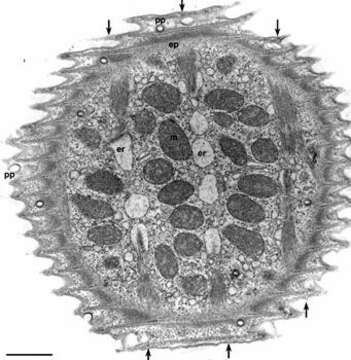
Electron micrograph of the surface of Vorticella convallaria. Like other peritrichs the pellicle is sculpted into ridges and grooves that circle the cell perpendicular to the aboral/adoral axis of the cell body. Pellicular pores (pp) penetrate the pellicle as cylindrical indentations of the plasma membrane (see Fig.3) that end as clathrin-coated pits. A system of alveoli underlies the plasma membrane and rod-like densities occupy the cytosolic tips of the pellicular ridges. A fibrous epiplasm (ep) covers the inside of the alveoli. Distinct myonemal bands run in the aboral to adoral direction (indicated by arrows) just inside the epiplasm. These bands are associated with the endoplasmic reticulum (er) by unique specializations of the ER (see Fig 17). In this EM preparation the ER has artifactually segmented into vesicles caused by the fixation process. Mitochondria (m) occupy the space between the bands. EM taken on 4/15/71 by R. Allen with Hitachi HU11A TEM. Neg. 9,250X. Bar = 1 micron. This image is available in Richard Allen's collection.
Included On The Following Pages:
- Life (creatures)
- Cellular (cellular organisms)
- Eukaryota (eukaryotes)
- SAR (Stramenopiles, Alveolates, Rhizaria)
- Alveolata (alveolates)
- Ciliophora (ciliates)
- Intramacronucleata
- Oligohymenophorea
- Peritrichia
- Sessilida
- Vorticellidae
- Vorticella (bell animalcules)
- Vorticella convallaria
This image is not featured in any collections.
Source Information
- license
- cc-by-nc
- author
- R. D. Allen
- provider
- micro*scope
- original
- original media file
- visit source
- partner site
- micro*scope
- ID