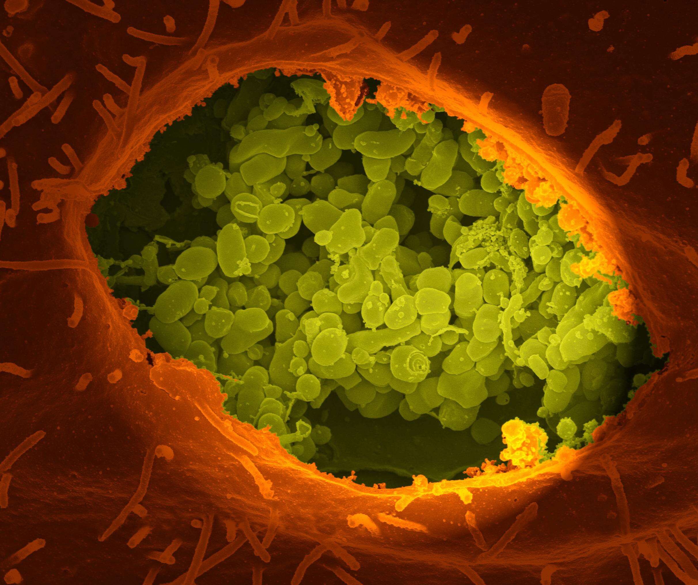
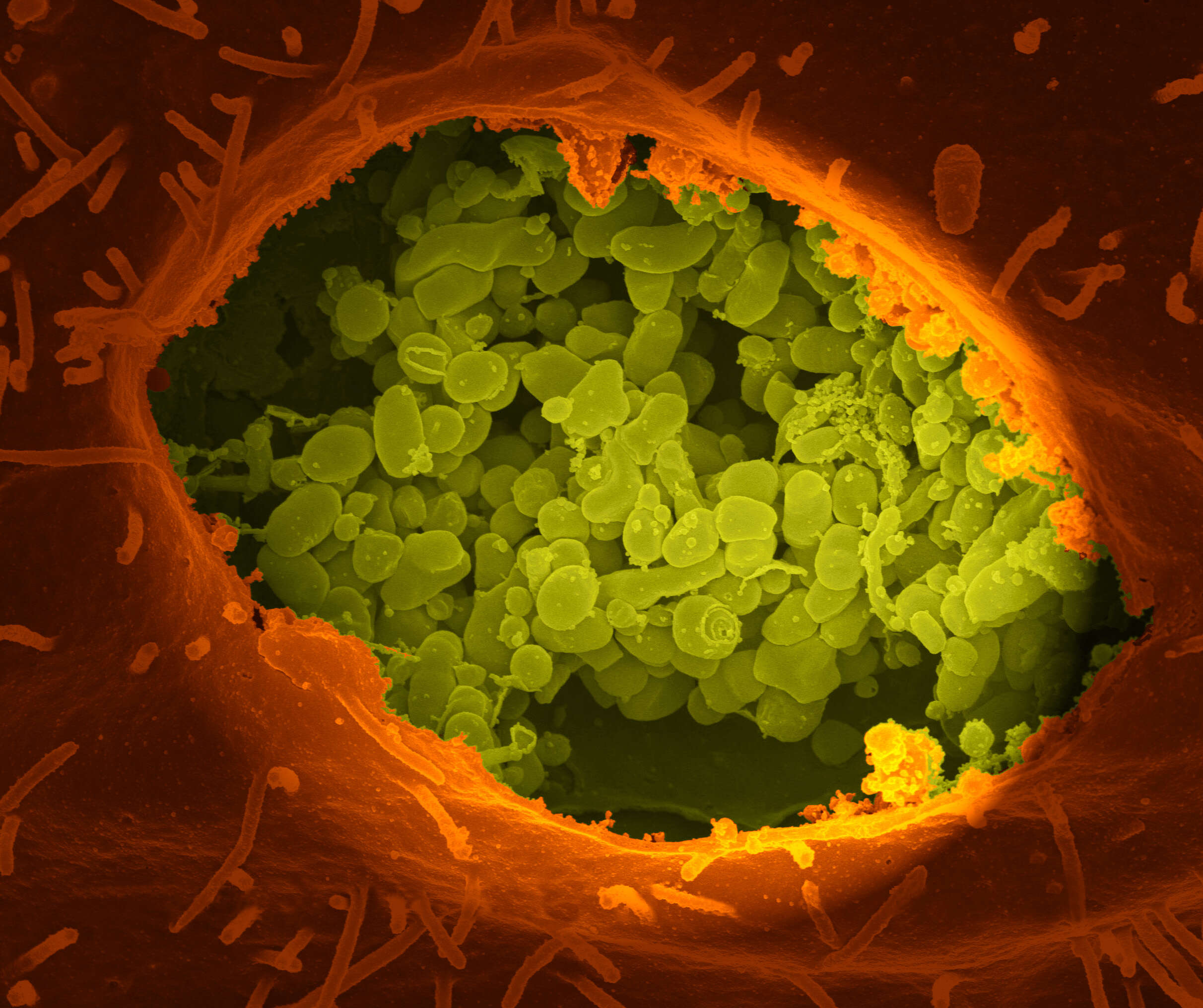
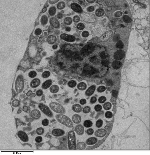
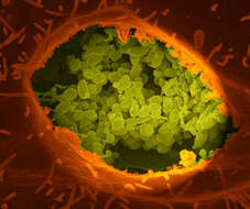
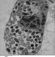
Summary.mw-parser-output table.commons-file-information-table,.mw-parser-output.fileinfotpl-type-information{border:1px solid #a2a9b1;background-color:#f8f9fa;padding:5px;font-size:95%;border-spacing:2px;box-sizing:border-box;margin:0;width:100%}.mw-parser-output table.commons-file-information-table>tbody>tr,.mw-parser-output.fileinfotpl-type-information>tbody>tr{vertical-align:top}.mw-parser-output table.commons-file-information-table>tbody>tr>td,.mw-parser-output table.commons-file-information-table>tbody>tr>th,.mw-parser-output.fileinfotpl-type-information>tbody>tr>td,.mw-parser-output.fileinfotpl-type-information>tbody>tr>th{padding:4px}.mw-parser-output.fileinfo-paramfield{background:#ccf;text-align:right;padding-right:0.4em;width:15%;font-weight:bold}.mw-parser-output.commons-file-information-table+table.commons-file-information-table,.mw-parser-output.commons-file-information-table+div.commons-file-information-table>table{border-top:0;padding-top:0;margin-top:-8px}@media only screen and (max-width:719px){.mw-parser-output table.commons-file-information-table,.mw-parser-output.commons-file-information-table.fileinfotpl-type-information{border-spacing:0;padding:0;word-break:break-word;width:100%!important}.mw-parser-output.commons-file-information-table>tbody,.mw-parser-output.fileinfotpl-type-information>tbody{display:block}.mw-parser-output.commons-file-information-table>tbody>tr>td,.mw-parser-output.commons-file-information-table>tbody>tr>th,.mw-parser-output.fileinfotpl-type-information>tbody>tr>td,.mw-parser-output.fileinfotpl-type-information>tbody>tr>th{padding:0.2em 0.4em;text-align:left;text-align:start}.mw-parser-output.commons-file-information-table>tbody>tr,.mw-parser-output.fileinfotpl-type-information>tbody>tr{display:flex;flex-direction:column}.mw-parser-output.commons-file-information-table+table.commons-file-information-table,.mw-parser-output.commons-file-information-table+div.commons-file-information-table>table{margin-top:-1px}.mw-parser-output.fileinfo-paramfield{box-sizing:border-box;flex:1 0 100%;width:100%}} Description: A dry fracture of a Vero cell exposing the contents of a vacuole where Coxiella burnetii are busy growing. Credit: NIAID. Date: 18 March 2005, 12:23. Source: Coxiella burnetii, the Bacteria That Cause Q Fever. Author: NIAID_Flickr.
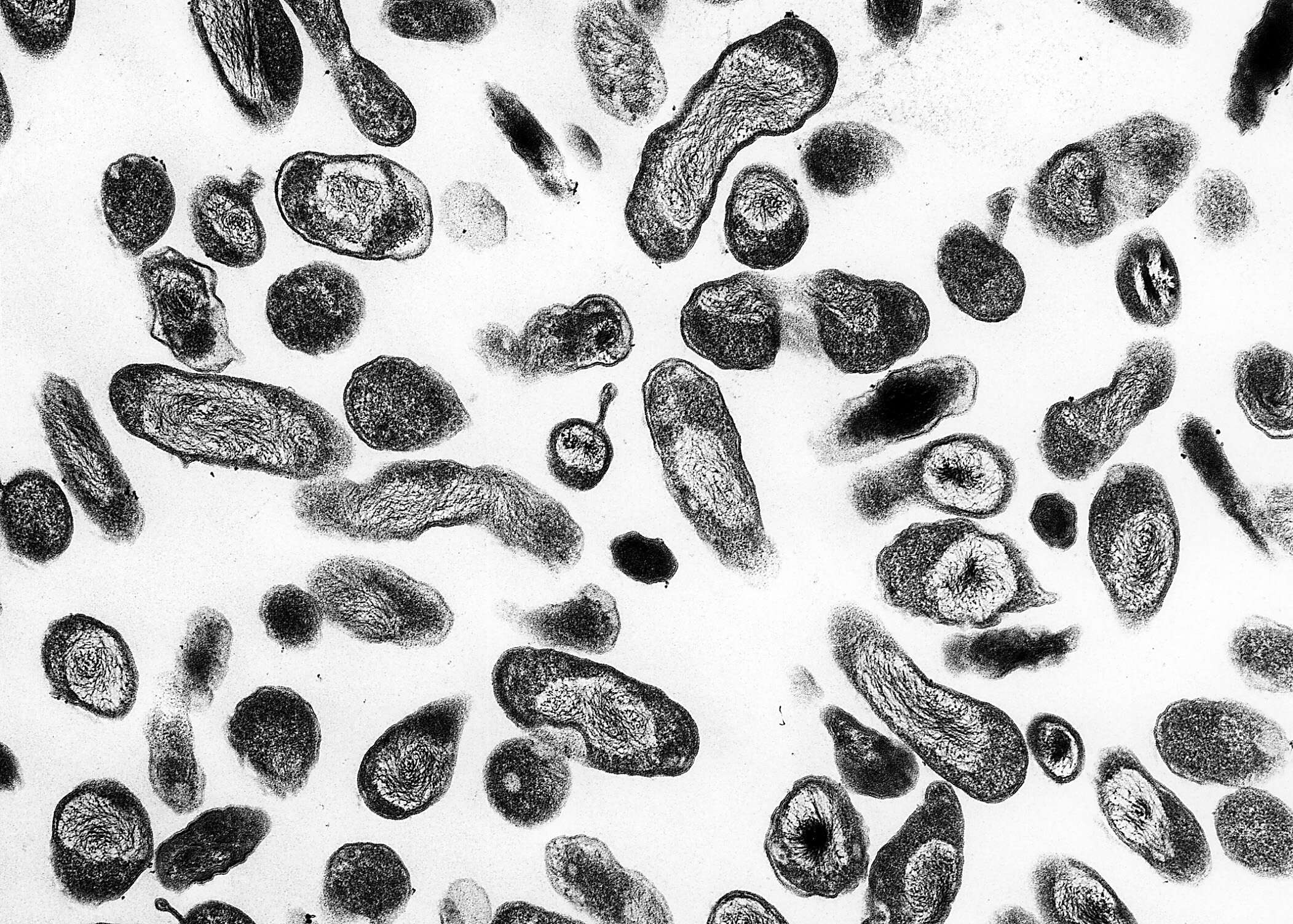
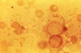
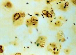
Description: English: This photomicrograph of an unknown tissue sample, revealed the presence of numerous, Gram-negative, Coxiella burnetii bacteria, which are the pathogens responsible for causing the disease, Q fever. Date: 1968. Source: https://phil.cdc.gov/Details.aspx?pid=19135. Author: CDC.
Description: English: A dry fracture of a Vero cell exposing the contents of a vacuole where Coxiella burnetii are busy growing. National Institute of Allergy and Infectious Diseases (NIAID). Date: 30 May 2013, 13:37:55. Source: National Institutes of Health (NIH). Author: National Institutes of Health (NIH).
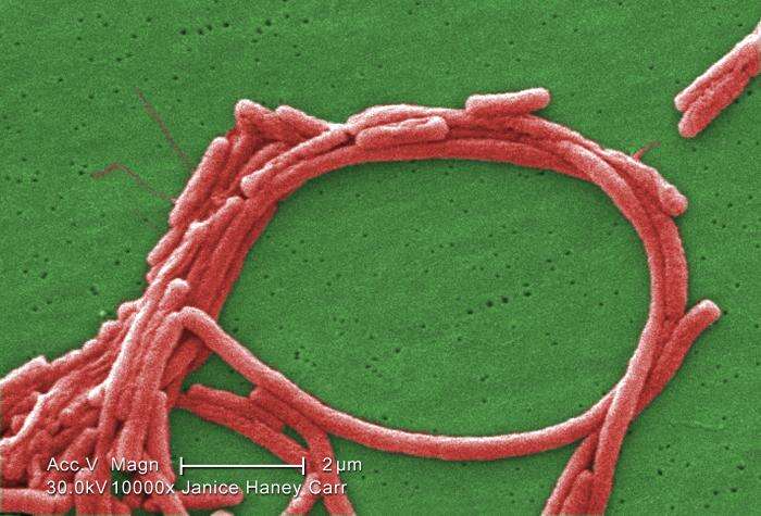
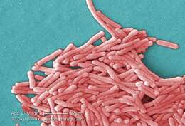
Description: English: colorized scanning electron micrograph (SEM) with moderately-high magnification of 8000X depicting a large grouping of Gram-negative Legionella pneumophila bacteria Русский: Цветная электросканограмма (х8000). Видны колонии Legionella pneumophila. Date: 2009. Source: : This media comes from the Centers for Disease Control and Prevention's Public Health Image Library (PHIL), with identification number #11150. Note: Not all PHIL images are public domain; be sure to check copyright status and credit authors and content providers. English | Slovenščina | +/−. Author: Janice Haney Carr; provided by CDC/ Margaret Williams, PhD; Claressa Lucas, PhD;Tatiana Travis, BS.
Description: ID#:934 Legionella pneumophila multiplying inside a cultured human lung fibroblast. Multiple intracellular bacilli, including dividing bacilli, are visible in longitudinal and cross section. Transmission electron micrograph. Date: 1979. Source: http://phil.cdc.gov/PHIL_Images/03101999/00015/AIDS19bb_lores.jpg. Author: CDC/Dr. Edwin P. Ewing, Jr. Permission(Reusing this file): Copyright Restrictions: None - This image is in the public domain and thus free of any copyright restrictions. As a matter of courtesy we request that the content provider be credited and notified in any public or private usage of this image.
Description: English: TEM image of cell infected with a number of Legionella pneumophila bacteria. Date: 19 October 2011. Source: TEM image of infected phagocytic cell Previously published: No. Author: Clares back.
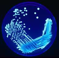
Description: English: A silver stain of Legionella pneumophila, the bacteria that causes Legionellosis. Although I got this image from a commercial website it is clearly labeled as from the CDC. This website routinely uses images from Wikipedia, which is a good thing, so no issue should be taken with using an presumably public domain image from their website. Date: 28 May 2010. Source: www.usmlerx.com. Author: William Cherry.
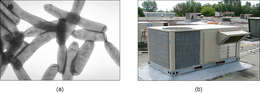
Description: English: Magnified 10000X, this digitally colorized scanning electron microscopic (SEM) image shows a group of Legionella pneumophila bacteria; some seemed to display flagella emanating from their cell walls, and some exhibited an elongated-rod morphology, which L. pneumophila are known to most frequently exhibit when grown in broth. They can also elongate when plate-grown cells age, and especially when they’ve been refrigerated, as in this case. Usually, L. pneumophila are stout, fat bacilli, which was the morphology displayed by the vast majority of these organisms. These bacteria originated on a 1 week-old culture plate (+/- 1 day), forming a single colony, at 37oC, on a buffered charcoal yeast extract (BCYE) medium with no antibiotics. The original sample was acid-treated for 15 min, to minimize fungal impurities, which would have inhibited the visualization of these organisms. Date: 2009. Source: https://phil.cdc.gov/Details.aspx?pid=11108. Author: Janice Haney Carr.
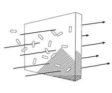
Description: Español: Gráfico del efecto barrera del Legiotex®. Date: 13 May 2013, 12:22:32. Source: Own work. Author: Legiotex. All images in this article were uploaded in the JPEG format even though it consists of non-photographic data. This information could be stored more efficiently or accurately in the PNG or SVG format. If possible, please upload a PNG or SVG version of this image without compression artifacts, derived from a non-JPEG source (or with existing artifacts removed). After doing so, please tag the JPEG version with {{Superseded|NewImage.ext}} and remove this tag. This tag should not be applied to photographs or scans. For more information, see {{BadJPEG}}.
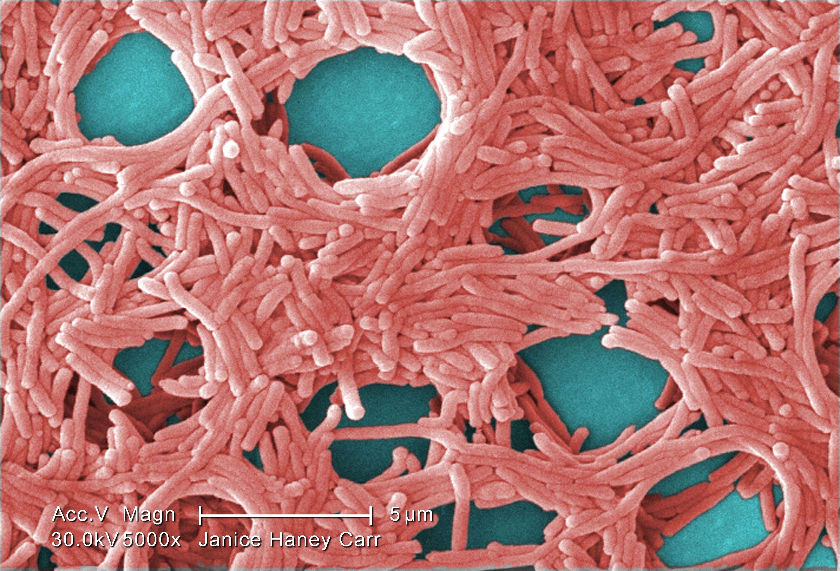
Description: English: Colorized scanning electron micrograph (SEM) with moderately-high magnification of 5000X, depicting a large grouping of Gram-negative Legionella pneumophila bacteria. Русский: Цветная электросканограмма, увеличение х5000. В поле зрения - Legionella pneumophila. Date: 2009. Source: : This media comes from the Centers for Disease Control and Prevention's Public Health Image Library (PHIL), with identification number #11148. Note: Not all PHIL images are public domain; be sure to check copyright status and credit authors and content providers. English | Slovenščina | +/−. Author: Janice Haney Carr; provided by CDC/ Margaret Williams, PhD; Claressa Lucas, PhD;Tatiana Travis, BS.
Description: English: Pronunciation recording of German noun "Legionelle", IPA: /leɡi̯oˈnɛlə/. Male voice, recorded by native German speaker from Berlin, Germany. Deutsch: Aussprachebeispiel des deutschen Substantivs "Legionelle", IPA: /leɡi̯oˈnɛlə/. Männliche Stimme, aufgenommen von deutschem Muttersprachler aus Berlin, Deutschland. Date: 26 September 2020. Source: Own work, recorded with Røde NT-USB and Audacity, converted with SoX (Sound eXchange audio editing software). Author: Jeuwre.


