11108 lores
Description:
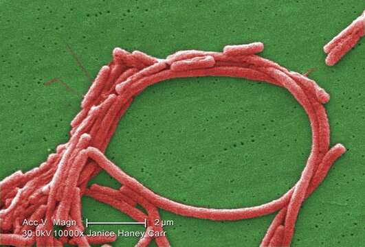
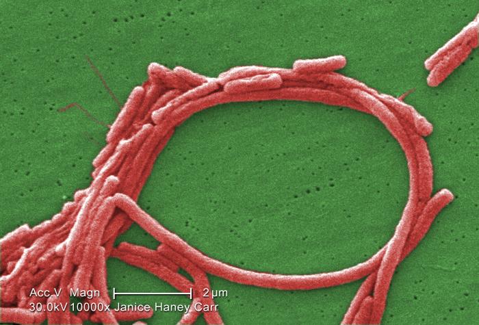
Description: English: Magnified 10000X, this digitally colorized scanning electron microscopic (SEM) image shows a group of Legionella pneumophila bacteria; some seemed to display flagella emanating from their cell walls, and some exhibited an elongated-rod morphology, which L. pneumophila are known to most frequently exhibit when grown in broth. They can also elongate when plate-grown cells age, and especially when they’ve been refrigerated, as in this case. Usually, L. pneumophila are stout, fat bacilli, which was the morphology displayed by the vast majority of these organisms. These bacteria originated on a 1 week-old culture plate (+/- 1 day), forming a single colony, at 37oC, on a buffered charcoal yeast extract (BCYE) medium with no antibiotics. The original sample was acid-treated for 15 min, to minimize fungal impurities, which would have inhibited the visualization of these organisms. Date: 2009. Source: https://phil.cdc.gov/Details.aspx?pid=11108. Author: Janice Haney Carr.
Included On The Following Pages:
- Life (creatures)
- Cellular (cellular organisms)
- Bacteria
- Proteobacteria (Purple Bacteria & relatives)
- Gammaproteobacteria
- Legionellales
- Legionellaceae
- Legionella
- Legionella pneumophila
This image is not featured in any collections.
Source Information
- license
- cc-publicdomain
- creator
- Janice Haney Carr
- source
- https://phil.cdc.gov/Details.aspx?pid=11108
- original
- original media file
- visit source
- partner site
- Wikimedia Commons
- ID