-
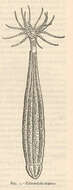
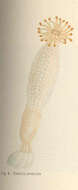
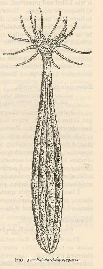
Edwardsia elegans.
-
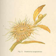
Anemonia sargassensis.
-
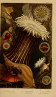
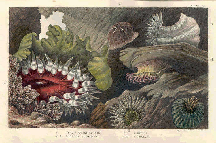
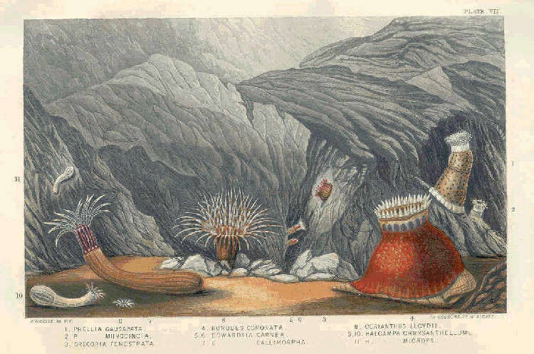
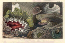
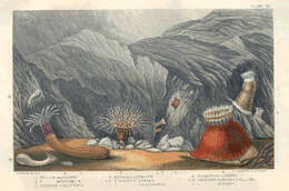
1. Tealia Crassicornis, 2. 3. Bunodes Cemmacea, 4. S. Ballii; 5. 6. S. Thallia.
-
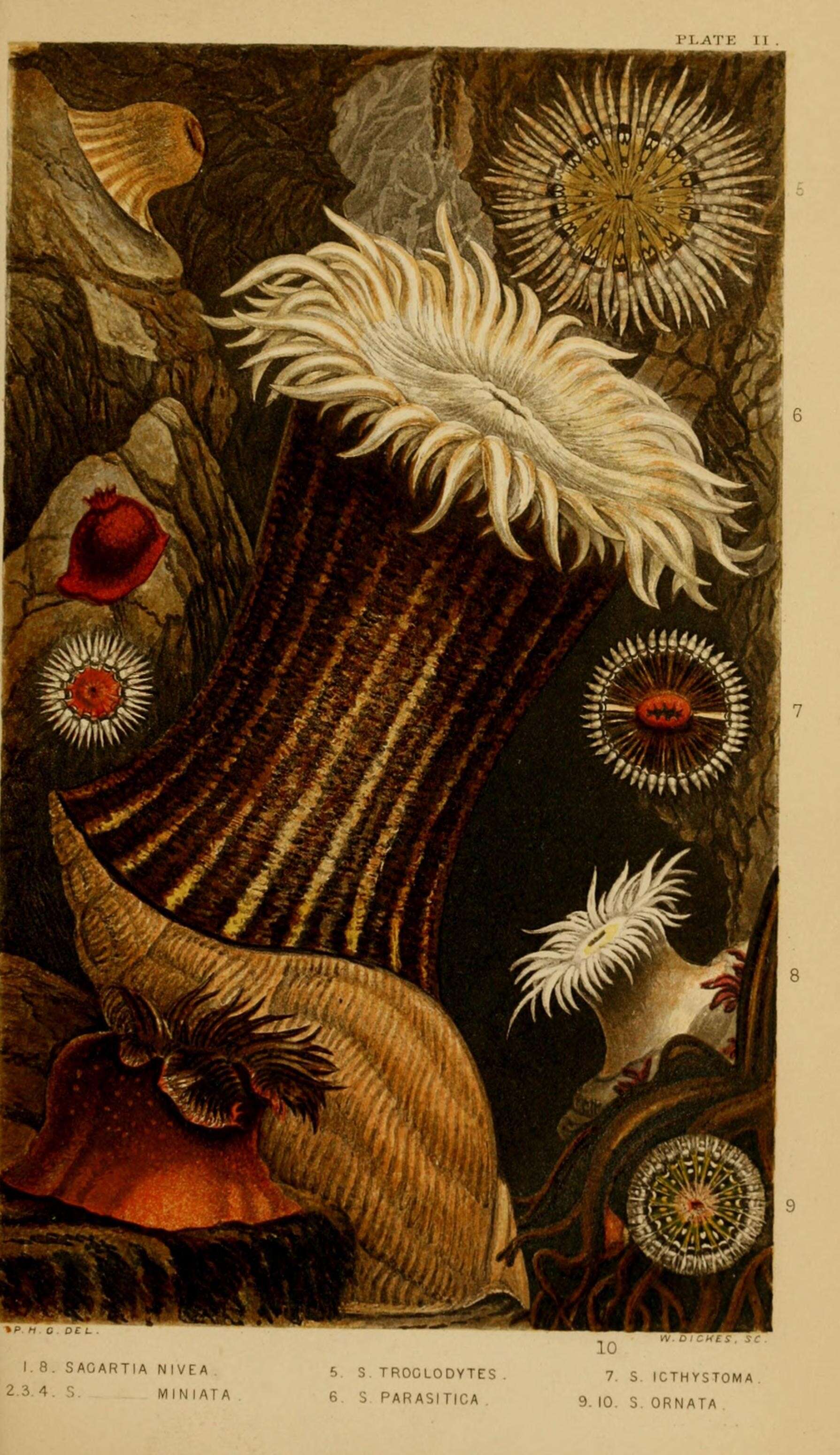
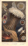
1. Sagartia Nivea, 2. 3. 4. S. Miniata,; 5. S. Troglodytes, 6. S. Parasitica, 7. S. Icthystoma .8. 9. S. Ornata.
-
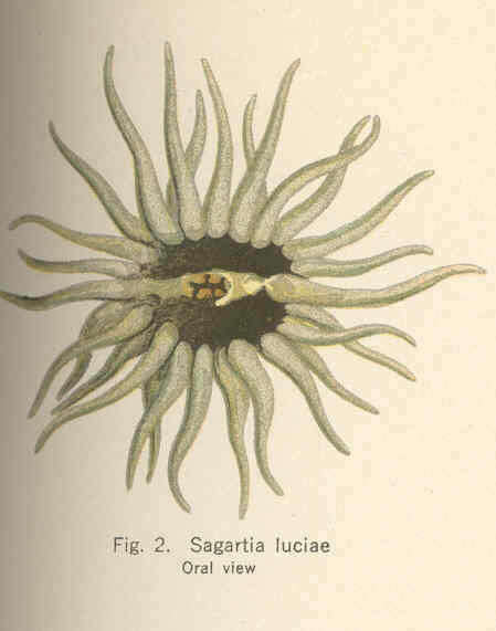
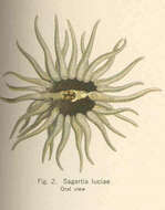
Sagartia luciae. Oral view.
-
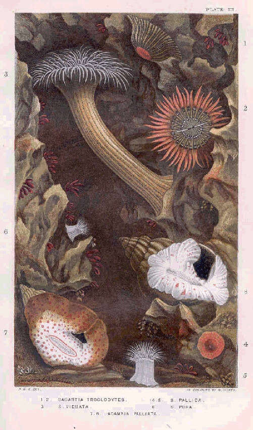
1. 2. Sagartia Troglodytes, 3. S. Viduata, 4. 5. S. Pallida, 6. S. Pura, 7. 8. Adamsia Palliata.
-
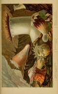
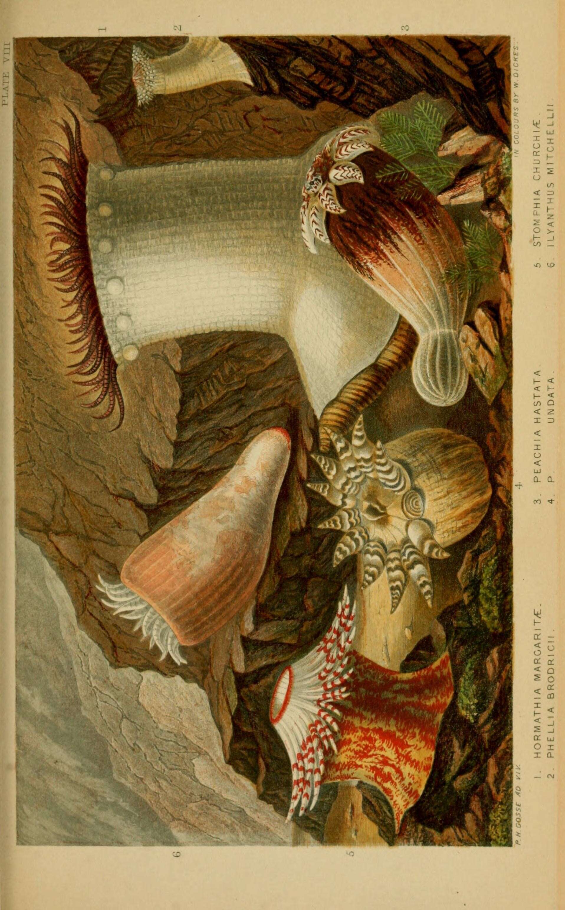
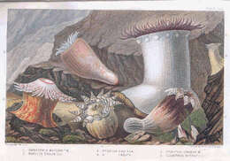
1. Hormathia Margarita, 2. Phellia Brodricii, 3. Peachia Hasata, 4. P. Undata, 5 Stomphia Churchiae, 6. Ilyanthus Mitchellii.
-
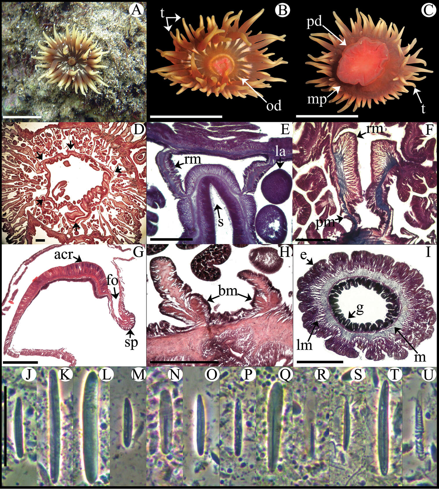
Ricardo González-Muñoz, Nuno Simões, José Luis Tello-Musi, Estefanía Rodríguez
Zookeys
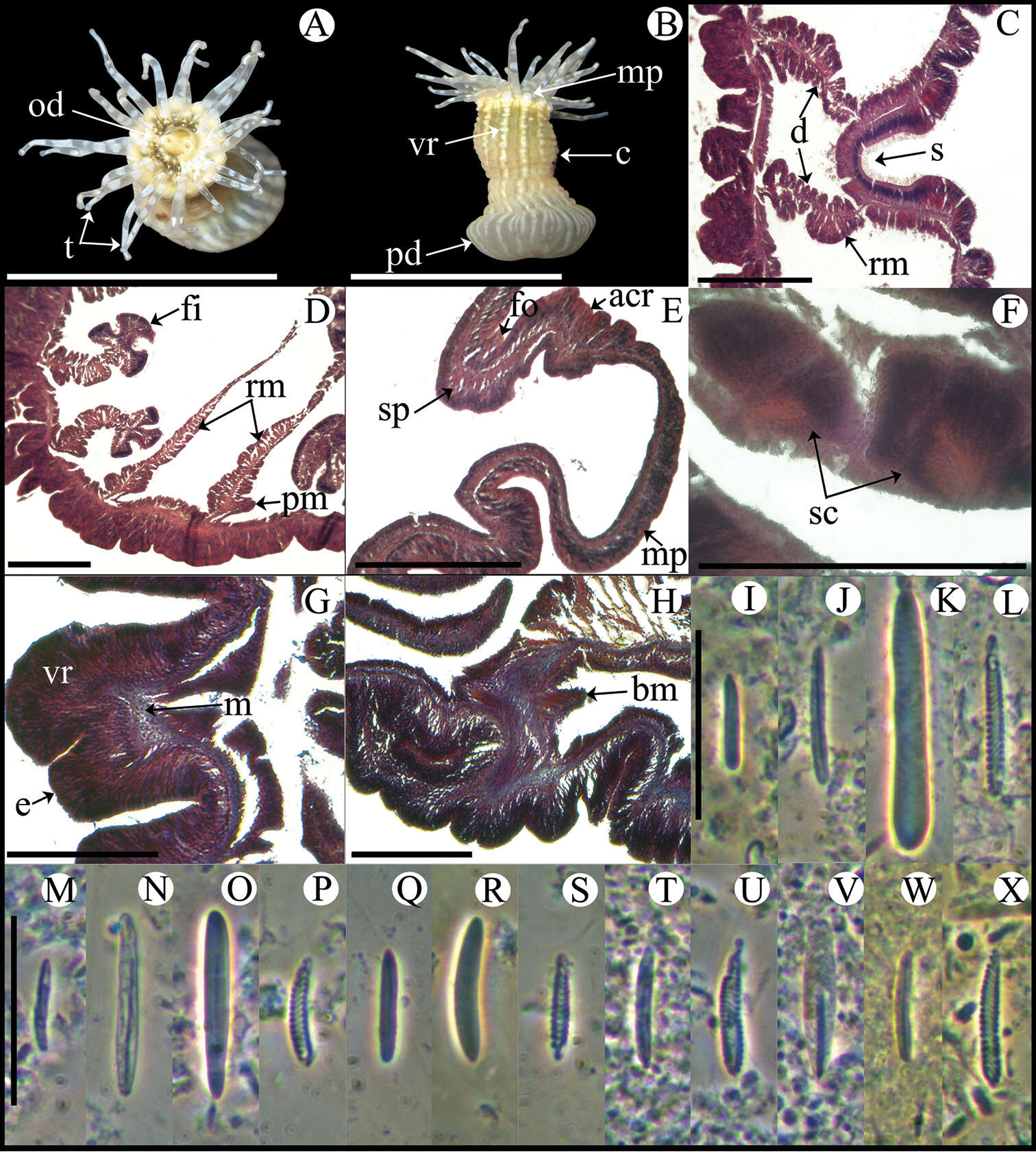
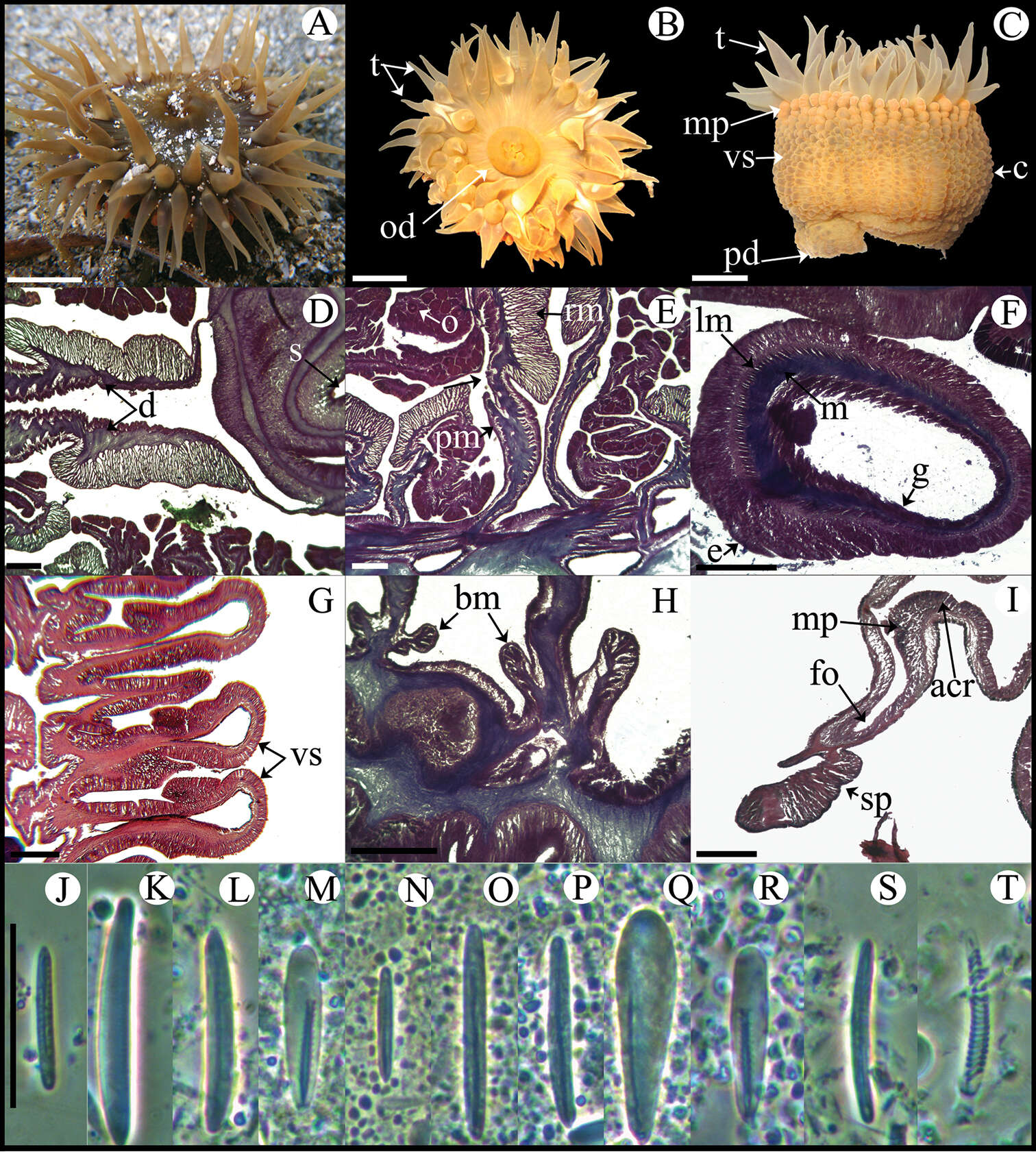
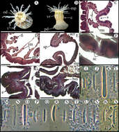
Figure 3.Anemonia sargassensis. A Live specimen in natural habitat B Oral view C Pedal disc view D Cross section through distal column showing mesenteries; arrows indicate siphonoglyphs E Detail of cross section through distal column showing a siphonoglyph F Detail of retractor and parietobasilar muscles G Longitudinal section through margin showing acrorhagi and marginal sphincter muscle H Longitudinal section through base showing basilar muscles I Cross section through tentacle J–U Cnidae.– acrorhagi: J small basitrich K basitrich L holotrich; actinopharynx: M small basitrich N microbasic p-mastigophore; column: O basitrich; filaments: P basitrich Q microbasic b-mastigophore R microbasic p-mastigophore; tentacle: S small basitrich T basitrich U spirocyst. Abbreviations.– acr: acrorhagi, bm: basilar muscle, e: epidermis, fo: fosse, g: gastrodermis, la: larvae, lm: longitudinal muscle, m: mesoglea, mp: marginal projection, od: oral disc, pd: pedal disc, pm: parietobasilar muscle, rm: retractor muscle, s: siphonoglyph, sp: sphincter, t: tentacle. Scale bars: A–C: 10 mm; D–I: 200 μm; J–U: 25 μm.
-
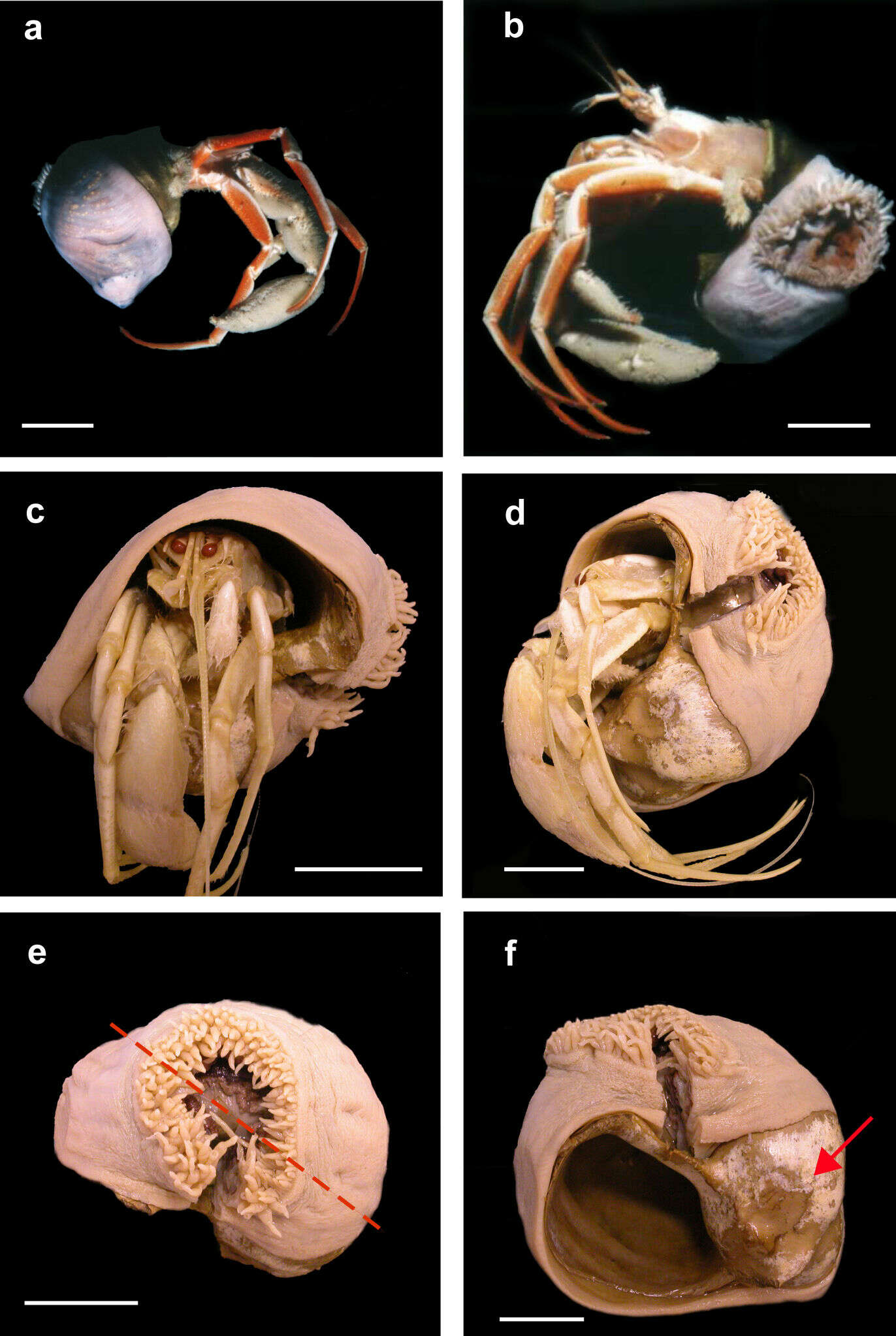
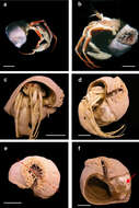
Andrea L. Crowther, Daphne G. Fautin, Carden C. Wallace
Zookeys
Figure 1.Stylobates birtlesi sp. n. holotype MTQ G57579 a, b soon after collection (photo: RA Birtles) c, d preserved specimen with Sympagurus trispinosus showing position of oral disc of anemone e preserved specimen: shortest tentacles beside longest ones (on right side of oral disc in this view); tentacles grade in length between longest and shortest around other side of oral disc (dashed line indicates directive axis) f preserved specimen without hermit crab showing aperture and part of carcinoecium not covered by anemone (arrow). Scale bars 20 mm.
-
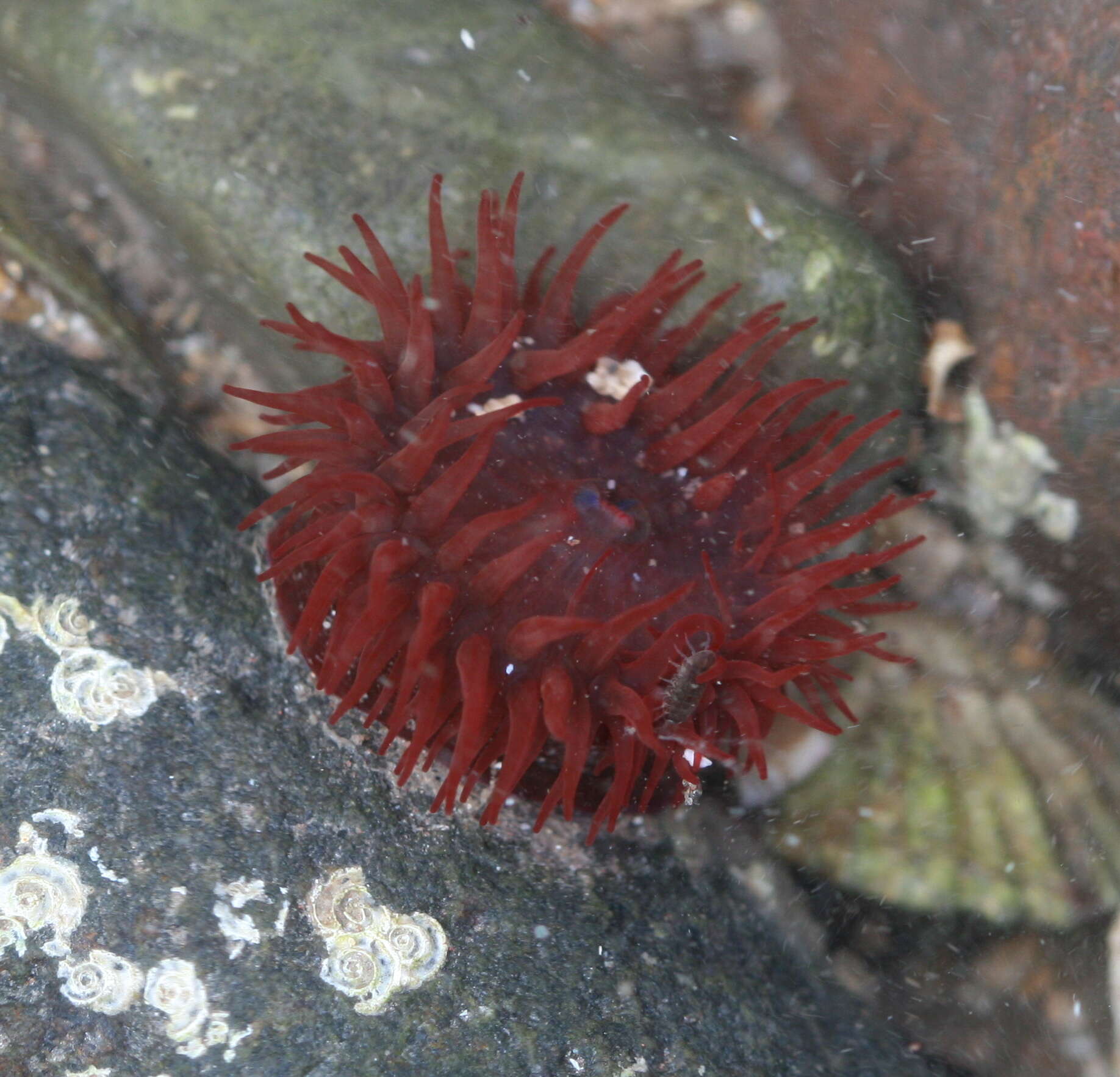
Tregonhawke, England, United Kingdom
-
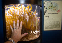
Condylactis gigantea is a tropical species of sea anemone that is found in coral reefs, shallow ocean waters, in shore areas in the Caribbean Sea - most specifically the West Indies - and the Western Atlantic Sea, ranging from southern Florida through the Florida keys. It is also commonly known as : Giant Caribbean Sea Anemone, Giant Golden Anemone, Condylactis Anemone, Haitian anemone, Pink tipped anemone, Purple tipped anemone, and Florida Condy. This species, can be easily seen growing in lagoons, or on inner reefs as either individuals or loose groups, but never as colonies.
-
-
-
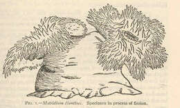
Metridium dianthus. Specimen in process of fission.
-
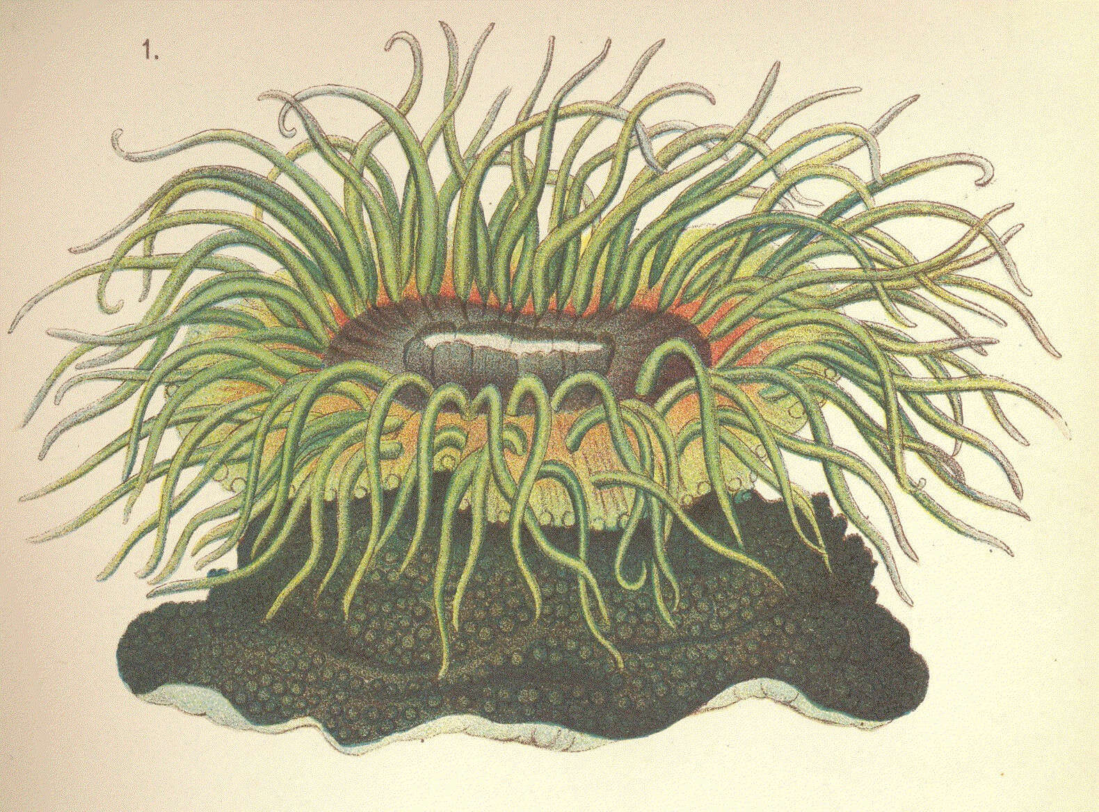
Phymactis veratra.
-
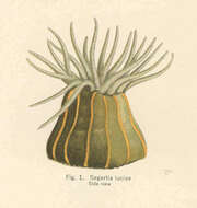
Sagartia luciae. Side view.
-
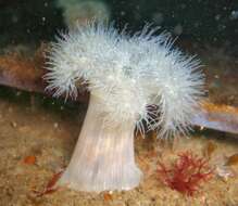
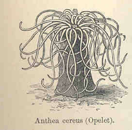
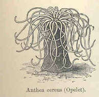
Anthea cereus (Opelet).
-
1. Hormathia Margarita, 2. Phellia Brodricii, 3. Peachia Hasata, 4. P. Undata, 5 Stomphia Churchiae, 6. Ilyanthus Mitchellii.
-
Ricardo González-Muñoz, Nuno Simões, José Luis Tello-Musi, Estefanía Rodríguez
Zookeys
Figure 4.Anthopleura pallida. A Oral view B Lateral view C Detail of directives and siphonoglyph D Cross section through proximal column E Longitudinal section through margin showing acrorhagi and marginal sphincter muscle F Detail of spermatic cysts G Longitudinal section through distal column showing one verruca H Longitudinal section through base showing basilar muscles I–X Cnidae.– acrorhagi: I small basitrich J basitrich K holotrich L spirocyst; actinopharynx: M small basitrich N basitrich O microbasic b-mastigophore P spirocyst; column: Q small basitrich R basitrich S spirocyst; filament: T basitrich U spirocyst V microbasic p-mastigophore; tentacle: W basitrich X spirocyst. Abbreviations.– acr: acrorhagi, bm: basilar muscle, c: column, d: directives, fo: fosse, mp: marginal projection, od: oral disc, pd: pedal disc, pm: parietobasilar muscle, rm: retractor muscle, s: siphonoglyph, sc: spermatic cyst, sp: sphincter, t: tentacles, vr: verruca. Scale bars: A–B: 10 mm; C–H: 200 μm; I–X: 25 μm.
-
Andrea L. Crowther, Daphne G. Fautin, Carden C. Wallace
Zookeys
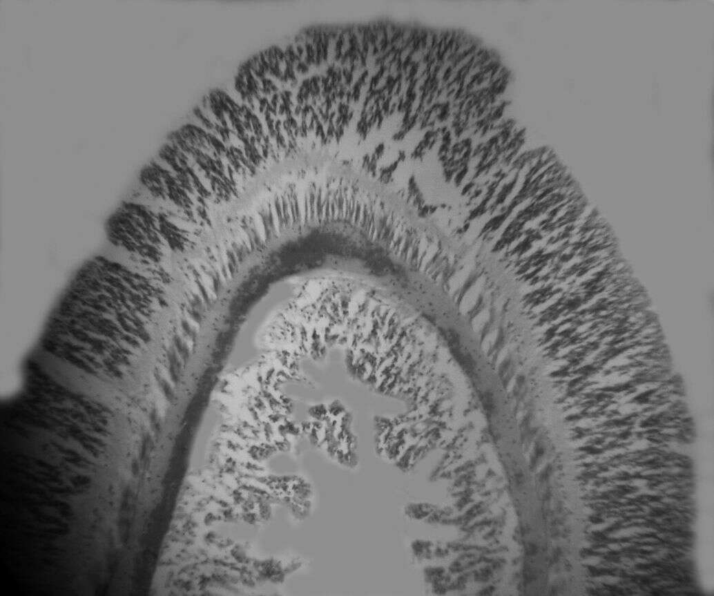
Figure 2.Longitudinal section through tentacle of Stylobates birtlesi sp. n. paratype MTQ G57580.
-
Dunbar, Scotland, United Kingdom
-
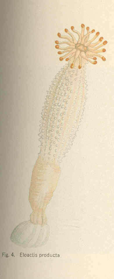
Eloactis producta.
-
Ricardo González-Muñoz, Nuno Simões, José Luis Tello-Musi, Estefanía Rodríguez
Zookeys
Figure 5.Bunodosoma cavernatum. A Live specimen in natural habitat B Oral view C Lateral view D Detail of directives; notice siphonoglyph E Cross section through proximal column showing oocytes F Cross section through tentacle G Longitudinal section through column showing vesicles H Longitudinal section though base showing basilar muscles I Longitudinal section through margin showing acrorhagi and marginal sphincter muscle J–T Cnidae.– acrorhagi: J basitrich K holotrich; actinopharynx: L basitrich M microbasic p-mastigophore; column: N small basitrich O basitrich; filament: P basitrich Q microbasic b-mastigophore R microbasic p-mastigophore; tentacle S basitrich T spirocyst. Abbreviations.– acr: acrorhagi, bm: basilar muscle, c: column, d: directives, e: epidermis, fo: fosse, g: gastrodermis, lm: longitudinal muscles, m: mesoglea, mp: marginal projection, o: oocyst, od: oral disc, pd: pedal disc, pm: parietobasilar muscle, rm: retractor muscle, s: siphonoglyph, sp: sphincter, t: tentacle, vs: vesicles. Scale bars: A–C: 10 mm; D–I: 200 μm; J–T: 25 μm.
-
Andrea L. Crowther, Daphne G. Fautin, Carden C. Wallace
Zookeys
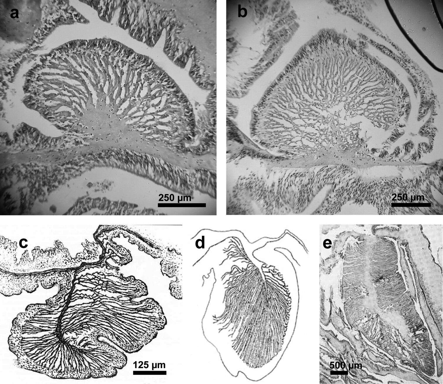
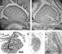
Figure 3.Endodermal circumscribed marginal sphincter muscles of Stylobates spp. a, b Palmate marginal sphincter muscle of Stylobates birtlesi sp. n. a paratype MTQ G57581 b paratype KUDIZ 003352 c-e Pinnate marginal sphincter muscles. c Stylobates aeneus (from Dunn et al. 1981) d Stylobates cancrisocia (from Carlgren 1928a) e Stylobates loisetteae (from Fautin 1987).