À propos
Éducation
Discuter
TraitBank
Se connecter
S’inscrire
Langue
Deutsch
English
Español
français
italiano
Nederlands
Piemontèis
Português do Brasil
suomi
Türkçe
Čeština
Ελληνικά
македонски
Українська
العربية
简体中文
繁體中文
noms dans le fil d’Ariane
vernaculaire
scientifique
À propos
Éducation
Discuter
TraitBank
Se connecter
S’inscrire
fr
Deutsch
English
Español
français
italiano
Nederlands
Piemontèis
Português do Brasil
suomi
Türkçe
Čeština
Ελληνικά
македонски
Українська
العربية
简体中文
繁體中文
noms dans le fil d’Ariane
vernaculaire
scientifique
Life
»
…
»
Alveolata
»
Dinoflagellés
»
…
»
Prorocentraceae
»
…
Life
»
Cellular
»
Eukaryota
»
SAR (Stramenopiles, Alveolates, Rhizaria)
»
Alveolata
»
Dinoflagellés
»
Prorocentrales
»
Prorocentraceae
»
Prorocentrum
«
Prorocentrum micans
recueillir
vue d’ensemble
données
média
articles
cartes
noms
licence
n’importe quelle licence
CC-BY
CC-BY-NC
CC-BY-NC-SA
No copyright
fournisseur
n’importe quel fournisseur
iNaturalist
Flickr Group
Harmful Phytoplankton Project
NMNH Marine Dinoflagellates
micro*scope
cc-publicdomain
fiable
cc-by-nc-sa-3.0
fiable
cc-by-nc-4.0
fiable
cc-by-nc-2.0
fiable
cc-by
fiable
cc-by-nc
fiable
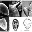
Plate 46
cc-publicdomain
NMNH Marine Dinoflagellates
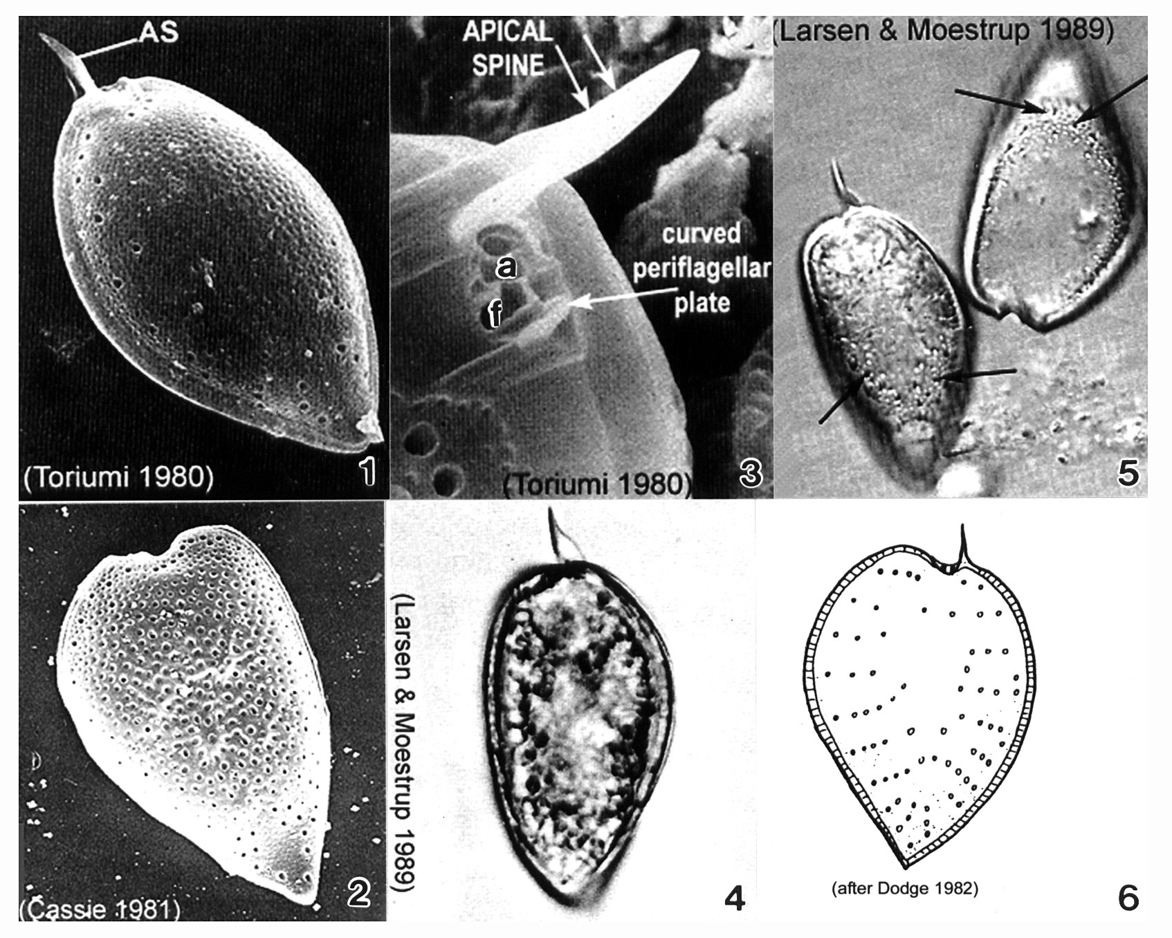
Plate 46. Prorocentrum micans. Figs. 1-3. SEM. Fig. 1. Right valve: cell tear-drop shaped; rounded anteriorly, pointed posteriorly, broadest in the middle. Apical spine (AS) winged. Rugose thecal surface. Intercalary band smooth and wide. Fig. 2. Heart-shaped cell. Apical spine missing. Fig. 3. Periflagellar area: small, shallow triangular depression on right valve. Flagellar (f) and auxiliary (a) pores present; curved periflagellar plate adjacent to f. Large winged AS directly opposite. Figs. 4-5. LM: Left valve. Winged AS visible. Fig. 5. Empty theca with visible trichocyst pores (arrows). Fig. 6 Line drawing: trichocyst pore arrangement.
Image de Prorocentraceae
cc-by-nc-sa-3.0
University of Liverpool
Harmful Phytoplankton Project
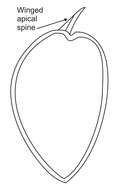
Fig 1: Prorocentrum micans Schematic diagram (ventral view) redrawn from Tomas et al. 1997.
"
cc-by-nc-4.0
paigepriester
iNaturalist
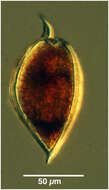
Procentrum micans
cc-by-nc-2.0
tintinnidguy
Flickr Group
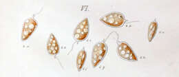
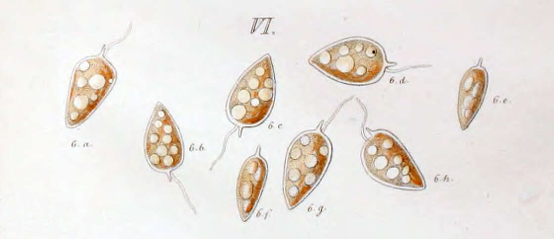
Prorocentrum micans first depiction by Ehrenberg in 1834
cc-by
tintinnidguy
Flickr Group
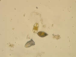
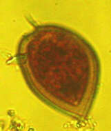
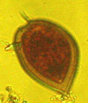
Lugol preservation
cc-by-nc
micro*scope
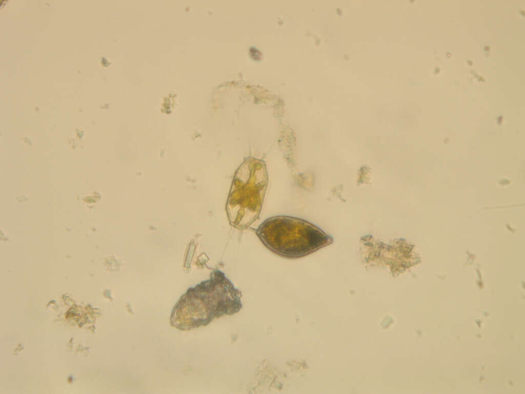
Members of this species have a fusiform body. The apex bears a prominent winged spine. In valve view the cell will have one arched and one convex side.