Plate 46
Description :
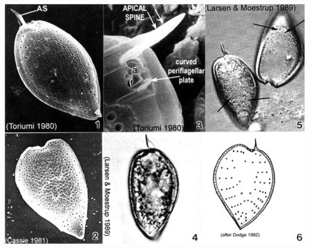
Plate 46. Prorocentrum micans. Figs. 1-3. SEM. Fig. 1. Right valve: cell tear-drop shaped; rounded anteriorly, pointed posteriorly, broadest in the middle. Apical spine (AS) winged. Rugose thecal surface. Intercalary band smooth and wide. Fig. 2. Heart-shaped cell. Apical spine missing. Fig. 3. Periflagellar area: small, shallow triangular depression on right valve. Flagellar (f) and auxiliary (a) pores present; curved periflagellar plate adjacent to f. Large winged AS directly opposite. Figs. 4-5. LM: Left valve. Winged AS visible. Fig. 5. Empty theca with visible trichocyst pores (arrows). Fig. 6 Line drawing: trichocyst pore arrangement.
Inclus dans les pages suivantes :
- Life
- Cellular
- Eukaryota
- SAR (Stramenopiles, Alveolates, Rhizaria)
- Alveolata
- Dinophyceae (Dinoflagellés)
- Prorocentrales
- Prorocentraceae
- Prorocentrum
- Prorocentrum micans
- Dinoflagellata
Cette image ne figure dans aucune collection.
Informations sur la provenance
- licence
- cc-publicdomain
- citation bibliographique
- Faust, Maria A. and Rose A. Gulledge. Identifying Harmful Marine Dinoflagellates. Smithsonian Contributions from the United States National Herbarium, volume 42: 1-144 (including 48 plates, 1 figure and 1 table).
- original
- fichier de média d’origine
- visiter la source
- site partenaire
- NMNH Marine Dinoflagellates
- ID