-
Centers for Disease Control/Division of Parasitic Diseases and Malaria
EOL staff
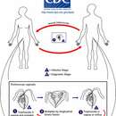
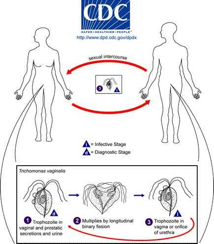
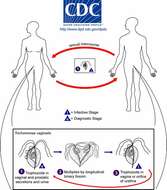
Life cycle of Trichomonas vaginalis, the cause of trichomoniasis in humansTrichomonas vaginalis resides in the female lower genital tract and the male urethra and prostate (1), where it replicates by binary fission (2). The parasite does not appear to have a cyst form, and does not survive well in the external environment. Trichomonas vaginalis is transmitted among humans, its only known host, primarily by sexual intercourse (3).From
Centers for Disease Control Parasites and Health website
-
Centers for Disease Control/Division of Parasitic Diseases and Malaria
EOL staff
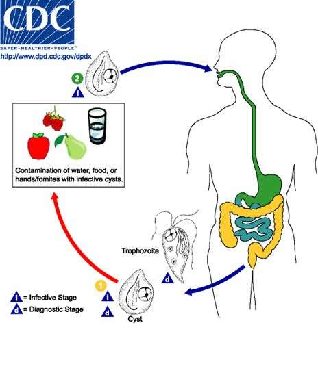
Life cycle of Chilomastix mesnili The resistant cyst stage in the life cycle of Chilomastix is responsible for transmission. Both cysts and trophozoites can be found in the feces (diagnostic stages) (1). Infection occurs by the ingestion of cysts in contaminated water or food or by the fecal-oral route (via hands or fomites, i.e., inanimate objects such as towels that transmit infectious organisms to a host) (2). In the large (and possibly small) intestine, excystation releases trophozoites.From
Centers for Disease Control Parasites and Health website.
-
Centers for Disease Control/Division of Parasitic Diseases and Malaria
EOL staff
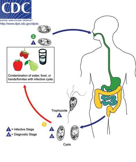
Life cycle of Enteromonas hominisBoth cysts (dormant stage) and trophozoites (active stage) of Enteromonas hominis are shed in feces. Infection occurs after the ingestion of cysts in fecal-contaminated food or water, or on fomites (inanimate objects or substances capable of transferring pathogens). In the large (and possibly small) intestine, excystation releases trophozoites. Enteromonas hominis resides in the large intestine, where it is regarded as a commensal (benefiting from its host but doing no harm) and is not known to cause disease.From
Centers for Disease Control Parasites and Health website.
-
Centers for Disease Control/Division of Parasitic Diseases and Malaria
EOL staff
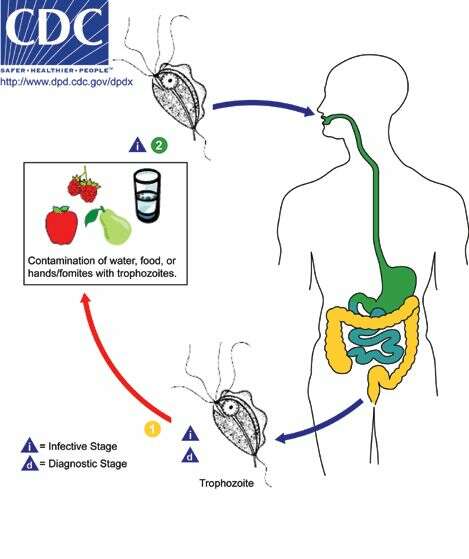
Life cycle of the flagellate Pentatrichomonas hominisPentatrichomonas hominis is a trichomonad flagellate with a worldwide distribution. Only trophozoites are shed in feces (1) as there is no known cyst stage for this species. Infection occurs after the ingestion of trophozoites in fecally-contaminated food or water or on fomites (i.e., other non-living objects or substances that can transmit them) (2). These organisms reside in the large intestine, where they are regarded as commensals (i.e., benefiting from but not harming their host) and are not known to cause disease in humans.From
Centers for Disease Control Parasites and Health website
-
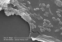
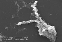
This scanning electron micrograph (SEM) of an untreated water specimen extracted from a wild stream mainly used to control flooding during inclement weather, revealed the presence of unidentified organisms, which included bacteria, protozoa, and algae. In this particular image, a protective biofilm had been inhabited by numbers of what appeared to be unidentified bacterial microorganisms.Created: 2009
-
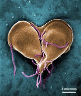
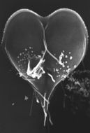
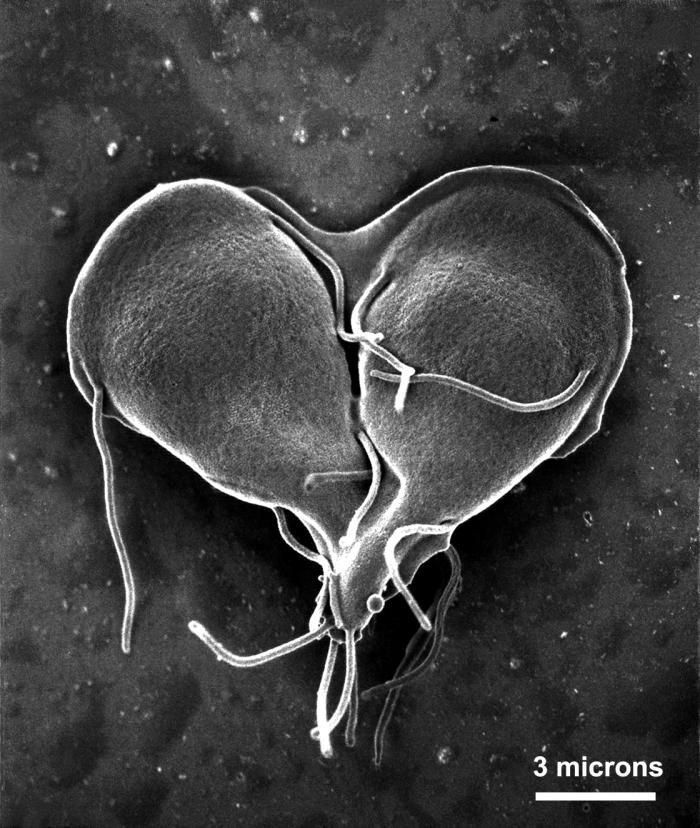
This scanning electron micrograph (SEM) depicted a Giardia lamblia protozoan that was about to become two, separate organisms, as it was caught in a late stage of cell division, producing a heart-shaped form.Created: 1999
-
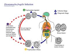
This is an illustration of the assumed life cycle of Dientamoeba fragilis, the cause of a protozoan parasitic infection.Created: 2002
-
This scanning electron micrograph (SEM) of an untreated water specimen extracted from a wild stream mainly used to control flooding during inclement weather, revealed the presence of unidentified organisms, which included bacteria, protozoa, and algae. Visible in this particular image were a number of different microorganisms including elongated diatoms, and an amorphic gelatinous biofilm mass, which had enveloped amoeboid and bacterial organisms. For a colorized version of this image, see PHIL 11712.Created: 2009
-
This is a scanning electron micrograph (SEM) of an in vitro Giardia lamblia culture, which had been cultivated in bile-free TYI-S-33 medium for 48 hrs, then incubated 24 hrs with 10 mg/ml bovine bile in order to stimulate cyst formation. This photograph contains both trophozoites, and a cluster of maturing cysts (bottom right). At far left, the two trophozoite-staged organisms are positionally situated opposite to one another, with the farthest left G. lamblia displaying its dorsal, or upper surface, and the protozoan to its immediate right, its ventral, or bottom surface.Created: 1999
-
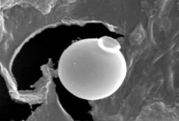
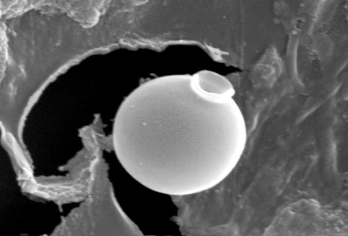
This scanning electron micrograph (SEM) of an untreated water specimen extracted from a wild stream mainly used to control flooding during inclement weather, revealed the presence of unidentified organisms, which included bacteria, protozoa, and algae. Clearly visible in the center of this image, was a exquisitely-formed unidentified round vescicle-shaped microorganism, which may have been algal, or diatomic. Shaped like an ancient Grecian urn, the almost perfectly rounded smooth, flawless surface was made even more beautiful given its delicate structure. For a colorized version of this image, see PHIL 11709.Created: 2009
-
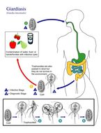
This is an illustration of the life cycle of Giardia lamblia (intestinalis), the causal agent of Giardiasis.Created: 2002
-
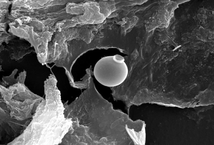
This scanning electron micrograph (SEM) of an untreated water specimen extracted from a wild stream mainly used to control flooding during inclement weather, revealed the presence of unidentified organisms, which included bacteria, protozoa, and algae. Clearly visible in the center of this image, was a exquisitely-formed unidentified round vescicle-shaped microorganism, which may have been algal, or diatomic. Shaped like an ancient Grecian urn, the almost perfectly rounded smooth, flawless surface was made even more beautiful given its delicate structure. For a colorized version of this image, see PHIL 11708.Created: 2009
-
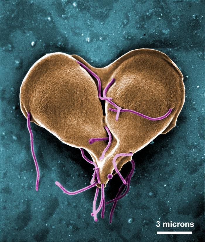
This digitally-colorized scanning electron micrograph (SEM) depicted a Giardia lamblia protozoan that was about to become two, separate organisms, as it was caught in a late stage of cell division, producing a heart-shaped form.Created: 1999
-
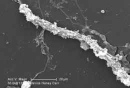
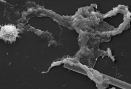
Magnified 1200X, this scanning electron micrograph (SEM) of an untreated water specimen extracted from a wild stream mainly used to control flooding during inclement weather, revealed the presence of unidentified organisms, which included bacteria, protozoa, and algae. In this particular image, an unidentified amorphous strand of mucoidal biofilm was featured, which appeared to have enmeshed numbers of amoeboid organisms.Created: 2009
-
This scanning electron micrograph (SEM) depicted a Giardia lamblia protozoan that was about to become two separate organisms, as it was caught in a late stage of cell division, producing a heart-shaped form. Note the intimate intertwining of two of the organisms eight flagella that will facilitate their motility.Created: 1986
-
This scanning electron micrograph (SEM) of an untreated water specimen extracted from a wild stream mainly used to control flooding during inclement weather, revealed the presence of unidentified organisms, which included bacteria, protozoa, and algae. Occupying most of the field of view, an unidentified amorphous mucoidal biofilm was featured, which appeared to have enmeshed numbers of amoeboid organisms, while on the left was a strangely-beautiful microorganism displaying an outer surface studded with numerous projections, making it appear like a microscopic sea urchin. See PHIL 11715 for a colorized version of this image.Created: 2009
-
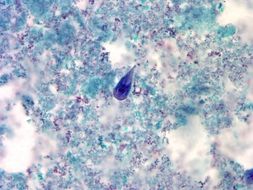
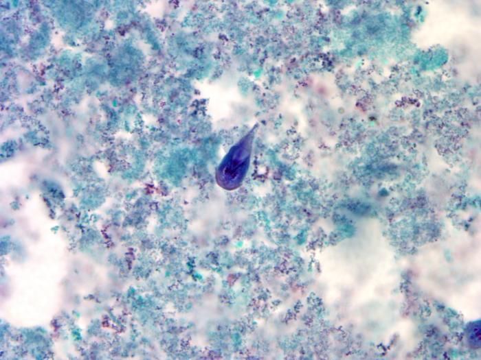
At a magnification of 1000X, this trichrome-stained photomicrograph revealed the morphologic characteristics of a blue-stained Giardia intestinalis protozoan trophozoite (center). In the small intestine, the protozoan cysts release trophozoites, with each cyst producing two trophozoites. Trophozoites multiply by longitudinal binary fission, remaining in the lumen of the proximal small bowel where they can be free, or attached to the mucosa by a ventral sucking disk. As the trophozoites mature, they are simultaneously migrating towards the colon, whereupon, they once again become thick-walled cysts, and are in this way, passed in the hosts stool into the environment. As cysts, these protozoan parasites can survive for many months until they are accidentally ingested by another unfortunate host.Created:
-
This scanning electron micrograph (SEM) of an untreated water specimen extracted from a wild stream mainly used to control flooding during inclement weather, revealed the presence of unidentified organisms, which included bacteria, protozoa, and algae. In this particular image, an unidentified amorphous mucoidal biofilm was featured, which appeared to have enmeshed numbers of amoeboid organisms.Created: 2009
-
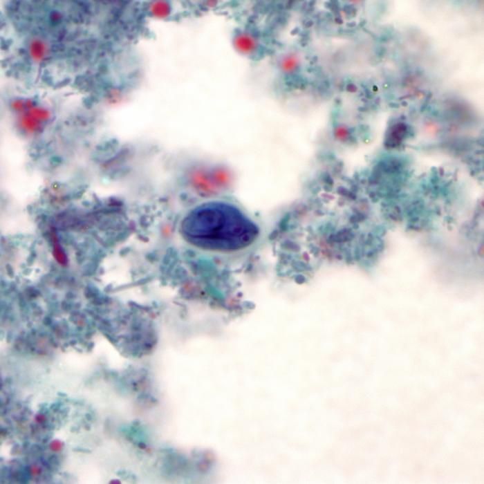
Using a trichrome stain, this photomicrograph revealed the morphologic characteristics of a blue-stained Giardia intestinalis protozoa cyst (center). The Giardia parasite lives in the intestine of infected humans or animals. Millions of cystic protozoa can be released in a bowel movement from an infected human or animal. Giardia is found in soil, food, water, or surfaces that have been contaminated with the feces from infected humans or animals. You can become infected after accidentally swallowing the parasite; you cannot become infected through contact with blood.Created:
-
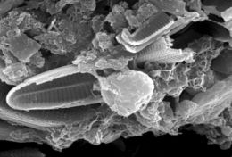
This scanning electron micrograph (SEM) of an untreated water specimen extracted from a wild stream mainly used to control flooding during inclement weather, revealed the presence of unidentified organisms, which included bacteria, protozoa, and algae. In this particular image, unidentified species of diatoms are seen to be caught up in an amorphous gelatinous biofilm, which had entrapped stream particulates as well.Created: 2009
-
This scanning electron micrograph (SEM) of an untreated water specimen extracted from a wild stream mainly used to control flooding during inclement weather, revealed the presence of unidentified organisms, which included bacteria, protozoa, and algae. In this particular image, unidentified species of diatoms are seen to be caught up in an amorphous gelatinous biofilm, which had entrapped stream particulates as well. In the center, youll note what may have been an amoeboid organism.Created: 2009
-
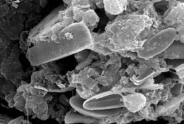
This scanning electron micrograph (SEM) of an untreated water specimen extracted from a wild stream mainly used to control flooding during inclement weather, revealed the presence of unidentified organisms, which included bacteria, protozoa, and algae. In this particular image, unidentified sheets of algae were wrapped in a mass of what appeared to be a mucoid amorphous biofilm. See PHIL 11713 for a colorized version of this image.Created: 2009
-
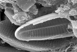
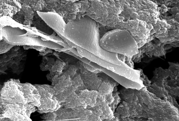
This scanning electron micrograph (SEM) of an untreated water specimen extracted from a wild stream mainly used to control flooding during inclement weather, revealed the presence of unidentified organisms, which included bacteria, protozoa, and algae. In this particular image, a number of unidentified oblong elliptical-shaped diatoms were featured, along side amorphically-shaped masses of organically-composed biofilm.Created: 2009
-
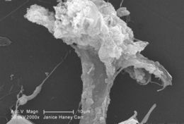
At a magnification of 2000X, this scanning electron micrograph (SEM) of an untreated water specimen extracted from a wild stream, which is mainly used to control flooding during inclement weather, revealed the presence of unidentified organisms, which included bacteria, protozoa, and algae. In this particular image, an expanding amorphous organic biofilm was featured within which numbers of amoeboid protozoa seemed to be embedded. For a colorized version of this image see PHIL 11714.Created: 2009