-

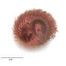
Infraciliature (posterior view, dorsal at top) of the colpodid ciliate, Maryna ovata GELEI, 1950. Cell size is highly variable (length 25-110 mm). The cells are mushroom or champagne cork-shaped. The conical anterior end is termed the calyx and the truncate cylindrical posterior part the uvula. There is a definite horseshoe-shaped sulcus oriented perpendicular to the long axis between the calyx and uvula. The infraciliature is complex, twisting around the calyx and running posteriorly down onto the uvula (as seen here). There is a semicircular array of longer terminal cilia along the posterior margin of the uvula (their kinetids are seen here). Some descriptions (e.g. Curds, C.R. British and Other Freshwater Ciliated Protozoa. Part I, p.182. Cambridge, Bath 1982.) erroneously describe the uvula as being anterior. This is probably due to the organism's habit of residing in its dwelling tube or lorica anterior end innermost leaving the uvula protruding. Interestingly, cells turn around in the dwelling tube in order to exit front end first. The cytostome is located in the sulcus between calyx and uvula. It is flanked on it right and left by dense polykinetids (seen to the viewer's left at 8 o'clock position here). The location of the large spherical macronucleus and single adjacent micronucleus is variable. The contractile vacuole is located in the posterior uvula. Many refractile yellow crystals are found in the cytoplasm. These impart a blackish color to the cells in vivo under low magnification. The tubular organic test is attached to the substrate and may be nearly 1000 μ long. The cell flees the test at the slightest disturbance so most cells are found swimming freely when examined under a coverslip. Maryna ovata feeds on bacteria and algae. This specimen stained with silver carbonate (see Foissner, W.Europ. J. Protistol.27,313-330;1991). Collected from a eutrophic pond near Boise, Idaho August 2004. Brightfield optics.
-
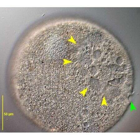
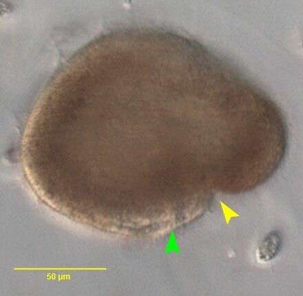
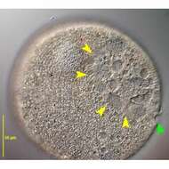
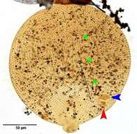
Posterior oblique view of moderately squashed specimen of Maryna umbrellata (Gelei,1950) Foissner, 1993. The yellow arrowheads mark the diagonal groove between anterior calix and the short, broad posterior uvula. The green arrowhead marks the oral aperture. Multiple collecting vesicles of the contractile vacuole system are seen in this image. Collected from an ephemeral puddle on a flood irrigated lawn in Boise, Idaho. July 2007.DIC.
-
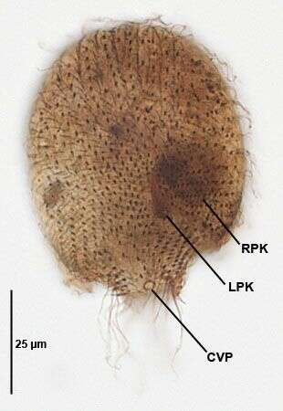
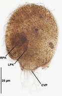
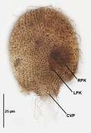
Infraciliature (left ventrolateral view) of the colpodid ciliate, Maryna ovata GELEI, 1950 (left ventrolateral view. LPK=left oral polykinetid.RPK=right oral polykinetid.The contractile vacuole pore (CVP) is located at the end of the uvula.This specimen stained with Protargol (Wilbert modification) (see Foissner, W.Europ. J. Protistol.27,313-330;1991). Collected from a eutrophic pond near Boise, Idaho August 2008. Brightfield.
-
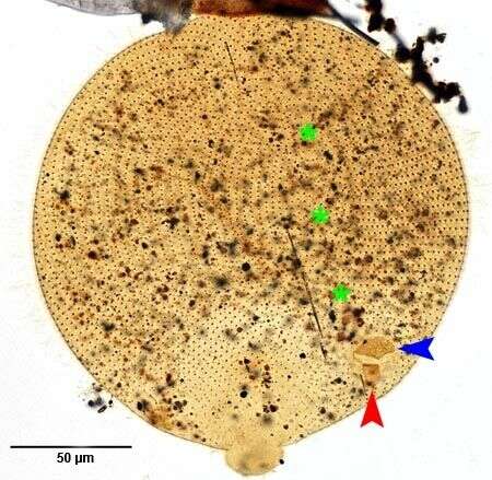
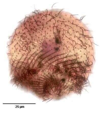
Right ventrolateral view of moderately squashed specimen of Maryna umbrellata (Gelei,1950) Foissner, 1993. The green asterisks mark the anterior preoral suture between right and left somatic kineties.The blue and red arrowheads mark the right and left oral polykinetids respectively.Collected from an ephemeral puddle on a flood irrigated lawn in Boise, Idaho. July 2007.Stained by the silver carbonate technique (see Foissner, W. Europ. J. Protistol., 27:313-330;1991).Brightfield.
-
Infraciliature (dorsolateral view) of the colpodid ciliate, Maryna ovata GELEI, 1950. Cell size is highly variable (length 25-110 mm). The cells are mushroom or champagne cork-shaped. The conical anterior end is termed the calyx and the truncate cylindrical posterior part the uvula. There is a definite horseshoe-shaped sulcus oriented perpendicular to the long axis between the calyx and uvula. The infraciliature is complex, twisting around the calyx and running posteriorly down onto the uvula (as seen here). Doubly ciliated dikinetids are seen here over the anterior calyx. There is a semicircular array of longer terminal cilia along the posterior margin of the uvula. Some descriptions (e.g. Curds, C.R. British and Other Freshwater Ciliated Protozoa. Part I, p.182. Cambridge, Bath 1982.) erroneously describe the uvula as being anterior. This is probably due to the organism's habit of residing in its dwelling tube or lorica anterior end innermost leaving the uvula protruding. Interestingly, cells turn around in the dwelling tube in order to exit front end first. The cytostome is located in the sulcus between calyx and uvula. It is flanked on it right and left by dense polykinetids (not seen in this view). The location of the large spherical macronucleus and single adjacent micronucleus is variable. The contractile vacuole is located in the posterior uvula. Many refractile yellow crystals are found in the cytoplasm. These impart a blackish color to the cells in vivo under low magnification. The tubular organic test is attached to the substrate and may be nearly 1000 μ long. The cell flees the test at the slightest disturbance so most cells are found swimming freely when examined under a coverslip. Maryna ovata feeds on bacteria and algae. This specimen stained with silver carbonate (see Foissner, W.Europ. J. Protistol.27,313-330;1991). Collected from a eutrophic pond near Boise, Idaho August 2004. Brightfield optics.
-
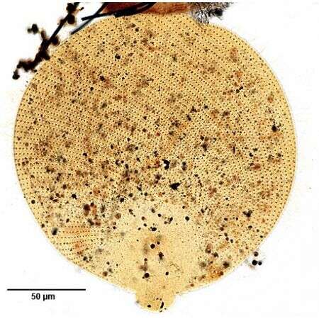
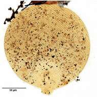
Left ventrolateral view of the infraciliature of a moderately squashed specimen of Maryna umbrellata (Gelei,1950) Foissner, 1993. Collected from an ephemeral puddle on a flood irrigated lawn in Boise, Idaho. July 2007.Stained by the silver carbonate technique (see Foissner, W. Europ. J. Protistol., 27:313-330;1991).Brightfield.
-

Portrait (right lateral view) of the colpodid ciliate, Maryna ovata GELEI, 1950 that has fled its organic test or dwelling tube. This cell is slightly squashed beneath the coverslip. Cell size is highly variable (length 25-110 mm). The cells are mushroom or champagne cork-shaped. The large conical anterior end is termed the calyx and the smaller truncate cylindrical posterior part the uvula. There is a definite horseshoe-shaped sulcus oriented perpendicular to the long axis between the calyx and uvula. The infraciliature is complex, twisting around the calyx and running posteriorly down onto the uvula. There is a semicircular array of longer terminal cilia along the posterior margin of the uvula (seen here). Some descriptions (e.g. Curds, C.R. British and Other Freshwater Ciliated Protozoa. Part I, p.182. Cambridge, Bath 1982.) erroneously describe the uvula as being anterior. This is probably due to the organism's habit of residing in its dwelling tube or lorica anterior end innermost leaving the uvula protruding. Interestingly, cells turn around in the dwelling tube in order to exit front end first. The cytostome is located in the sulcus between calyx and uvula (seen here to viewer's right). It is flanked on it right and left by dense polykinetids. The location of the large spherical macronucleus and single adjacent micronucleus is variable. The contractile vacuole is located in the posterior uvula (just visible here). Many refractile yellow crystals are found in the cytoplasm (seen here in the posterior calyx). These impart a blackish color to the cells under low brightfield magnification. The tubular organic test is attached to the substrate and may be nearly 1000 μ long. The cell flees the test at the slightest disturbance so most cells are found swimming freely when examined under a coverslip as seen here. Maryna ovata feeds on algae and bacteria. Collected from a eutrophic pond near Boise, Idaho August 2004. DIC optics.
-
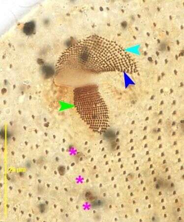
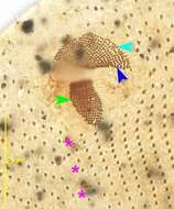
Ventral view of the oral infraciliature of Maryna umbrellata (Gelei,1950) Foissner, 1993. The light blue arrowhead marks the right oral polykinetid. The dark blue arrowhead marks a marginal row of dikinetids along its proximal margin. The green arrowhead marks the left oral polykinetid. The right edg of the vestibulum is visible between the two polykinetids. The pink asterisks mark the postoral suture. Collected from an ephemeral puddle on a flood irrigated lawn in Boise, Idaho. July 2007.Stained by the silver carbonate technique (see Foissner, W. Europ. J. Protistol., 27:313-330;1991).Brightfield.
-
Portrait of the colpodid ciliate, Maryna ovata GELEI, 1950. Cell size is highly variable (length 25-110 mm). The cells are mushroom or champagne cork-shaped. The conical anterior end is termed the calyx and the truncate cylindrical posterior part the uvula. There is a definite horseshoe-shaped sulcus oriented perpendicular to the long axis between the calyx and uvula. The infraciliature is complex, twisting around the calyx and running posteriorly down onto the uvula. There is a semicircular array of longer terminal cilia along the posterior margin of the uvula. Some descriptions (e.g. Curds, C.R. British and Other Freshwater Ciliated Protozoa. Part I, p.182. Cambridge, Bath 1982.) erroneously describe the uvula as being anterior. This is probably due to the organismâs habit of residing in its dwelling tube or lorica anterior end innermost leaving the calyx protruding (seen here). Interestingly, cells turn around in the dwelling tube in order to exit front end first. The cytostome is located in the sulcus between calyx and uvula. It is flanked on it right and left by dense polykinetids. The location of the large spherical macronucleus and single adjacent micronucleus is variable. The contractile vacuole is located in the posterior uvula (just visible here). Many refractile yellow crystals are found in the cytoplasm. These impart a blackish color to the cells under low magnification (seen here). The tubular organic test is attached to the substrate and may be nearly 1000 mm long. The cell flees the test at the slightest disturbance so most cells are found swimming freely when examined under a coverslip. This specimen was photographed without a coverslip resulting in some degradation of the image. Maryna ovata feeds on bacteria and algae. Collected from a eutrophic pond near Boise, Idaho August 2004. Brightfield optics.
-
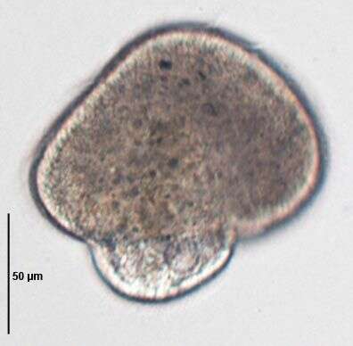
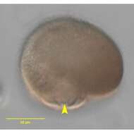
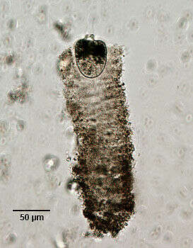
Lateral view of Maryna umbrellata (Gelei,1950) Foissner, 1993 in vivo. The yellow arrowhead marks the oral aperture. The green arrowhead marks the uvula.Collected from an ephemeral puddle on a flood irrigated lawn in Boise, Idaho. July 2007.Brightfield.
-
Infraciliature (right dorsolateral view) of the colpodid ciliate, Maryna ovata GELEI, 1950 (left ventrolateral view. LPK=left oral polykinetid.RPK=right oral polykinetid.The contractile vacuole pore (CVP) is located at the end of the uvula.This specimen stained with Protargol (Wilbert modification) (see Foissner, W.Europ. J. Protistol.27,313-330;1991). Collected from a eutrophic pond near Boise, Idaho August 2008. Brightfield.
-
Posterolateral view of Maryna umbrellata (Gelei,1950) Foissner, 1993 in vivo. The yellow arrowhead marks the posterior contractile vacuole in the uvula.Collected from an ephemeral puddle on a flood irrigated lawn in Boise, Idaho. July 2007.Brightfield.
-
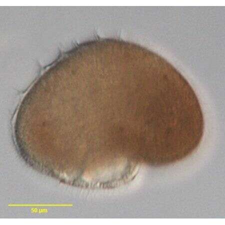
Lateral view of Maryna umbrellata (Gelei,1950) Foissner, 1993 in vivo.Collected from an ephemeral puddle on a flood irrigated lawn in Boise, Idaho. July 2007.Stained by the silver carbonate technique (see Foissner, W. Europ. J. Protistol., 27:313-330;1991).DIC.
-
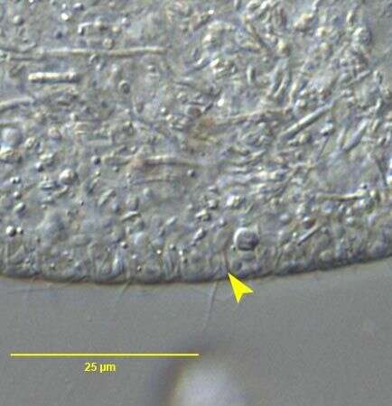
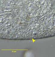
Optical section of cortex of Maryna umbrellata (Gelei,1950) Foissner, 1993. The yellow arrowhead marks one of the approximately 3-4 µm long slender rod-shaped extrusomes. Although the layer of subcortical extrusomes produces a distinct "fringe" in living specimens, individual extrusomes are very difficult to see without DIC and high magnification.Collected from an ephemeral puddle on a flood irrigated lawn in Boise, Idaho. July 2007.DIC.
-
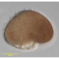
in vivo view of Maryna umbrellata (Gelei,1950) Foissner, 1993. Collected from an ephemeral puddle on a flood irrigated lawn in Boise, Idaho.July 2007.Brightfield.