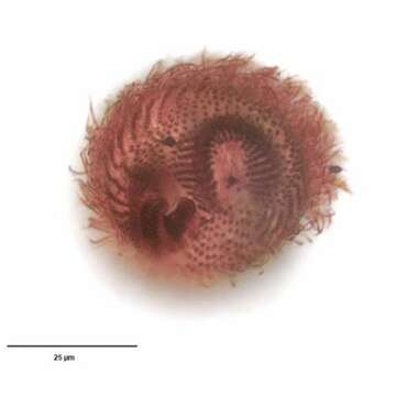
Infraciliature; posterior view
Description :
Infraciliature (posterior view, dorsal at top) of the colpodid ciliate, Maryna ovata GELEI, 1950. Cell size is highly variable (length 25-110 mm). The cells are mushroom or champagne cork-shaped. The conical anterior end is termed the calyx and the truncate cylindrical posterior part the uvula. There is a definite horseshoe-shaped sulcus oriented perpendicular to the long axis between the calyx and uvula. The infraciliature is complex, twisting around the calyx and running posteriorly down onto the uvula (as seen here). There is a semicircular array of longer terminal cilia along the posterior margin of the uvula (their kinetids are seen here). Some descriptions (e.g. Curds, C.R. British and Other Freshwater Ciliated Protozoa. Part I, p.182. Cambridge, Bath 1982.) erroneously describe the uvula as being anterior. This is probably due to the organism's habit of residing in its dwelling tube or lorica anterior end innermost leaving the uvula protruding. Interestingly, cells turn around in the dwelling tube in order to exit front end first. The cytostome is located in the sulcus between calyx and uvula. It is flanked on it right and left by dense polykinetids (seen to the viewer's left at 8 o'clock position here). The location of the large spherical macronucleus and single adjacent micronucleus is variable. The contractile vacuole is located in the posterior uvula. Many refractile yellow crystals are found in the cytoplasm. These impart a blackish color to the cells in vivo under low magnification. The tubular organic test is attached to the substrate and may be nearly 1000 μ long. The cell flees the test at the slightest disturbance so most cells are found swimming freely when examined under a coverslip. Maryna ovata feeds on bacteria and algae. This specimen stained with silver carbonate (see Foissner, W.Europ. J. Protistol.27,313-330;1991). Collected from a eutrophic pond near Boise, Idaho August 2004. Brightfield optics.
Inclus dans les pages suivantes :
- Life
- Cellular (Organismes cellulaires)
- Eukaryota (eucaryotes)
- SAR (Stramenopiles, Alveolates, Rhizaria)
- Alveolata
- Ciliophora
- Intramacronucleata
- Colpodea
- Colpodida
- Marynidae
- Maryna
- Maryna ovata
Cette image ne figure dans aucune collection.
Informations sur la provenance
- licence
- cc-by-nc
- auteur
- William Bourland
- fournisseur
- micro*scope
- original
- fichier de média d’origine
- visiter la source
- site partenaire
- micro*scope
- ID