Description
(
anglais
)
fourni par Zookeys
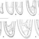
Female. Body moderately long (L=5.6–8.2 mm) and plump (a=51–72.4), assuming a spiral to C shape when heat relaxed. Cuticle consisting of several layers under light microscope: 11–14 µm thick at guiding ring level; 7–8 µm along the body; 13–15 µm on tail posterior to anus. Lateral pores number 10–14 in pharyngeal region: a single pore in front of guide ring, rarely two or none; 3–5 in odontostyle and 1–3 in odontophore regions; 3–4 dorsal pores and 7–10 ventral pores; numerous lateral body pores. Usually the fifth ventral pore (sometimes the fourth) differs in size (Figs 1A, 4F and 6H) compared to the other ventral pores. Lip region continuous, anteriorly almost flat, 7–9 μm high. Labial papillae prominent. Amphid aperture assumed to be a minute pore, difficult to be observed under light microscope. Pouch-like amphidial fovea with convoluted fine dendritic branches (receptors), extending to 1/2 - 2/3 the distance between anterior end and guiding ring, fovea slightly longer (15–18 μm, n=5) than wide (14–16 μm, n=4) with no distinct margins. Fusus (sensillium pouch) at 57±1.9 (55–60) μm from anterior end. Guiding ring 7–9 μm wide. Odontostyle long and very slender, 2 μm wide at the base. Odontophore with weakly developed flanges. In all females a small (2–3 μm long) rudimentary odontostyle tip (vestigium) present, directed forward, and observed in the slender pharynx at 300.5±40.3 (224–350) μm from anterior end; in two specimens the vestigium located in odontophore area. Slender pharynx often coiled in its posterior part. In this region 5–7 glandular bodies are observed in all females. Nerve ring surrounding odontophore base, rarely surrounding mid-odontophore, or just behind it, second nerve ring at a distance of 85.2±6.6 (78–98) μm behind the first one. Hemizonid flat, 10–11 μm long. Pharyngeal bulb about 1/4 of the neck length. Normal arrangement of pharyngeal glands, the nuclei of dorsal and ventrosublateral glands approximately the same size, their diameters 3.4±0.4 (3–4) μm, n=7 and 3.9±0.2 (3.5–4) μm, n=11, respectively. Cardia small, broadly rounded, wider than long, variable in size: 20.1±1.8 (10–23) × 10.1±1.8 (7–12) μm . Reproductive system amphidelphic, varying in dimensions due to the stage of maturity of female. Vagina extending about half body width. Pars distalis vaginae with characteristic shape (Fig. 2F, G), 26–28 µm and pars proximalis vaginae 32–38 µm long, respectively; muscular walls of the latter almost parallel. Uteri very long, anterior uterus 494.6±52 (430–563) µm long, posterior uterus 510.0±88.7 (357–643) µm long, differentiated, filled with sperm cells in all females examined; well developed sphincter between uterus and pars dilatata oviductus also containing numerous sperm cells. Anterior and posterior oviduct of similar size, measured in four specimens: 275–348 μm, and 283–330 μm. Anterior ovarium 263.4±51.8 (210–347) μm long, n=7, posterior ovarium 234.3±35.8 (183–309) μm long, n=5; in older mature specimens the length is about 3 times greater (1055–1060 μm for anterior and 1020 μm for posterior ovary). One egg in anterior pars dilatata oviductus measuring 227 × 87.5 μm and one uterine egg measuring 225 × 77.5 μm. A weakly developed ovijector present, 112.0±12 (95–125) μm long. In one female a rudimentary adanal pair of supplementary papillae was observed (Fig. 9E). Prerectum variable in length; rectum 0.7±0.1 (0.6–0.8) body width at anus. A short post-intestinal sac present. Tail bluntly conoidal, rounded to almost hemispherical; ventral side straight or slightly convex, the dorsal curvature greater. Two pairs of lateral pores.
Male. Body C shaped when heat relaxed, posterior part more strongly coiled ventrally. Similar to females in general morphology except for genital system. Lateral pores number 10–15 in pharyngeal region: a single pore in front of guide ring, 3–5 in odontostyle and 1–2 in odontophore regions; 2–5 dorsal pores, mostly 3–4, and 7–10 ventral pores. Cuticle in post-labial region at the guiding ring level 10.5–13.5 µm thick, 6.5–9 µm along body, 9–10 µm in post-cloacal area. Second nerve ring at 80.7±14 (50–100) μm behind the first one (n=14). In all males a small vestigium (2–3 μm, in one specimen 6 μm long), directed forward (in two specimens directed rearward), is observed in the slender pharynx at 300.5±40.3 (224–350) μm from anterior end; in two specimens the vestigium detected in odontophore area. Two to eleven glandular bodies observed in all males in posterior part of the slender pharynx and pharyngeal bulb. In two specimens lens-like hemizonion at a distance of 242 and 271 μm from anterior end observed. Pharyngeal bulb slightly less than 1/4 of neck length (22.9±1.6 (20.9–26.7%). Ventromedian supplements composed of one adanal pair and a row of 13–17 irregularly spaced single ones, the first three appear as double in some specimens. Spicules comparatively slender, of almost equal width along the length, curved to almost at right angle. Lateral guiding piece not bifid, with uneven internal walls. Post-cloacal papilla well developed. Tail short, bluntly conoidal, ventral side almost straight, dorsal side convex. Two or three pairs of lateral caudal pores.
Juveniles. Four developmental stages clearly present (Fig. 11) as determined from the position of the replacement odontostyle and the principal morphometric characters of body, odontostyle and replacement odontostyle lengths, and developing gonad (genital primordium) size. The habitus of juveniles not changing considerably during successive stages, assuming J or C shape. In first stage juvenile, lip region somewhat different from the next stages, it is rounded with a very weak depression after the second circle of labial papillae, the latter slightly protruding and changing the lip region outline. Amphidial fovea in first two stages has no clearly visible receptors, only small refractive elements discernable. Both the tail and body width at anus is increasing in length and c’ ratio is decreasing. Tail shape in J1 is conoidal, ventrally almost straight or slightly concave, dorsally convex, which gives asymmetrical appearance, in successive stages it gradually becomes rounded but always with the dorsal curvature more strongly expressed.
- licence
- cc-by-3.0
- droit d’auteur
- Saša Širca, Gregor Urek, Stela Lazarova, Milka Elshishka, Vlada Peneva
- citation bibliographique
- Širca S, Urek G, Lazarova S, Elshishka M, Peneva V (2011) Longidorus carniolensis sp. n. (Nematoda, Longidoridae) from vineyard soil in Slovenia ZooKeys 141: 1–27
- auteur
- Saša Širca
- auteur
- Gregor Urek
- auteur
- Stela Lazarova
- auteur
- Milka Elshishka
- auteur
- Vlada Peneva
Distribution
(
anglais
)
fourni par Zookeys
Longidorus carniolensis n. sp were detected in 6 out of 10 soil samples from locations of Drašiči and Krmačina. Population density was 4–15 specimens of all developmental stages per 200 cm3 of soil sample.
- licence
- cc-by-3.0
- droit d’auteur
- Saša Širca, Gregor Urek, Stela Lazarova, Milka Elshishka, Vlada Peneva
- citation bibliographique
- Širca S, Urek G, Lazarova S, Elshishka M, Peneva V (2011) Longidorus carniolensis sp. n. (Nematoda, Longidoridae) from vineyard soil in Slovenia ZooKeys 141: 1–27
- auteur
- Saša Širca
- auteur
- Gregor Urek
- auteur
- Stela Lazarova
- auteur
- Milka Elshishka
- auteur
- Vlada Peneva