-
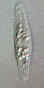
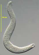
Portrait of the pleurostomatid ciliate, Loxophyllyum meleagris (Mueller,1773) Dujardin, 1841. Glides with ribbon-like movement over substrate. Oral region is slit-like and oriented to the right in this image. Wart-like aggregates of extrusomes are seen at intervals along the dorsal (left) surface. Macronucleus is multinodal in this species. From standing temporary fresh water puddlenear Boise, Idaho. Phase contrast illumination.
-
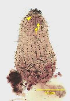
Dorsal infraciliature of the haptorid ciliate, Fuscheria terricola (BERGER,FOISSNER & ADAM,1983). The longer first and shorter second dorsal brush rows of clavate cilia are indicated by the arrows. The arrowhead indicates the circumoral kinety.The densely stained extrusome bundle of the cytopharynx is seen partially ejected anteriorly. Stained by the silver carbonate technique (see Foissner, W. Europ. J. Protistol., 27:313-330;1991). Brightfield.
-

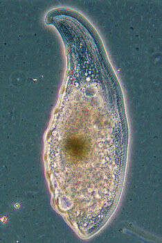
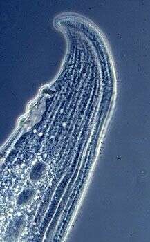
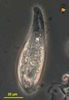
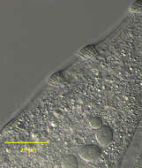
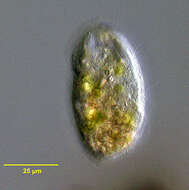
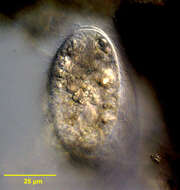
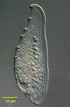
Detail view (dorsal surface) of the large pleurostomatid ciliate, Loxophyllum meleagris (Mueller,1773) Dujardin, 1841. The strongly laterally compressed cell is scimitar-shaped in outline. . The cell is slightly contractile and highly flexible. The rounded anterior end is curved dorsally. The posterior is bluntly tapered. The right side is more densely ciliated than the left. Somatic kineties are longitudinal. The dorsal edge bears characteristic nodular protrusions called extrusome warts (seen well here). The slit-like cytostome is located along the anteroventral edge. There is one posterior contractile vacuole which has a long collecting canal extending anteriorly along the dorsal edge of the cell. The macronucleus (part of which is seen well here) is moniliform. There are multiple inconspicuous micronuclei (not seen here). L.meleagris swims slowly, gliding gracefully over the substrate. L.meleagris feeds on other ciliates and even metazoans such as rotifers. Differentiated from the similar L. helus by its much larger size. Collected from a freshwater agricultural irrigation ditch near McCall, Idaho 9/21/03. DIC.
-
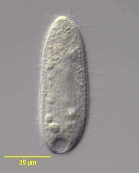
Portrait of the haptorid ciliate, Fuscheria terricola (BERGER,FOISSNER & ADAM,1983). DIC.
-
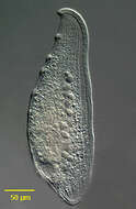
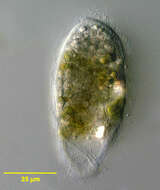
Portrait (right side) of the large pleurostomatid ciliate, Loxophyllum meleagris (Mueller,1773) Dujardin, 1841. The strongly laterally compressed cell is scimitar-shaped in outline. . The cell is slightly contractile and highly flexible. The rounded anterior end is curved dorsally. The posterior is bluntly tapered. The right side is more densely ciliated than the left. Somatic kineties are longitudinal. The dorsal edge bears characteristic nodular protrusions called extrusome warts. The slit-like cytostome is located along the anteroventral edge. There is one posterior contractile vacuole which has a long collecting canal extending anteriorly along the dorsal edge of the cell. The macronucleus is moniliform. There are multiple inconspicuous micronuclei (not seen here). L.meleagris swims slowly, gliding gracefully over the substrate. L.meleagris feeds on other ciliates and even metazoans such as rotifers. Differentiated from the similar L. helus by its much larger size. Collected from a freshwater agricultural irrigation ditch near McCall, Idaho 9/21/03. DIC.
-
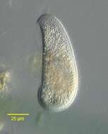
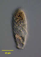
Bryophyllum vorax (Stokes, 1888) Kahl, 1931. Collected from a freshwater pond near Boise, Idaho. DIC
-
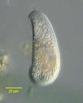
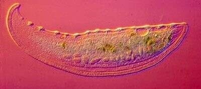
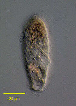
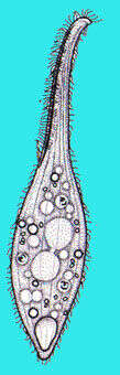
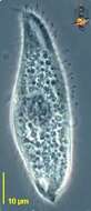
This image of the anterior end shows the curved oral region with faintly visible extrusomes that are used to capture protists as food. The surface is folded along the lines of the kineties. The contractile vacuole, upper left, has a long feeding canal, and the macronucleus is in the form of a series of linked beads. Phase contrast.
-
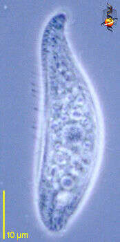
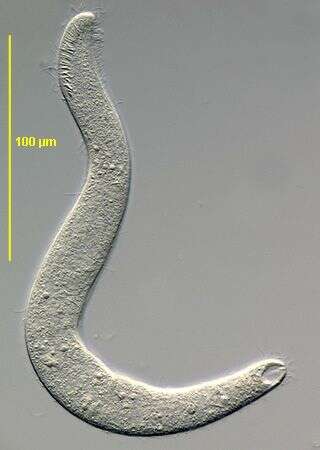
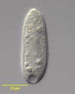
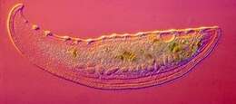
Portrait (left side) of the haptorid ciliate, Perispira ovum (Stein, 1859). The cell body is cylindrical to ovoid. The anterior end is slightly truncate. An unciliated cortical ridge makes a complete right-hand spiral the length of the body (seen here running obliquely across mid body). The slit-like cytostome, supported by fine trichites, is located at the anterior end of the cortical ridge. The uniform longitudinal somatic kineties spiral slightly. Densely packed food vacuoles and highly refractile cytoplasmic crystals often obscure the ellipsoid macronucleus. There is a single large terminal contractile vacuole posteriorly. Swims slowly. Collected from anoxic bottom sediments of slow flowing freshwater stream near Boise, Idaho march 2004. DIC optics
-
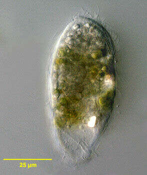
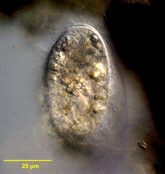
Interference contrast image of a single living cell. the warts on the oral face are distinctive.
-
Portrait (right side) of the haptorid ciliate, Perispira ovum (Stein, 1859). The cell body is cylindrical to ovoid. The anterior end is slightly truncate. An unciliated cortical ridge makes a complete right-hand spiral the length of the body. The slit-like cytostome, supported by fine trichites, is located at the anterior end of the cortical ridge. The uniform longitudinal somatic kineties spiral slightly. Densely packed food vacuoles and highly refractile cytoplasmic crystals often obscure the ellipsoid macronucleus. There is a single large terminal contractile vacuole posteriorly. Swims slowly. Collected from anoxic bottom sediments of slow flowing freshwater stream near Boise, Idaho march 2004. DIC optics
-
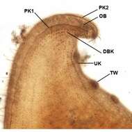
Infraciliature (left side) of Loxophyllum meleagris (Mueller,1773) Dujardin, 1841. The oral bulge (OB is bordered by one left perioral kinety (PK1) and two right perioral kineties (only PK2 visible here). The dorsal edge bears characteristic nodular protrusions called extrusome or trichocyst warts (TW). A row of unciliated kinetids is seen at the base of each trichocyst wart (UK)The slit-like cytostome is located in the center of the oral bulge. DK= dorsal brush kinetids. Collected from a freshwater canal in Boise,Idaho 10/27/08. Protargol.Brightfield.
-
Portrait (right side) of the haptorid ciliate, Perispira ovum (Stein, 1859). The cell body is cylindrical to ovoid. The anterior end is slightly truncate. A narrow unciliated cortical ridge makes a complete right-hand spiral the length of the body. The slit-like cytostome, supported by fine trichites (seen well in this image), is located at the anterior end of the cortical ridge. The uniform longitudinal somatic kineties spiral slightly. Densely packed food vacuoles and highly refractile cytoplasmic crystals often obscure the ellipsoid macronucleus. There is a single large terminal contractile vacuole posteriorly. Swims slowly. Collected from anoxic bottom sediments of slow flowing freshwater stream near Boise, Idaho March 2004. DIC optics.
-
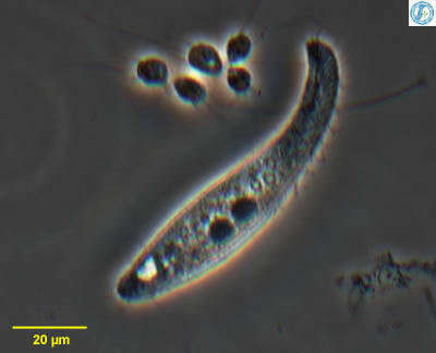
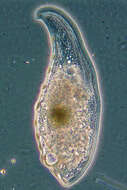
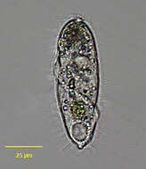
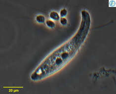
Litonotus (light-o-note-us) is one of the more commonly encountered predatory ciliates. Scyth-shaped, and flattened. The mouth is located on the convex curve of the anterior part of the body (upper right in the picture). The food is captured in part by the action of rod-shaped extrusomes which can be seen just inside the cell adjacent to the mouth. The large structure near the centre of the cell is the macronucleus. The light region towards the rear is the contractile vacuole. Cilia cover the body in sparse kineties. Phase contrast.
-
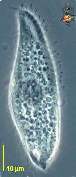
Portrait (lateral view) of the haptorid ciliate, Perispira ovum (Stein, 1859). The cell body is cylindrical to ovoid. The anterior end is slightly truncate. An unciliated cortical ridge makes a complete right-hand spiral the length of the body. The slit-like cytostome, supported by fine trichites, is located at the anterior end of the cortical ridge. The uniform longitudinal somatic kineties spiral slightly. Densely packed food vacuoles and highly refractile cytoplasmic crystals often obscure the ellipsoid macronucleus. There is a single large terminal contractile vacuole posteriorly. Swims slowly. Collected from anoxic bottom sediments of slow flowing freshwater stream near Boise, Idaho December 2004. Brightfield optics, closed condenser.
-
Litonotus (light-o-note-us) is one of the more commonly encountered predatory ciliates. It is flattened, and glides along the substrate, exploring detritus with the anterior convex margin - which is where the mouth is located. There are extrusomes just internal to the margin of the cell, and these can be discharged to kill potential prey - usually other ciliates. Phase contrast.
-
Portrait (left side) of the haptorid ciliate, Perispira ovum (Stein, 1859). The cell body is cylindrical to ovoid. The anterior end is slightly truncate. An unciliated cortical ridge makes a complete right-hand spiral the length of the body. The slit-like cytostome, supported by fine trichites, is located at the anterior end of the cortical ridge. The uniform longitudinal somatic kineties spiral slightly. Densely packed food vacuoles and highly refractile cytoplasmic crystals often obscure the ellipsoid macronucleus. There is a single large terminal contractile vacuole posteriorly. Swims slowly. Collected from anoxic bottom sediments of slow flowing freshwater stream near Boise, Idaho December 2004. DIC optics
-
Litonotus (light-o-note-us) is one of the more commonly encountered predatory ciliates. It is flattened, and glides along the substrate, exploring detritus with the anterior convex margin - which is where the mouth is located. There are extrusomes just internal to the margin of the cell, and these can be discharged to kill potential prey - usually other ciliates. Phase contrast.
-
Infraciliature (posterior apical view) of the haptorid ciliate, Perispira ovum (Stein, 1859). The cell body in vivo is cylindrical to ovoid. The longitudinal somatic kineties spiral slightly. An unciliated cortical ridge, bordered on either side by by a file of closely spaced kinetids, makes a complete right-hand spiral the length of the body. The posterior portion of this structure is seen here to the viewer's left. The three files of clavate cilia (dorsal brush) are seen at the viewr's upper right. The right-most kinety of the dorsal brush has longer cilia than the two kineties to it's left. Collected from anoxic bottom sediments of slow flowing freshwater stream near Boise, Idaho December 2004. Stained by the silver carbonate technic (see Foissner, W. Europ. J. Protistol., 27:313-330;1991).Brightfield.
-
-
Infraciliature (ventrolateral view) of the haptorid ciliate, Perispira ovum (Stein, 1859). The cell body in vivo is cylindrical to ovoid. The longitudinal somatic kineties spiral slightly. An unciliated cortical ridge, bordered on either side by by a file of closely spaced kinetids, makes a complete right-hand spiral the length of the body. The anterior portion of this structure is seen here. The cytostome is located at the anterior end of the spiral ridge. The cytostome is supported by trichites (not seen here).Collected from anoxic bottom sediments of slow flowing freshwater stream near Boise, Idaho December 2004. Stained by the silver carbonate technic (see Foissner, W. Europ. J. Protistol., 27:313-330;1991).Brightfield.
-
Litonotus, predatory ciliate. Flattened with mouth located along convex outer edge of the front part of the cell. Numerous extrusomes lie under the mouth. With two large macronuclei located on either side of a smaller micronucleus., From Lake Donghu, China. Phase contrast micrograph.
-
Portrait of the haptorid ciliate, Perispira ovum (Stein, 1859). The cell body is cylindrical to ovoid. The anterior end is slightly truncate. An unciliated cortical ridge (not well seen here) makes a complete right-hand spiral the length of the body. The slit-like cytostome, supported by fine trichites, is located at the anterior end of the cortical ridge. The uniform longitudinal somatic kineties spiral slightly. There are thre files of clavate (club-shaped) cilia forming a dorsal brush. The right-most of these (visible here to the viewer's upper right) hhas longer cilia than the two kineties to it's right. The cytoplasm in this individual is densely packed chloroplasts from ingested euglenae. These are probably "kleptoplasts". Kleptoplasts are plastids from ingested prey that are maintained in the cytoplasm and not digested. Perispira ovum may also sequester mitochondria from its prey in similar fashion (Johnson, PW et al. J. Euk. Microbiol.42:323-335,1995). There is a single large terminal contractile vacuole posteriorly. Collected from anoxic bottom sediments of slow flowing freshwater stream near Boise, Idaho march 2004. DIC.
-
Right side view of Litonotus, a common pleurostomatid ciliate genus with many species. Oral aperture is slit-like and lined with extrusomes. Two round centrally located macronuclei. Posterior contractile vacuole. Parallel kineties on right surface (seen well in this image) distinguish this genus from the similar Amphileptus in which longitudinal kineties converge on each other anteriorly and posteriorly. From freshwater pond near Boise, Idaho. Oblique illumination
-
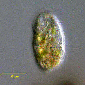
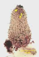
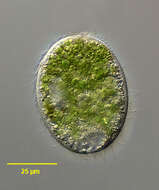
Collected from a non-flooded Petri dish culture of topsoil from a public park in Boise, Idaho. November 2006.DIC.