-
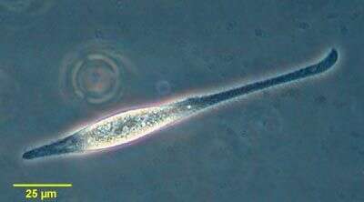
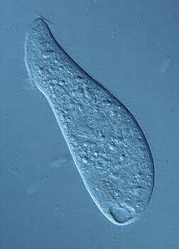
Litonotus is a predatory ciliate. The mouth extends along the convex margin running from the middle to the front of the cell (at the top of the picture). It is equipped with organelles that are ejected from the cell and are used to immobilize and kill prey. The two dark structures near the middle of the cell are macronuclei.
-
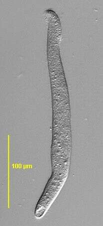
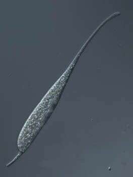
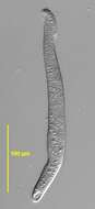
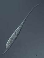
Portrait of Arcuospathidium (FOISSNER,1984). Collected from a non-flooded Petri dish culture of topsoil from a public park in Boise, Idaho. November 2006.DIC.
-
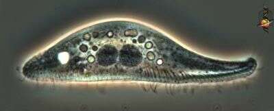
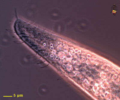
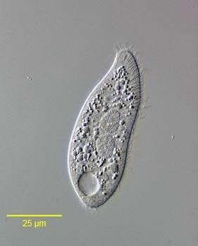
This Litonotus cell as been viewed from the side. The face that is applied to the substrate is ciliated. This face is usually called the right side of the cell because it lies to the right of the mouth. The mouth in this image extends from the anterior (to the right of the picture) to about half way down the ventral (or right) face of the cell. Numerous extrusomes abut onto the membrane of the mouth. There are two rounded macronuclear nodes in the pcture and two small micronuclei (1 o'clock relative to the right-hand nucleus and 10 o'clock relative to the left-hand nucleus). Phase contrast microscopy.
-
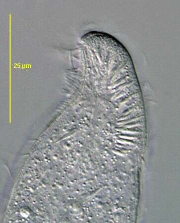
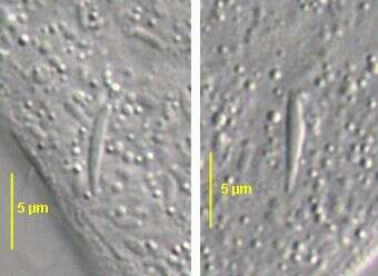
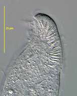
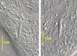
Ventral view of the oral bulge of Arcuospathidium sp. (FOISSNER,1984). Collected from a non-flooded Petri dish culture of topsoil from a public park in Boise, Idaho. November 2006.DIC.
-
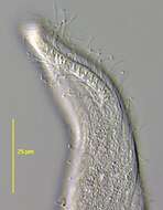
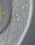
Detail of the anterior (mouth) end of the cell. The mouth is drawn out along the flatened antero-lateral margin of the cell. Extrusomes lie under the membrane and these are used in food capture. Phase contrast microscopy.
-
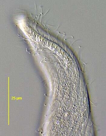
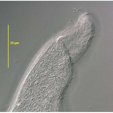
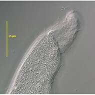
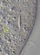
Lateral view of the anterior end of of Arcuospathidium sp. (FOISSNER,1984). Collected from a non-flooded Petri dish culture of topsoil from a public park in Boise, Idaho. November 2006.DIC.
-
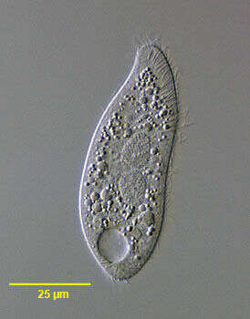
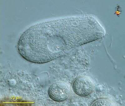
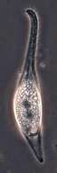
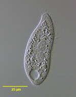
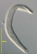
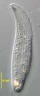
Optical section of the common pleurostomatid ciliate, Litonotus (Wresniowski 1870). Ccollected from a freshwater pond near Boise, Idaho. April 2005. DIC
-
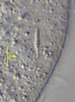
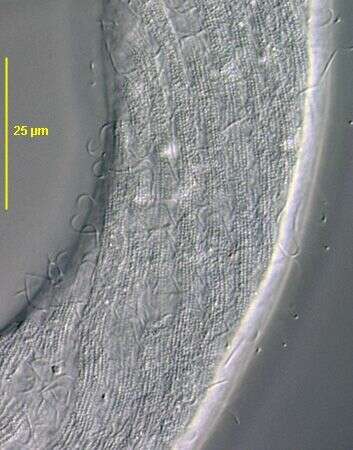
Optical section of the anterior end of of Arcuospathidium sp. (FOISSNER,1984) showing the longer dorsal brush row of cilia and abundant ellipsoid subpellicular mitochondria. Collected from a non-flooded Petri dish culture of topsoil from a public park in Boise, Idaho. November 2006.DIC.
-
-
-
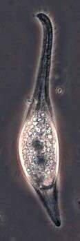
Found in a sample with Fucus taken from close to Tvarminne Zoological Station on April 3rd, 2012. Bruce Taylor tells us that this may be Litonotus cygnus, we'd be pleased with any feedback.
-
-
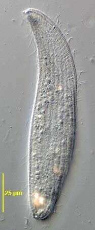
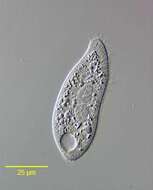
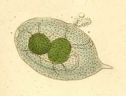
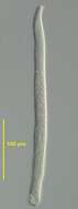
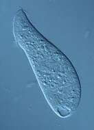
Portrait of Litonotus cygnus (Mueller, 1773) Foissner, 1995, a pleurostomatid ciliate. Markedly extensile, this individual is contracted. The slit-like oral aperture on the convex surface extends along most of the length of the neck region. Extrusomes are visible at the base of the neck region on the ventral surface. Two round centrally located macronuclei. Posterior contractile vacuole. Parallel kineties on right surface distinguish this genus from the similar Amphileptus in which longitudinal kineties converge on each other anteriorly and posteriorly. From freshwater pond near Boise, Idaho. Phase contrast.
-
-
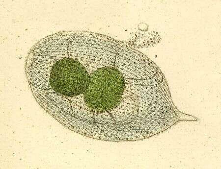
Originally described by Ehrenberg under the name Trachelius lamella.
-
-
Left side of Acineria incurvata DUJARDIN,1841, a pleurostomatid ciliate found in heavily polluted freshwater and marine habitats. Collected from effluent of a protein skimmer at a commercial saltwater aquarium in Boise,Idaho. January 2007.
-
Collected from a non-flooded Petri dish culture of topsoil from a public park in Boise, Idaho. November 2006.DIC.
-
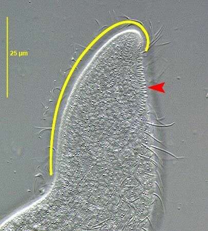
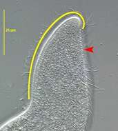
Acineria incurvata DUJARDIN,1841 a pleurostomatid ciliate found in heavily polluted freshwater and marine habitats. The yellow line parallels the distinctive oral bulge which recurves dorsally and to the left at its anterior end. The bulge has a helical structure like a two-stranded string.The pattern is faintly visible here.The dorsal brush consists of a single file of obliquely oriented dikinetids bearing non-motile clavate cilia (red arrowhed). Collected from effluent of a protein skimmer at a commercial saltwater aquarium in Boise,Idaho. January 2007.DIC.
-
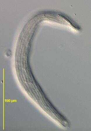
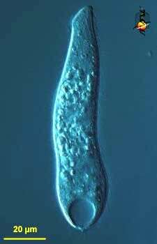
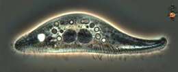
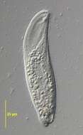
Spathidium (spa-thid-ee-um), a predatory ciliate. the mouth is the slightly expanded region at the front (top) of the cell and this is underlain with extrusomes which assist in the capture of food. The structure at the back end is the contractile vacuole. These guys usually eat other ciliates. Differential interference contrast. Material from Nymph Creek and Nymph Lake, thermal sites within Yellowstone National Park, photograph by Kathy Sheehan and David Patterson.
-
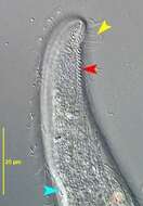
Acineria incurvata DUJARDIN,1841 a pleurostomatid ciliate found in heavily polluted freshwater and marine habitats. The yellow arrowhead indicates cilia of perioral kinety 2 which borders the oral bulge on the right side. The distinctive oral bulge recurves dorsally and to the left at its anterior end. The dorsal brush consists of a single file of obliquely oriented dikinetids bearing non-motile clavate cilia (red arrowhed). The light blue arrowhead indicates the posterior end of the oral bulge.Collected from effluent of a protein skimmer at a commercial saltwater aquarium in Boise,Idaho. January 2007.DIC.
-
Spathidium (spa-thid-ee-um) is a predatory ciliate. The front end looks as if it is flattened, this is the mouth. Immediately inside the mouth are short extrusomes which are used to kill and capture food. May form cysts. Differential interference contrast.
-
Right side of Acineria incurvata DUJARDIN,1841, a pleurostomatid ciliate found in heavily polluted freshwater and marine habitats. Collected from effluent of a protein skimmer at a commercial saltwater aquarium in Boise,Idaho. January 2007.DIC.
-
Differential interference contrast image, mouth to top.