-
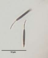
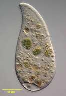
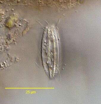
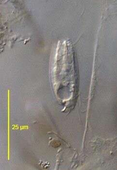
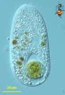
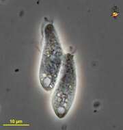
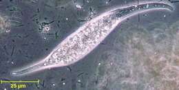
Surface view of Pithothorax processus a small haptorid ciliate found in polysaprobic habitats. The body is a slightly flattened cylinder. The pellicle is rigid with longitudinal ribbing. The oral aperture is at the anterior apex surrounded by projections of the pellicular ridges. There is a curved funnel-shaped posterior process from which a long caudal cilium protrudes (seen here). The round macronucleus is anterior and the contractile vacuole is located in the posterior 1/3 at the periphery (seen here). The somatic ciliature is confined to the anterior and posterior quarters of the body. From stagnant organically enriched freshwater pond near Boise, Idaho. DIC optics.
-
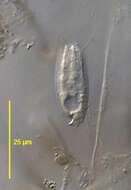
Stained by the silver carbonate technique (see Foissner, W.Europ. J. Protistol.27:313-330;1991).Brightfield.
-
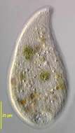
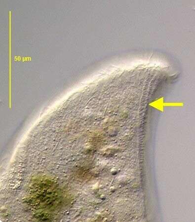
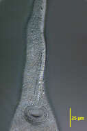
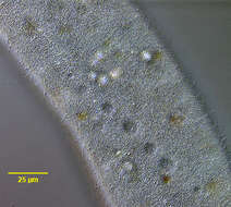
Detail of the oral aperture and ventral proboscis of Monilicaryon monilatus Monilicaryon monilatus (Stokes, 1886) Jankowski, 1967. Similar in overall appearance to Dileptus anser. M. monilatus differs by having a shorter proboscis relative to the length of the body (1/3 to 1/4) and by lacking the row of obliquely oriented closely spaced kinetids on the ventral aspect of the left side of the proboscis (this feature requires demonstration by DIC or protargol staining). M. monilatus has two single files of kinetids extending from either side of the oral aperture anteriorly along the ventral aspect of the proboscis separated by a strip bearing extrusomes (See Foissner W., Berger H and Kohmann F. Taxonomische und ökologische Revision der Ciliaten des Saprobiensystems- Band IV: Gymnostomatea, Loxodes, Suctoria. Informationsberichte Bayer. Landesamtes für Wasserwirtschaft. 1/95:185-202, 1995). In this image the two parallel kineties along the right side of the extrusome strip are visible. Collected from a freshwater pond near Boise, Idaho. DIC.
-

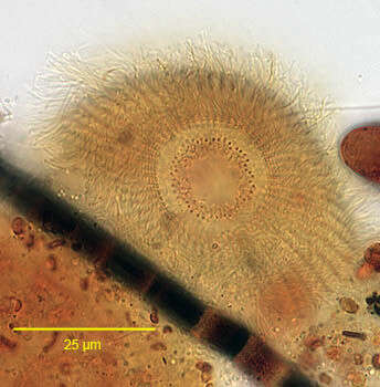
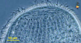
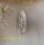
Anterolateral view of the infraciliature of the haptorid ciliate,Askenasia volvox (Eichwald,1852) Kahl, 1930. The cell is spherical posteriorly and the anterior is a truncate cone.The cytostome is at the anterior apex.The cytostome is surrounded by an undulating line of granules (seen only in silver impregnated specimens).Somatic kineties are arranged in three (anterior,middle and posterior)girdles seen in this image.The anterior cilia are directed forward and the middle girdle cilia are longer,curving backwards in a "sickle" configuration.These cilia produce the saltatory locomotion typical of this genus.The middle ciliary girdle kineties have a zig-zag configuration (best seen here to the viewer's right).The posterior girdle consists of long stiff bristles (the three kinetids of these bristles are seen immediately posterior to the middle ciliary girdle kineties).The posterior of the cell is unciliated.Stained by the silver carbonate technique (see Foissner, W. Europ. J. Protistol., 27:313-330;1991). From a freshwater pond near Boise, Idaho.Brightfield.
-
Enchelys (ench-el-is), anterior end of this predatory ciliate, picture taken to show the rows of cilia or kineties which extend from the front of the cell to the posterior. Phase contrast.
-
Saggital optical section of Pithothorax processus (Kahl, 1926), a small haptorid ciliate found in polysaprobic habitats. The body is a slightly flattened cylinder. The pellicle is rigid with longitudinal ribbing. The oral aperture is at the anterior apex surrounded by projections of the pellicular ridges. There is a curved funnel-shaped posterior process from which a long caudal cilium protrudes (seen here). The round macronucleus is anterior and the contractile vacuole is located in the posterior 1/3 at the periphery (seen here). The somatic ciliature is confined to the anterior and posterior quarters of the body. From stagnant organically enriched freshwater pond near Boise, Idaho. DIC optics.
-
-
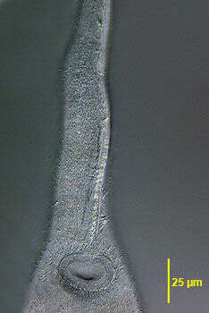
Detail of the oral aperture and ventral proboscis of Monilicaryon monilatus Monilicaryon monilatus (Stokes, 1886) Jankowski, 1967. Similar in overall appearance to Dileptus anser. M. monilatus differs by having a shorter proboscis relative to the length of the body (1/3 to 1/4) and by lacking the row of obliquely oriented closely spaced kinetids on the ventral aspect of the left side of the proboscis (this feature requires demonstration by DIC or protargol staining). M. monilatus has two single files of kinetids extending from either side of the oral aperture anteriorly along the ventral aspect of the proboscis (See Foissner W., Berger H and Kohmann F. Taxonomische und ökologische Revision der Ciliaten des Saprobiensystems- Band IV: Gymnostomatea, Loxodes, Suctoria. Informationsberichte Bayer. Landesamtes für Wasserwirtschaft. 1/95:185-202, 1995). In this image the area between these kineties appears more refractile due to the presence of extrusomes. Collected from a freshwater pond near Boise, Idaho. DIC.
-
Anterolateral view of the infraciliature of the haptorid ciliate,Askenasia volvox (Eichwald,1852) Kahl, 1930. The cell is spherical posteriorly and the anterior is a truncate cone.The cytostome is at the anterior apex.The cytostome is surrounded by an undulating line of granules (seen here).Somatic kineties are arranged in three (anterior,middle and posterior)girdles seen in this image.The anterior cilia are directed forward and the middle girdle cilia are longer,curving backwards in a "sickle" configuration.These cilia produce the saltatory locomotion typical of this genus.The middle ciliary girdle kineties have a zig-zag configuration seen here.The posterior girdle consists of long stiff bristles.The posterior of the cell is unciliated.Stained by the silver carbonate technique (see Foissner, W. Europ. J. Protistol., 27:313-330;1991). From a freshwater pond near Boise, Idaho.Brightfield.
-
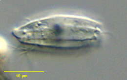
Enchelys (ench-el-is) -cylindrical predatory ciliate, body fairly flexible, mouth slit zone at anterior end, underlain by a number of extrusomes. The cell has just eaten a dinoflagellate. Differential interference contrast.
-
Surface view of Pithothorax processus (Kahl, 1926) a small haptorid ciliate found in polysaprobic habitats. The body is a slightly flattened cylinder. The pellicle is rigid with longitudinal ribbing. The oral aperture is at the anterior apex surrounded by projections of the pellicular ridges. There is a curved funnel-shaped posterior process from which a long caudal cilium protrudes (seen here). The round macronucleus is anterior and the contractile vacuole is located in the posterior 1/3 at the periphery (seen here). The somatic ciliature is confined to the anterior and posterior quarters of the body. From stagnant organically enriched freshwater pond near Boise, Idaho. DIC optics.
-
2 of the three dorsal brush rows are seen in this image.DIC.
-
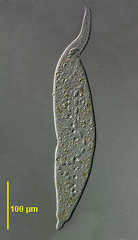
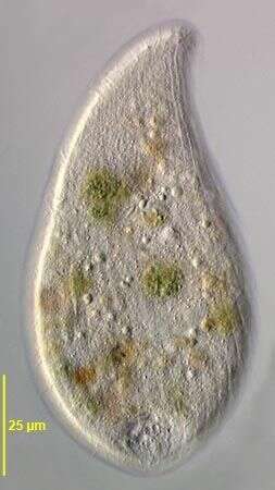
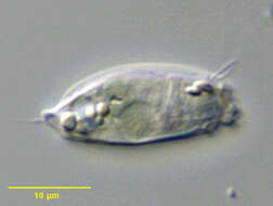
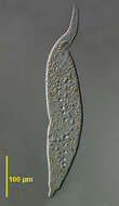
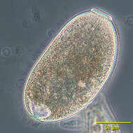
Portrait of the large ciliate, Monilicaryon monilatus (Stokes, 1886) Jankowski, 1967. This cell is slightly compressed. Similar in overall appearance to Dileptus anser. M. monilatus differs by having a shorter proboscis relative to the length of the body (1/3 to 1/4) and by lacking the row of obliquely oriented closely spaced kinetids on the ventral aspect of the left side of the proboscis (this feature requires demonstration by DIC or protargol staining). M. monilatus has two single files of kinetids extending from either side of the oral aperture anteriorly along the ventral aspect of the proboscis separated by a strip bearing extrusomes (See Foissner W., Berger H and Kohmann F. Taxonomische und ökologische Revision der Ciliaten des Saprobiensystems- Band IV: Gymnostomatea, Loxodes, Suctoria. Informationsberichte Bayer. Landesamtes für Wasserwirtschaft. 1/95:185-202, 1995). The moniliform macronucleus is seen here. There are 20-40 small dorsal contractile vacuoles each emptying through a single excretory pore. There is often a larger contractile vacuole at the base of the tail. Collected from a freshwater pond near Boise, Idaho. DIC.
-
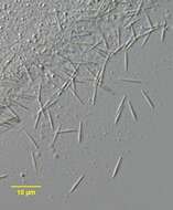
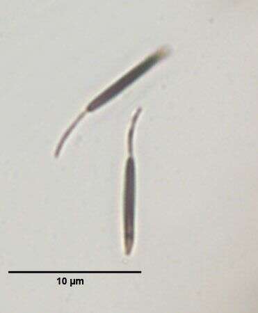
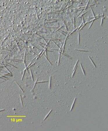
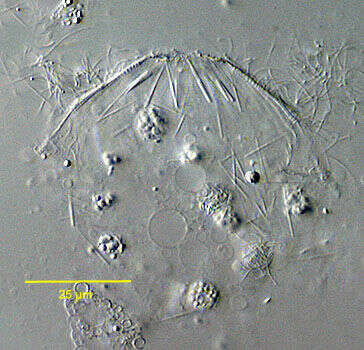
Extrusomes of the haptorid ciliate,Askenasia volvox (Eichwald,1852) Kahl, 1930. The cell is disrupted by coverslip pressue.The cytostome is at the anterior apex.The needle-loke extrusomes (some in bundles)are seen here scattered through the cytoplasm.In the living organism they are in a longitudinal bundle posterior to the anterior apical oral dome. From a freshwater pond near Boise, Idaho.DIC.
-
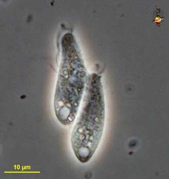
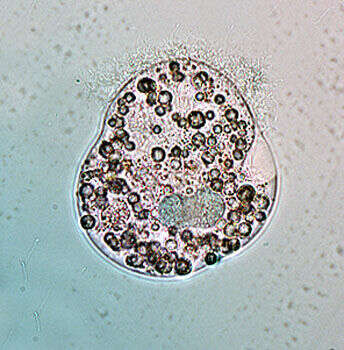
Enchelys (en-chill-iss) small predatory ciliate, here two cells are linked in conjugation (during which time DNA is exchanged - it+s a kind of sexual activity but without reproduction). Clear vacuoles at the rear are contractile vacuoles. Phase contrast micrograph.
-
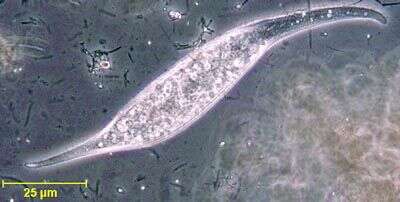
In vivo portrait of Pithothorax processus (Kahl, 1926), a small haptorid ciliate found in polysaprobic habitats. The body is a slightly flattened cylinder. The pellicle is rigid with longitudinal ribbing. The oral aperture is at the anterior apex surrounded by projections of the pellicular ridges. There is a curved funnel-shaped posterior process from which a long caudal cilium protrudes (seen here). The round macronucleus is anterior and the contractile vacuole is located in the posterior 1/3 at the periphery (seen here). The somatic ciliature is confined to the anterior and posterior quarters of the body. From stagnant organically enriched freshwater pond near Boise, Idaho. June 2005. DIC optics.
-
-
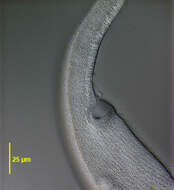
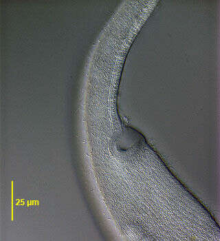
Detail of the dorsal surface of Monilicaryon monilatus (Stokes, 1886) Jankowski, 1967 showing the single excretory pore of several of the 20 to 40 small contractile vacuoles(See Foissner W., Berger H and Kohmann F. Taxonomische und ökologische Revision der Ciliaten des Saprobiensystems- Band IV: Gymnostomatea, Loxodes, Suctoria. Informationsberichte Bayer. Landesamtes für Wasserwirtschaft. 1/95:185-202, 1995). There is often a larger contractile vacuole at the base of the tail. Collected from a freshwater pond near Boise, Idaho. DIC.
-
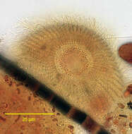
Macronucleus (stained green in this preparation)of the haptorid ciliate,Askenasia volvox (Eichwald,1852) Kahl, 1930. The cell is spherical posteriorly and the anterior is a truncate cone.The short C-shaped macronucleus is in the posterior cell perpendicular to the long axis.Stained by the methyl green-pyronin technique (see Foissner, W. Europ. J. Protistol., 27:313-330;1991). From a freshwater pond near Boise, Idaho.Brightfield.
-
Enchelydium KAHL,1930, species undetermined.Phase contrast.
-
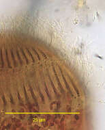
Portrait of Pithothorax processus (Kahl, 1926), a small haptorid ciliate found in polysaprobic habitats. The body is a slightly flattened cylinder. The pellicle is rigid with longitudinal ribbing. The oral aperture is at the anterior apex surrounded by pointed projections of the pellicular ridges. There is a funnel shaped posterior process from which a long caudal cilium protrudes (visible here). The round macronucleus is anterior. The somatic ciliature is confined to the anterior and posterior quarters of the body. Although this specimen was stained by the silvercarbonate technic (see Foissner, W. Europ. J. Protistol., 27:313-330;1991, the somatic kinetids did not impregnate. Short rod shaped partially discharged extrusomes (stained brown here) are visible anteriorly. From stagnant organically enriched freshwater pond near Boise, Idaho. Brighfield.
-
-
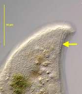
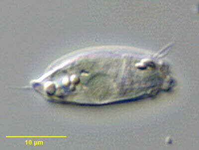
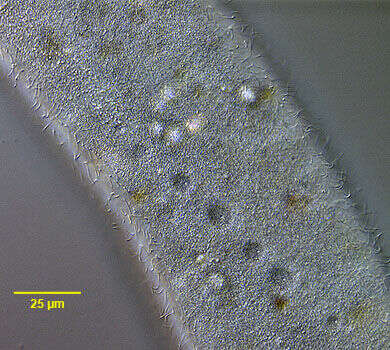
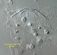
Amphileptus (am-fee-lep-tus) claparedii. The body of the members of the genus Amphileptus is laterally compressed and elongate. The oral aperture is a slit on the convex edge of the neck region, and extends less than halfway down the body. Ciliation is present on both lateral surfaces although there is a tendency to some reduction on the left surface. Ciliation on the right surface is extensive and forms longitudinal rows which converge on each other in the anterior region. Trichocysts are common - particularly in neck. Macronucleus in 2 to 4 spherical parts with single micronucleus placed between macronuclei. Many contractile vacuoles occur along both dorsal and ventral edges. Lives in fresh water ponds and lakes. Slightly squashed specimen of Amphileptus claparadeii. Macronuclei and the contractile vacuoles located at the edges are visible. Measuring 230 microns. Differential interference contrast.
-
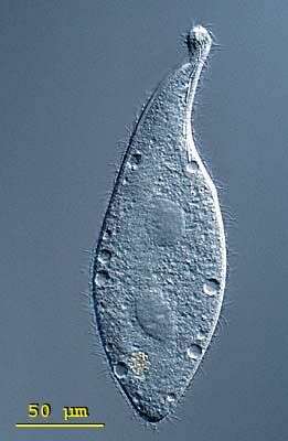
Portrait of Amphileptus, a widely distributed haptorid ciliate with posterior drawn out to a tapered point and elongate anterior neck region. Prominent refractile trichites are clustered at the anterior end of the neck region. Right side longitudinal kineties converge on one another anteriorly and posteriorly in a diamond shaped pattern unlike the parallel kineties of the similar genus Litonotus. The mouth is slit-like and difficult to see, located on the convex aspect of the neck region. Two spherical macronuclei. Multiple contractile vacuoles along dorsal and ventral margins. Some species attach to and prey on stalked peritrich ciliates. From freshwater pond near Boise, Idaho. Phase contrast.