Centers for Disease Control/Division of Parasitic Diseases and Malaria
EOL staff
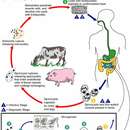
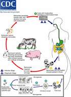
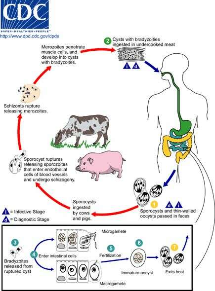
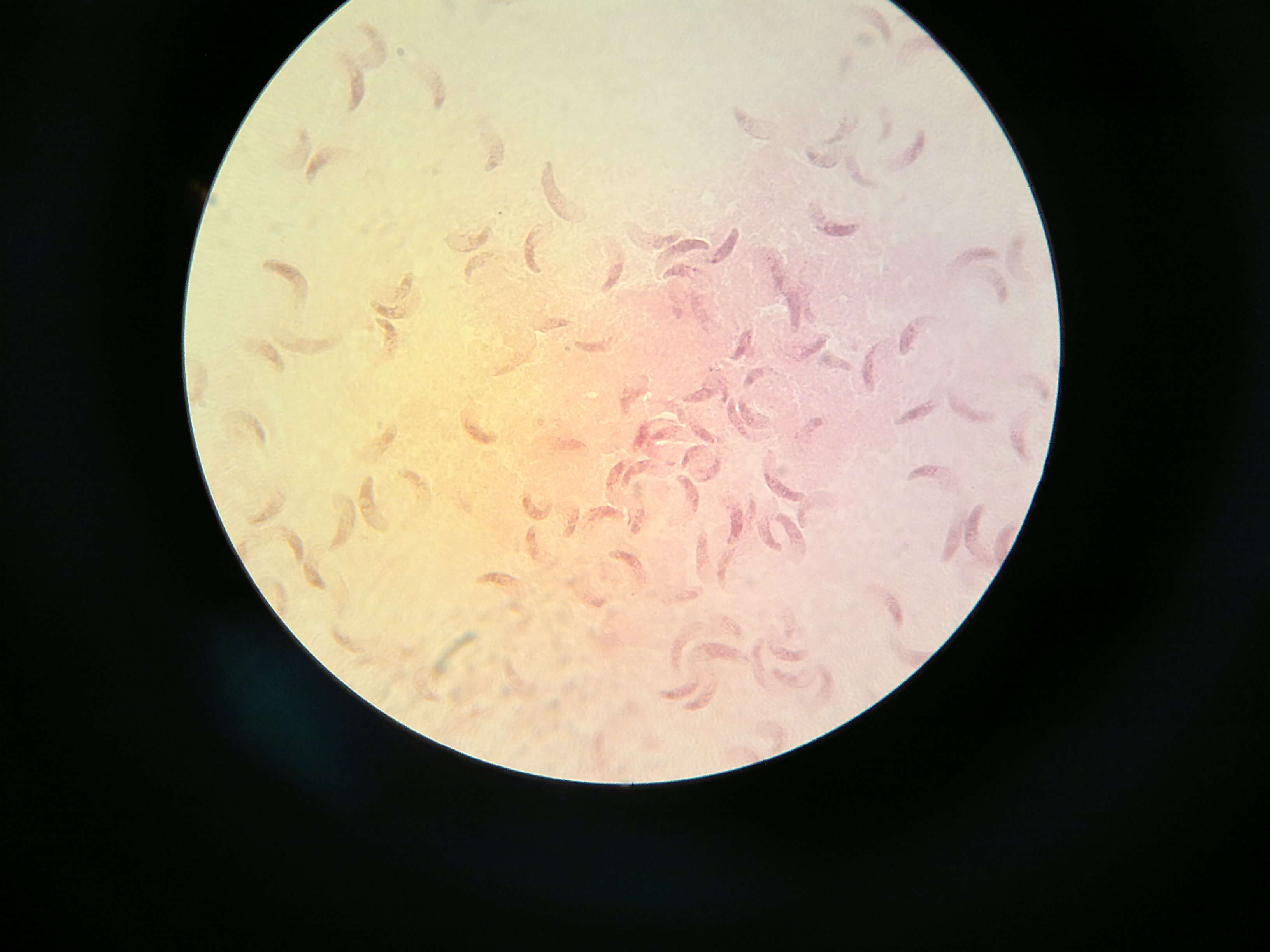
Life cycle of Sarcocystic hominis and S. suihominisBoth sporulated oocysts (containing two sporocysts) and individual sporocysts can be passed in stool (1). Sporocysts contain four sporozoites and a refractile residual body. Sporocysts ingested by the intermediate host (cattle for S. hominis and pigs for S. suihominis) rupture, releasing sporozoites. Sporozoites enter endothelial cells of blood vessels and undergo schizogony, resulting in first-generation schizonts. Merozoites derived from the first-generation invade small capillaries and blood vessels, becoming second-generation schizonts. The second generation merozoites invade muscle cells and develop into sarcocysts containing bradyzoites, which are the infective stage for the definitive host (2). Humans become infected when they eat undercooked meat containing these sarcocysts. Bradyzoites are released from ruptured cysts in the small intestine (3) and invade the lamina propria of the intestinal epithelium (4). There, they differentiate into macro- and microgametocytes. Fusion of male and female gametes (5) results in the formation of oocysts (6). Oocysts sporulate in the intestinal epithelium and are shed from the host in feces (7). Due to the fragile nature of the oocyst wall, individual sporocysts may also be detected in feces.From Centers for Disease Control Parasites and Health website
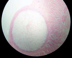
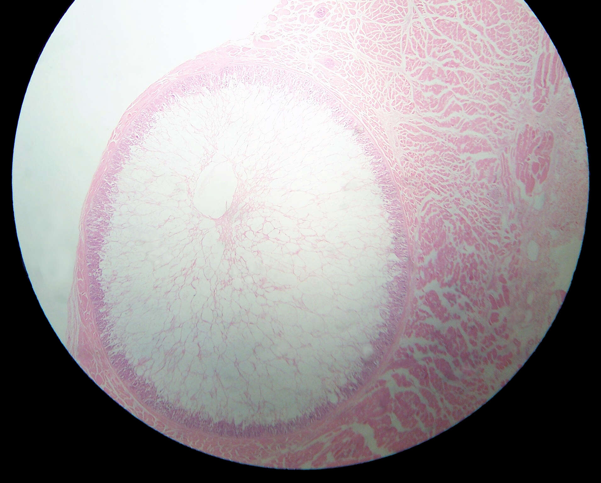
Description: Sarcocystis in a sheep oesophagus. Photograph taken from teaching slides at the University of Edinburgh. The cyst is approximately 4mm across. Date: 26 November 2007. Source: Own work. Author: Adam Cuerden.