-
-
-
-
-
-
-
-
-
-
-
-
-
-
-
-
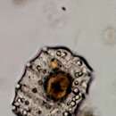
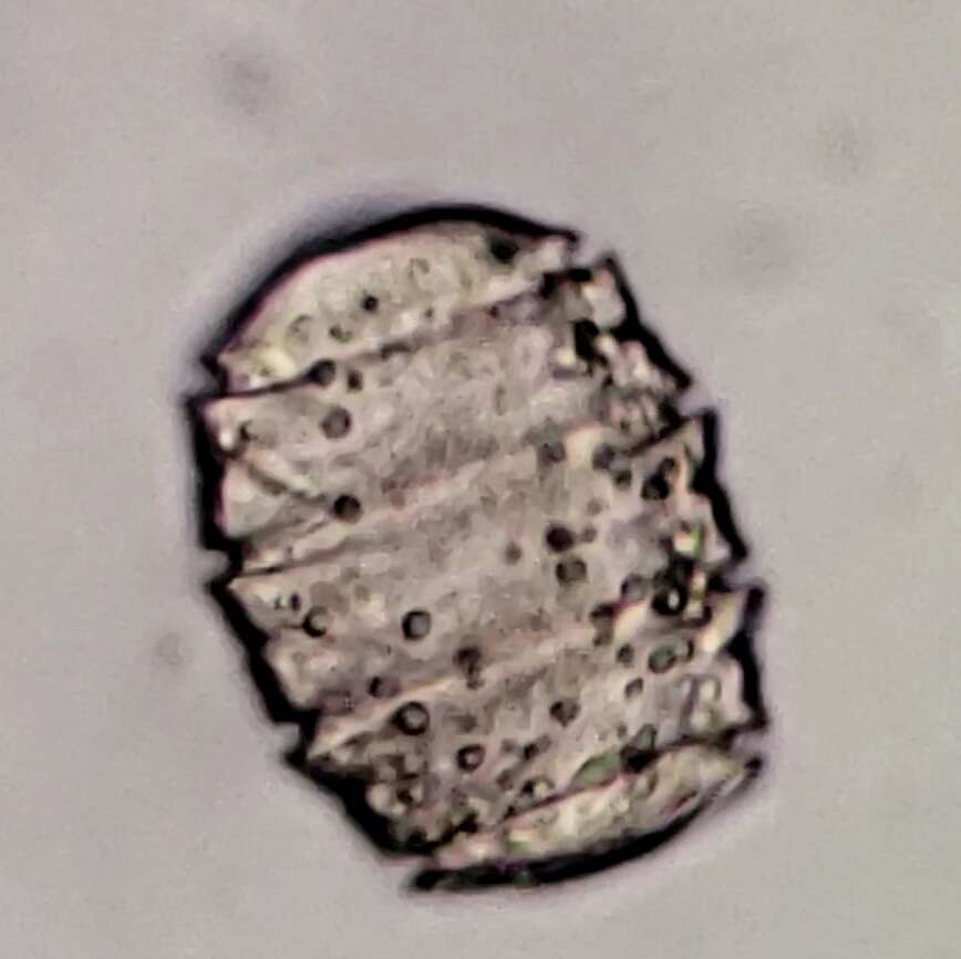
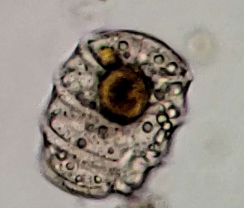
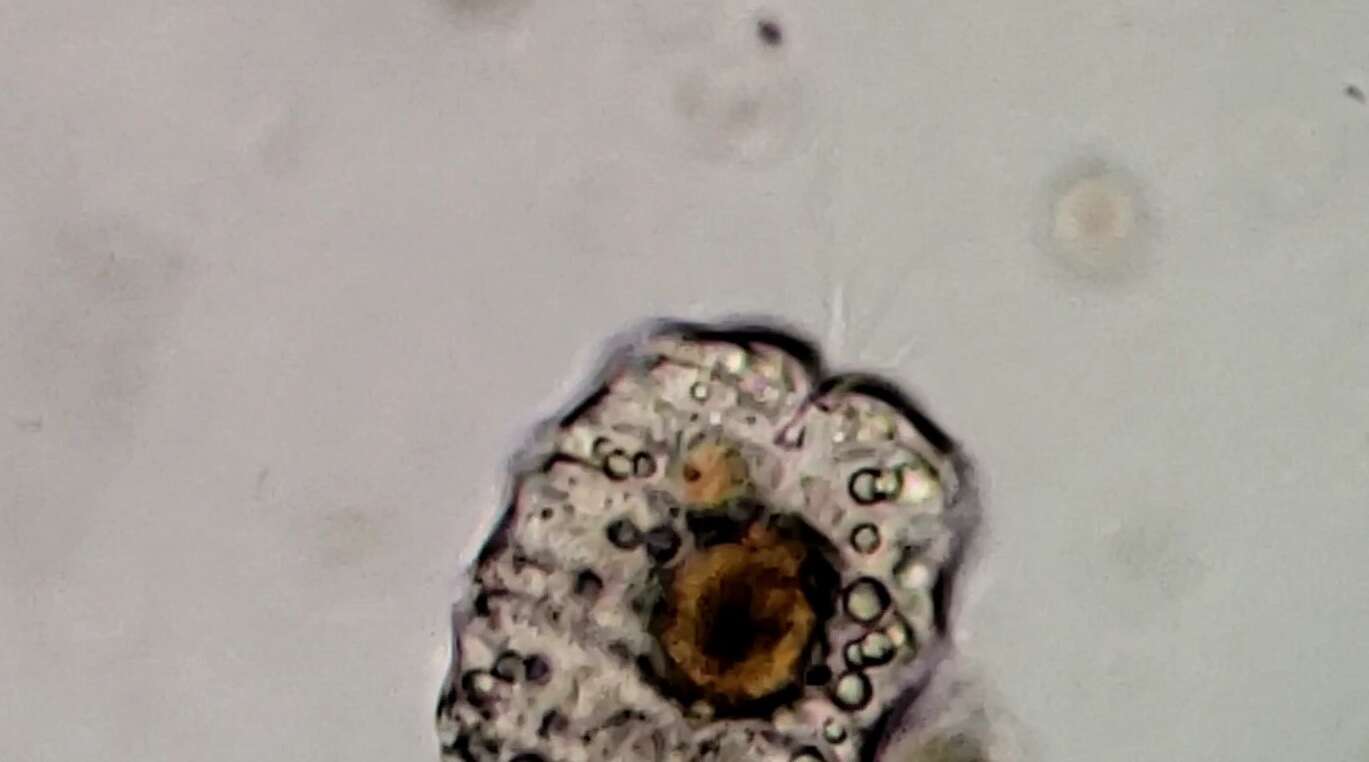
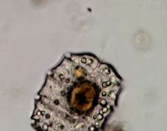
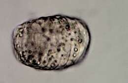
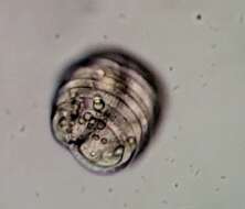
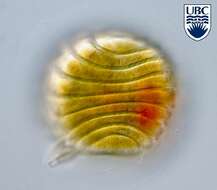
Polykrikos (polly-cry-coz) lebourae Herdman 1923. The image shows a cell in right lateral view. The cell has many cingula. The cell contains no plastids, however a food particle is present.
-
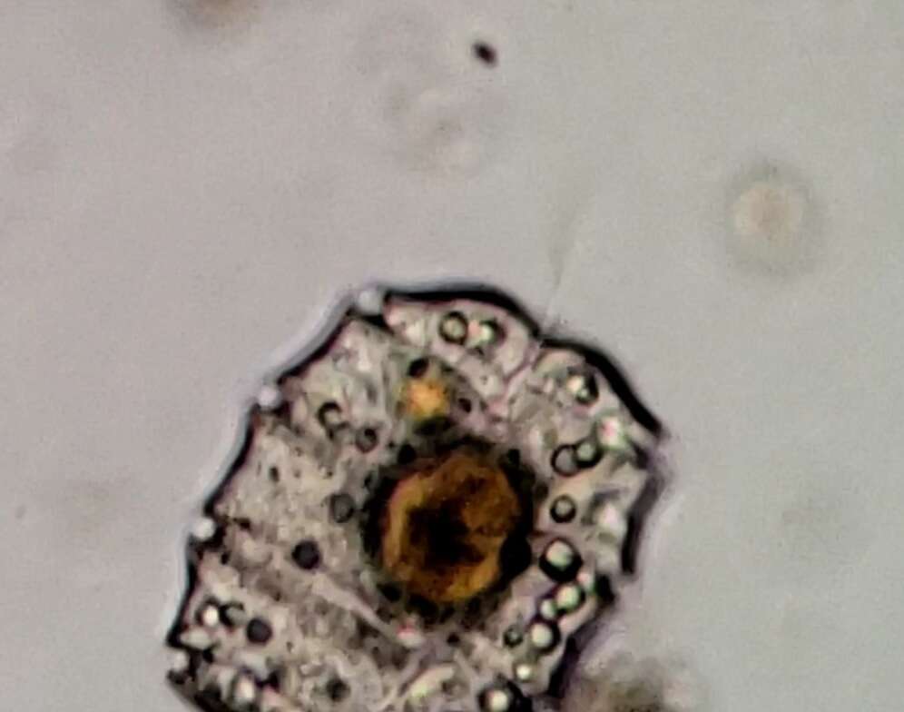
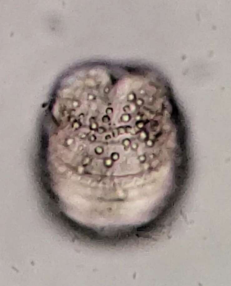
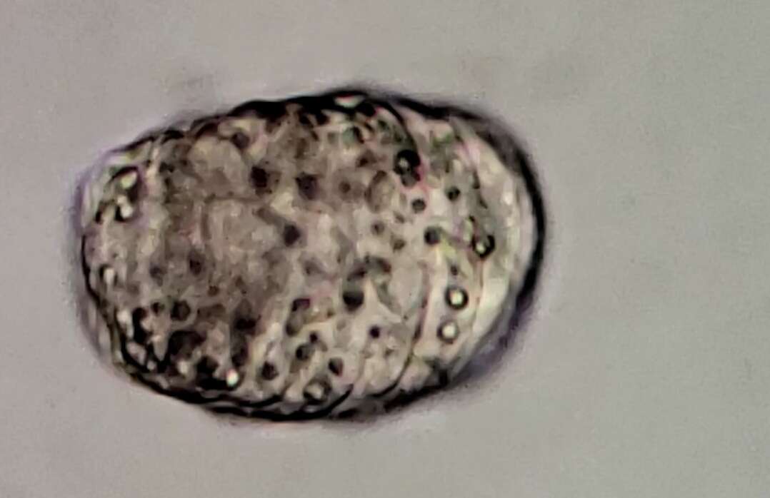
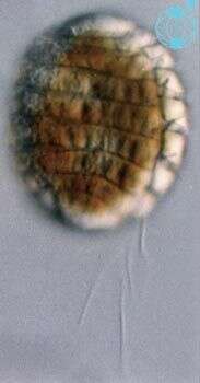
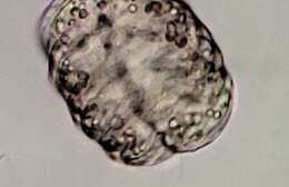
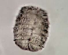
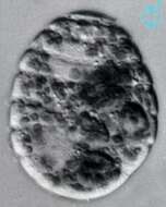
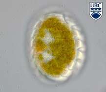
Polykrikos (polly-cry-coz) lebourae Herdman 1923. The image shows a cell in lateral view. The cell has many cingula. There are two nuclei present, one in the posterior of the cell, the other near the anterior end.
-
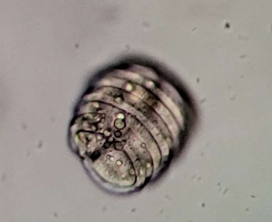
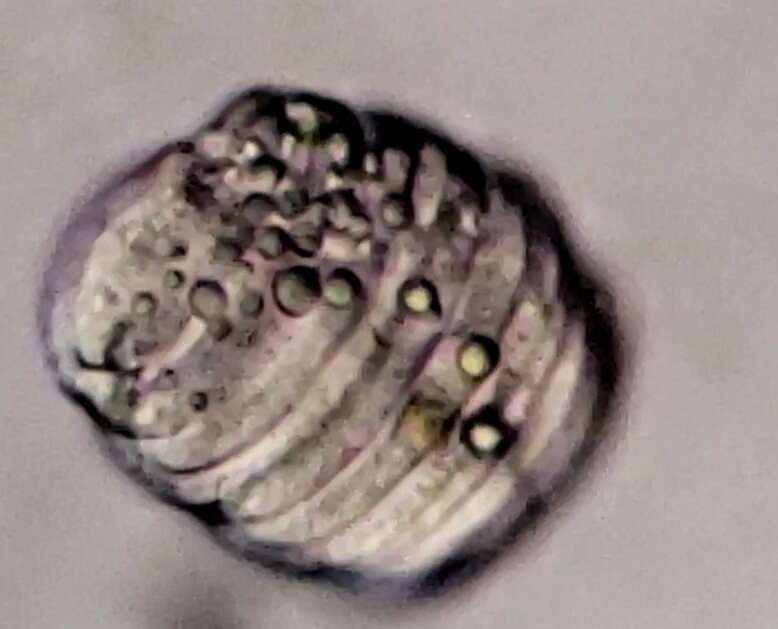
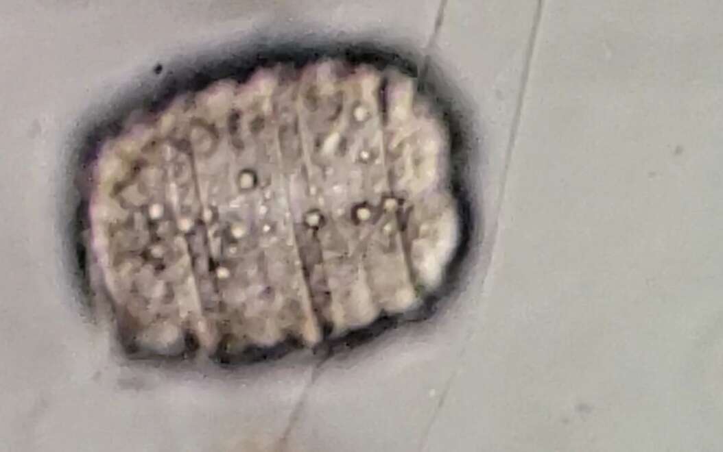
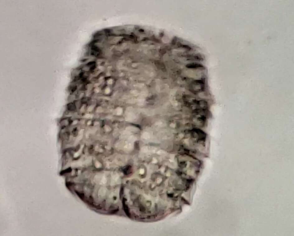
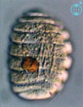
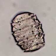
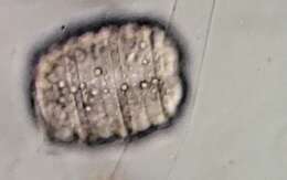
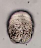
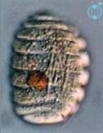
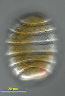
Polykrikos (polly-cry-coz) lebourae Herdman 1923. The image shows a cell in right lateral view. The cell has many cingula.
-
Polykrikos lebourae Herdman 1923
-
Polykrikos lebourae Herdman 1923
-
Polykrikos lebourae Herdman 1923
-
Polykrikos lebourae Herdman 1923
-
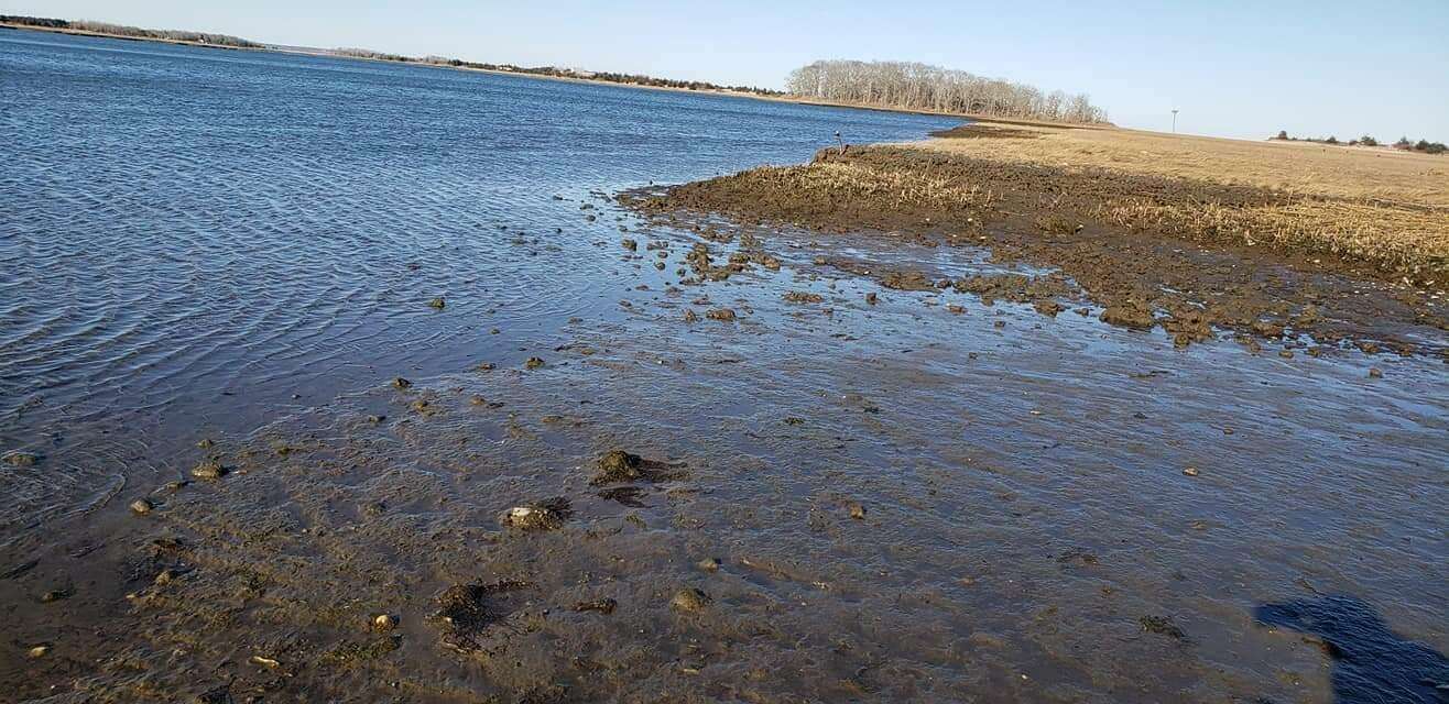
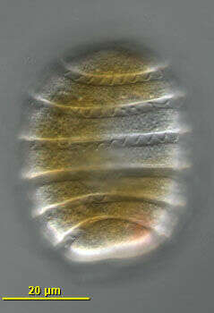
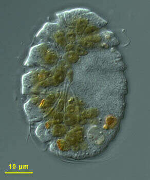
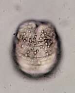
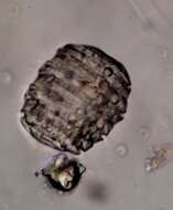
Dorso-lateral view of the numerous transverse flagella lie in the cingular grooves of this phagotrophic gymnodinioid dinoflagellate. Isolated by Bob Moore from Little Sippiwissett marsh near Woods Hole, Massachusetts, USA. Differential interference contrast optics.
-
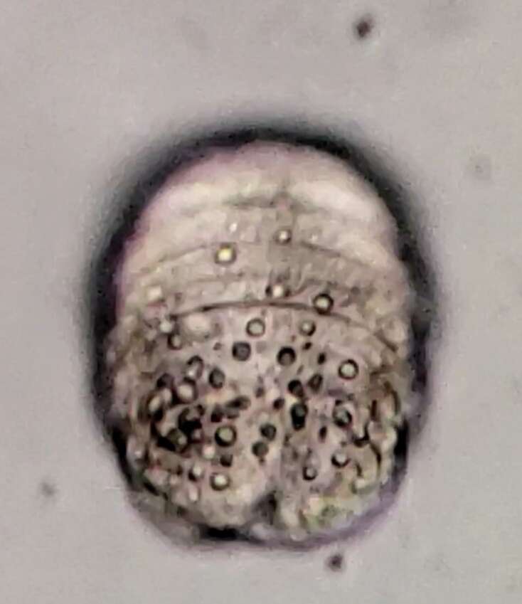
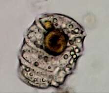
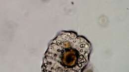
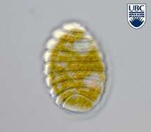
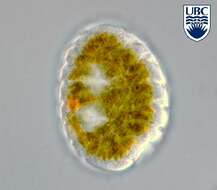
Differential interference contrast image of this phagotrophic dinoflagellate isolated from Chappaquoit beach, Massachusetts. The brown inclusions are partly digested elements from food - this species was observed consuming other dinoflagellates. This species may have more than one nucleus, but in this cell there is a singler nucleus (middle of right hand side of cell). There are also large extrusomes within the cell. Image by all.