-
Wayne N. Mathis, Alessandra Rung, Marion Kotrba
Zookeys
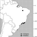
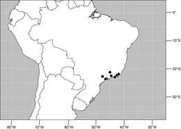
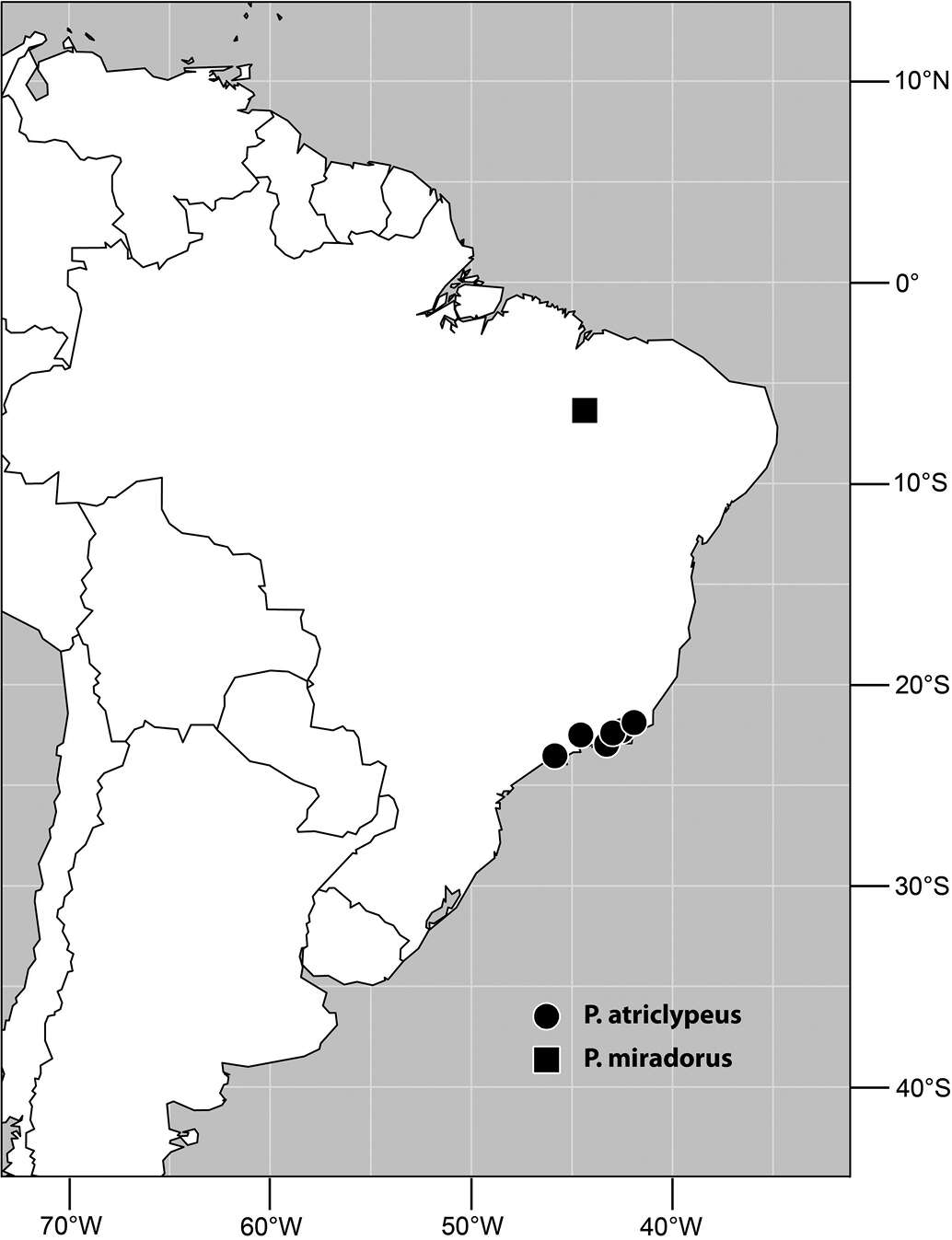
Figure 34.Distribution of Planinasus miradorus sp. n. (square) and Planinasus atriclypeus (dots).
-
Wayne N. Mathis, Alessandra Rung, Marion Kotrba
Zookeys
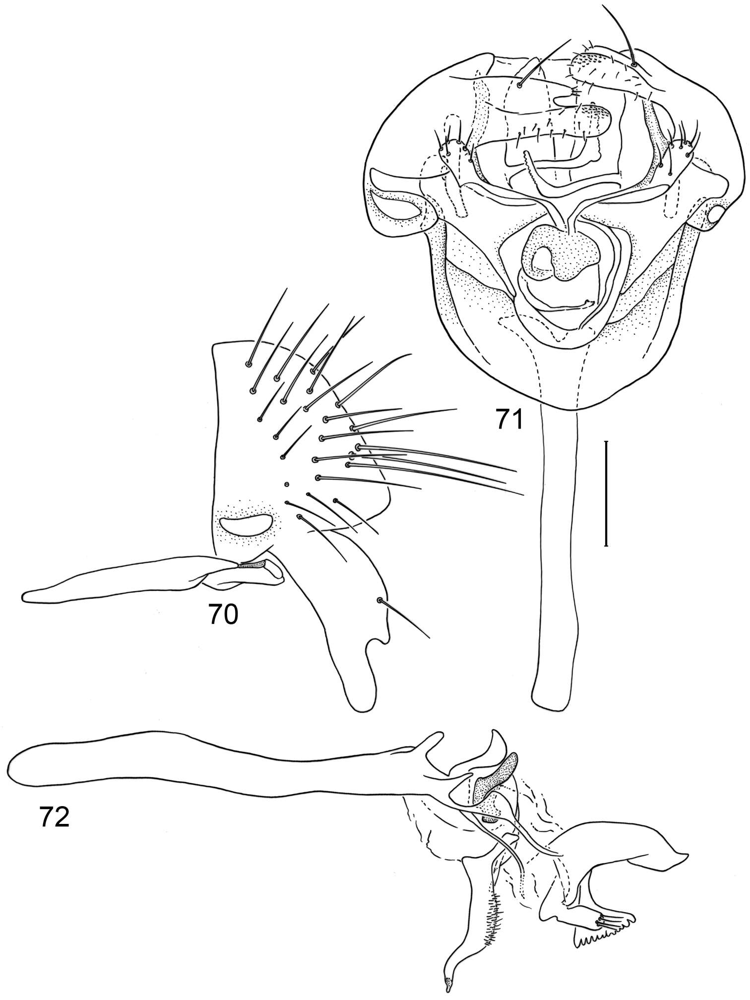
Figures 70–72.Illustrations of Planinasus atriclypeus sp. n. (male). 70 epandrium, surstylus, hypandrium, and pregonite, lateral view 71 structures of internal male terminalia, ventral view 72 internal structures of male terminalia, lateral view, lateral view. Scale bar = 0.1 mm.
-
Wayne N. Mathis, Alessandra Rung, Marion Kotrba
Zookeys
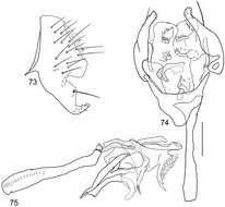
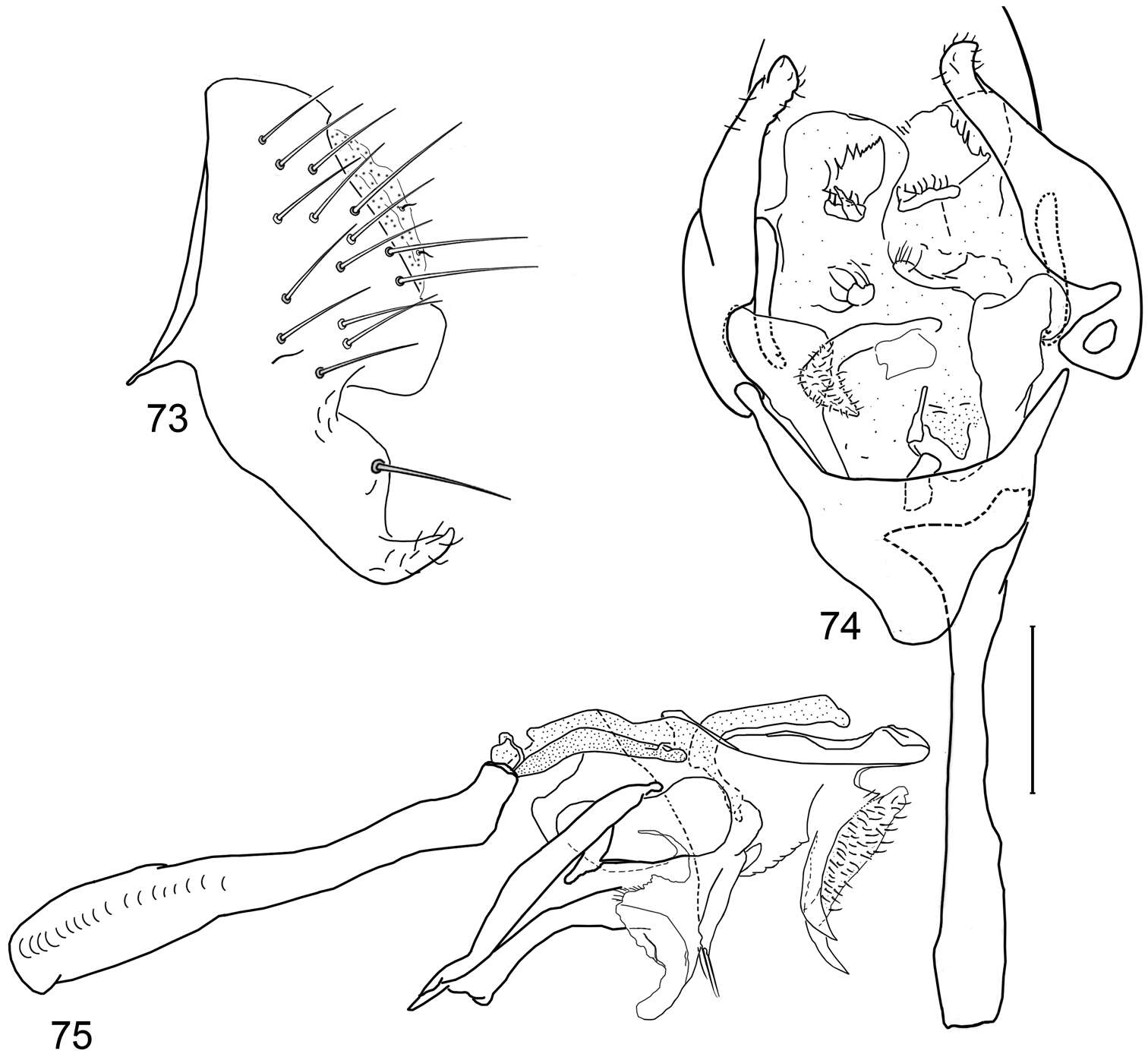
Figures 73–75.Illustrations of Planinasus atrifrons sp. n. (male). 73 epandrium and surstylus, lateral view 74 structures of internal male terminalia, ventral view 75 internal structures of male terminalia, lateral view. Scale bar = 0.1 mm.
-
Wayne N. Mathis, Alessandra Rung, Marion Kotrba
Zookeys
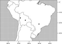
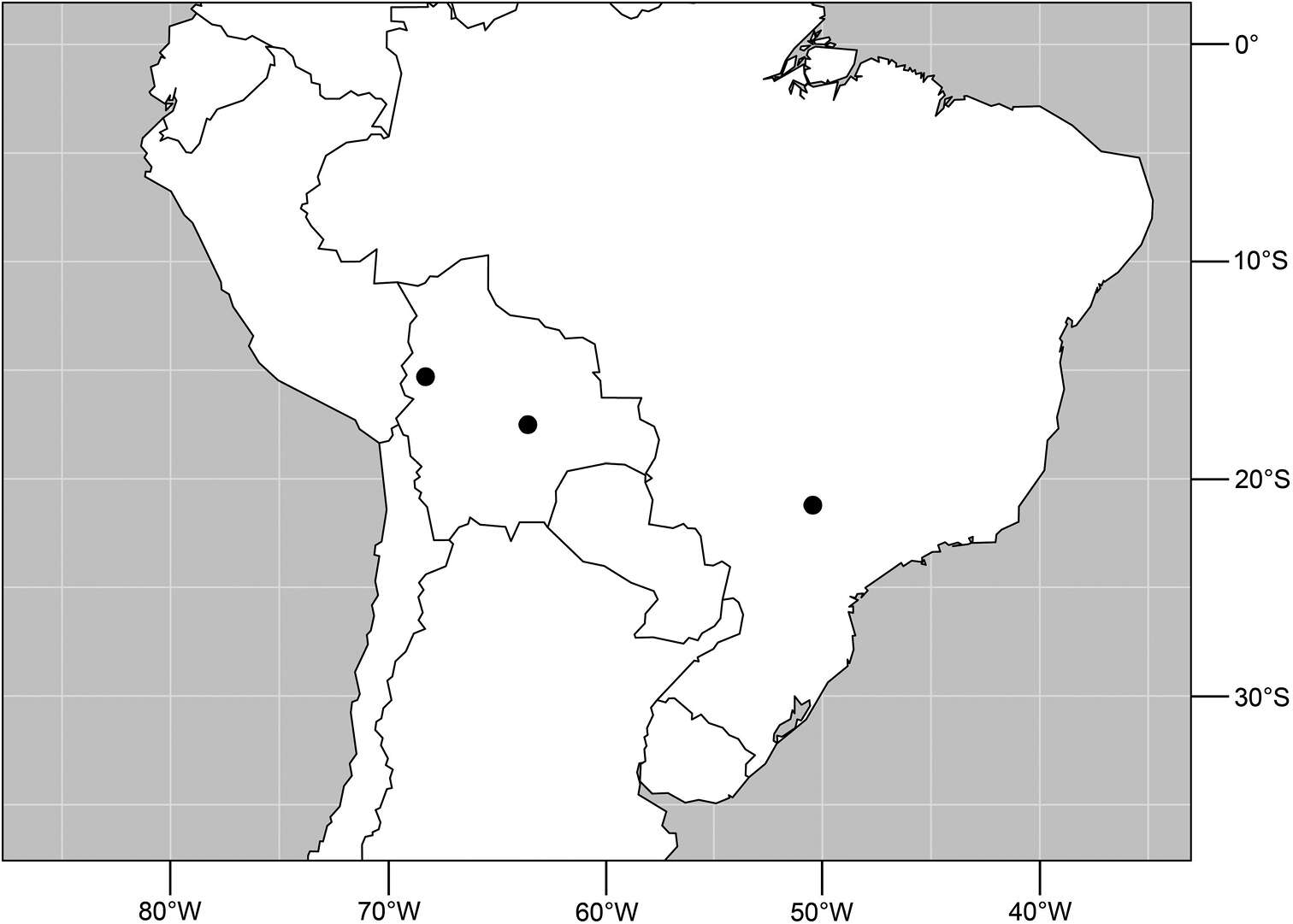
Figure 76.Distribution of Planinasus atrifrons sp. n.
-
Wayne N. Mathis, Alessandra Rung, Marion Kotrba
Zookeys
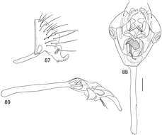
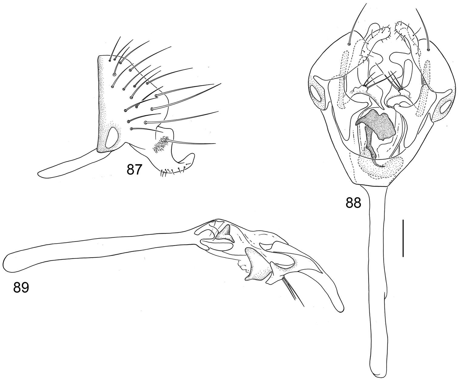
Figures 87–89.Illustrations of Planinasus nigrifacies sp. n. (male). 87 epandrium, surstylus, hypandrium, lateral view 88 structures of internal male terminalia, ventral view 89 internal structures of male terminalia, lateral view. Scale bar = 0.1 mm.
-
Wayne N. Mathis, Alessandra Rung, Marion Kotrba
Zookeys
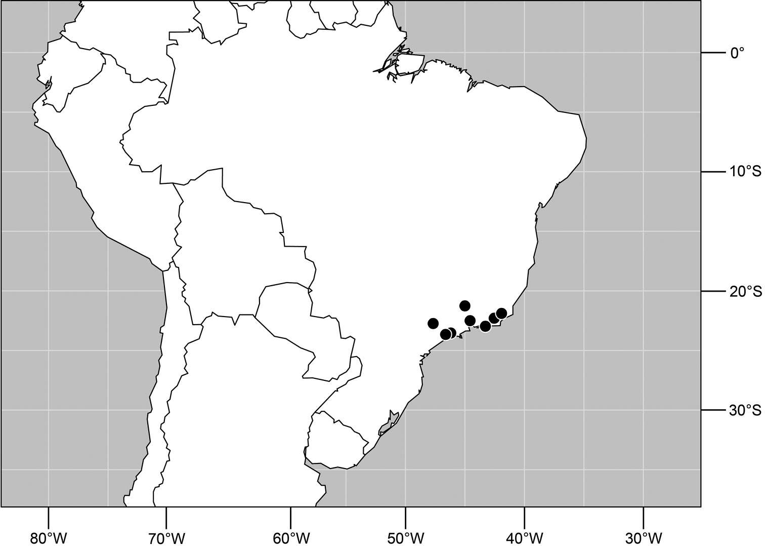
Figure 90.Distribution of Planinasus nigrifacies sp. n.
-
Wayne N. Mathis, Alessandra Rung, Marion Kotrba
Zookeys
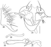
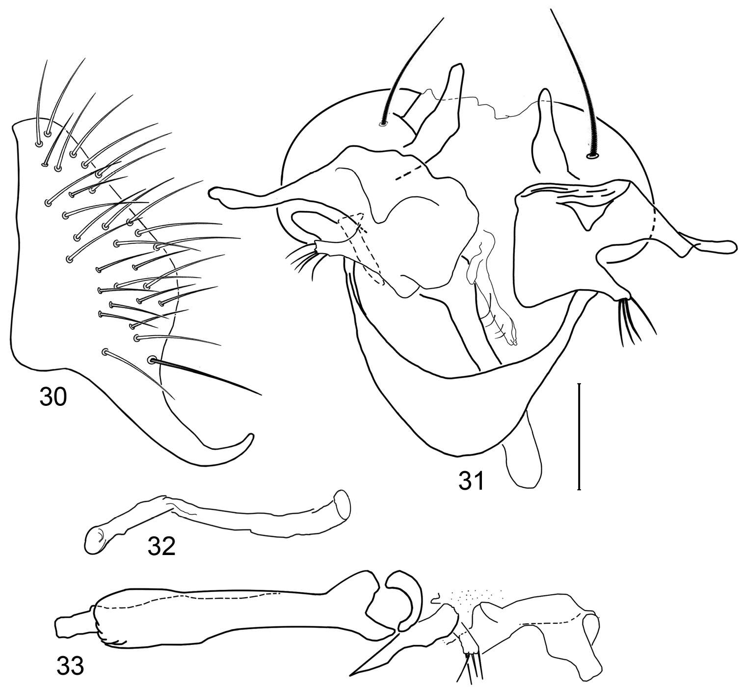
Figures 30–33.Illustrations of Planinasus miradorus sp. n. (male). 30 epandrium, surstylus, , lateral view 31 epandrium, hypandrium, and internal structures of male terminalia, ventral view 32 ejaculatory apodeme, lateral view 33 internal structures of male terminalia, lateral view. Scale bar = 0.1 mm.
-
Wayne N. Mathis, Alessandra Rung, Marion Kotrba
Zookeys
Figure 34.Distribution of Planinasus miradorus sp. n. (square) and Planinasus atriclypeus (dots).
-
Wayne N. Mathis, Alessandra Rung, Marion Kotrba
Zookeys
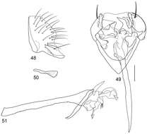
Figures 48–51.Illustrations of Planinasus xanthops sp. n. (male). 48 epandrium, surstylus, lateral view 49 structures of internal male terminalia, ventral view 50 ejaculatory apodeme, lateral view 51 phallus, phallapodeme, pre- and postgonite, lateral view. Scale bar = 0.1 mm.
-
Wayne N. Mathis, Alessandra Rung, Marion Kotrba
Zookeys
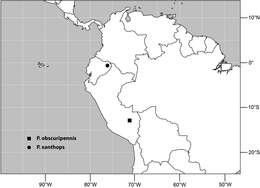
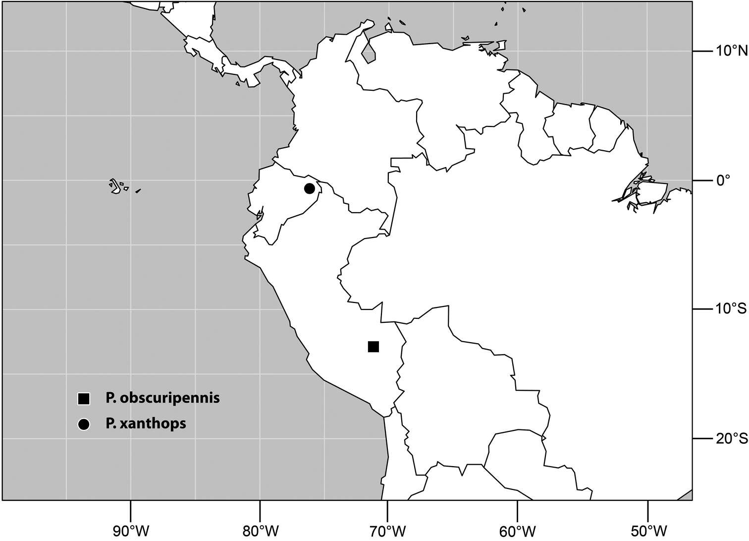
Figure 52.Distribution of Planinasus xanthops sp. n. (dot) and Planinasus obscuripennis sp. n. (square).