-
Christer Hansson, Jean-Paul Lachaud, Gabriela Pérez-Lachaud
Zookeys
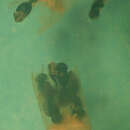
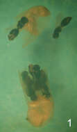
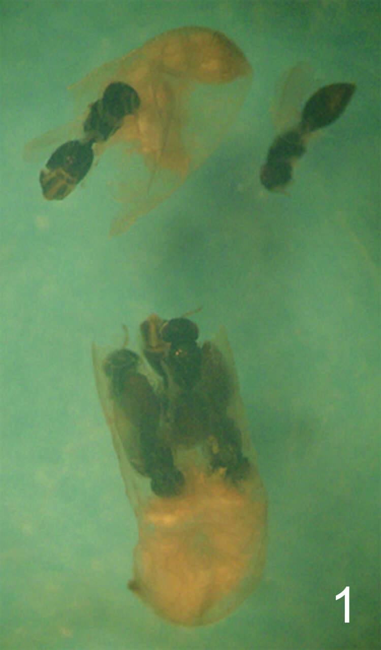
Figure 1. Camponotus sp. ca. textor larva parasitized by Horismenus myrmecophagus. H. myrmecophagus develops as a gregarious endoparasitoid. The ant larva has been cut open (its head is at the bottom of the picture). Several pupae of the eulophid parasitoid may be observed, some of them still inside the ant larva.
-
Ekaterina Shevtsova, Christer Hansson
Zookeys
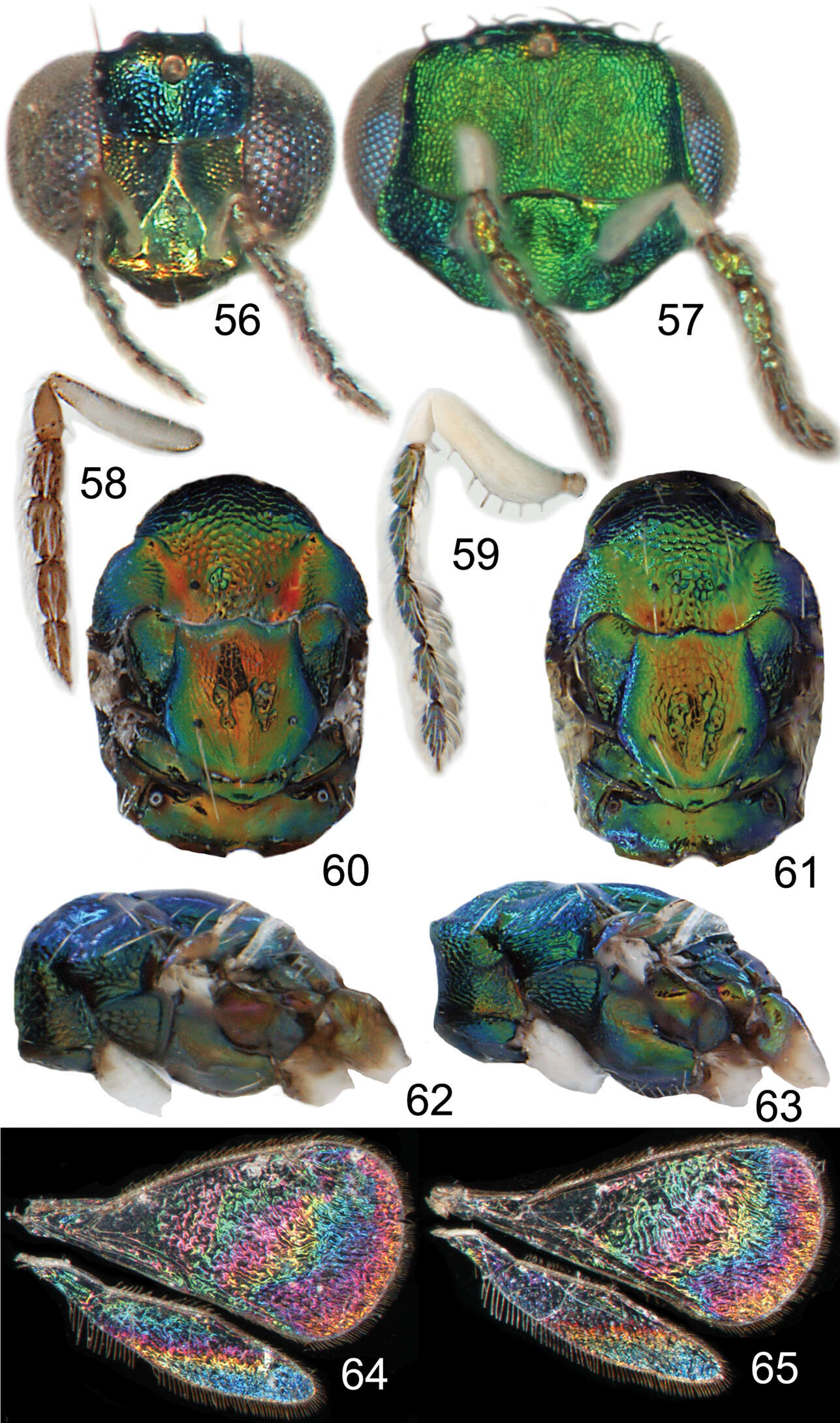
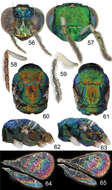
Figures 56–65. Achrysocharoides maieri sp. nov.: 56 Head frontal, female 57 Ditto, male 58 Antenna lateral, female 59 Ditto, male 60 Mesosoma dorsal, female 61 Ditto, male 62 Mesosoma lateral, female 63 Ditto, male 64 Wing interference pattern (WIP), female 65 Ditto, male.
-
Christer Hansson, Ekaterina Shevtsova
Zookeys
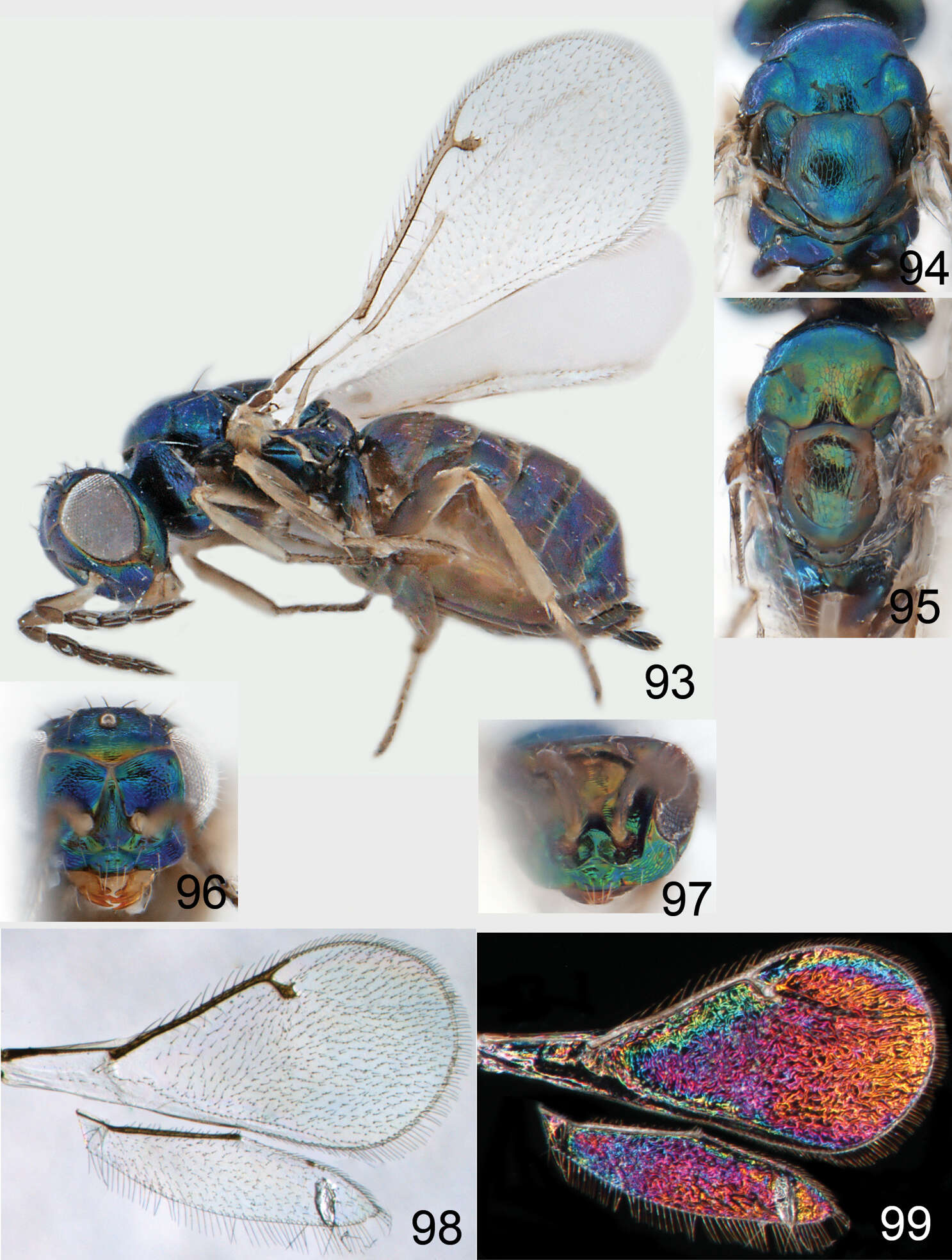
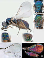
Figures 93–99.Omphale cornula: 93 habitus in lateral view, female, length of specimen 1.5 mm 94 thoracic dorsum, female 95 thoracic dorsum, male 96 head in frontal view, female 97 head in frontal view, male 98 transparent wings, female 99 wing interference patterns, female.
-
Zoya Yefremova, Graciela González-Santarosa, J. Refugio Lomeli-Flores, Néstor Bautista-Martínez
Zookeys
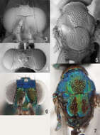
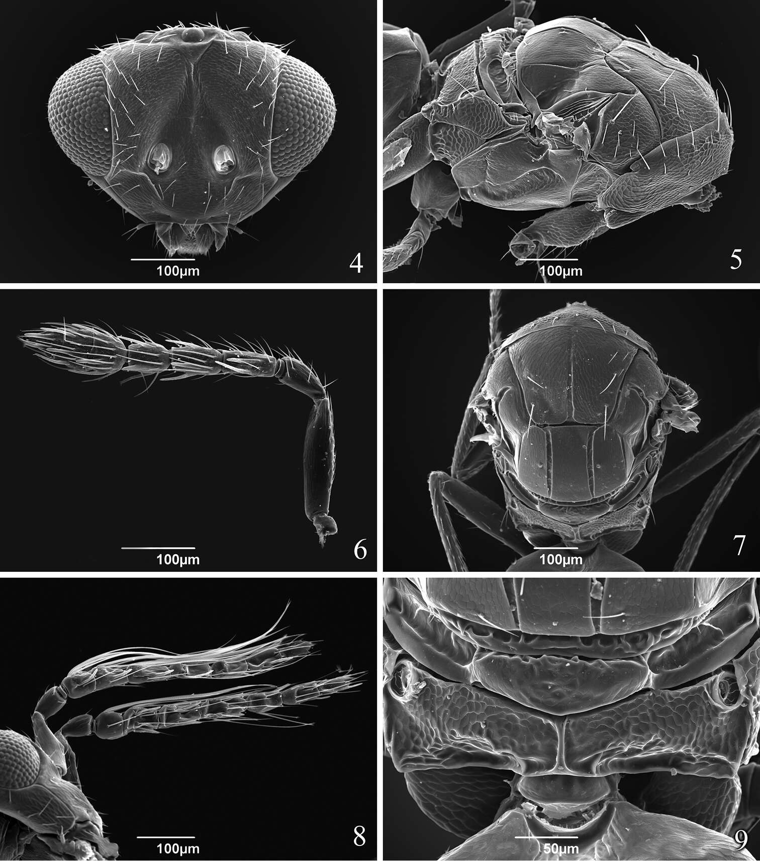
Figures 4–9.Tamarixia aguacatensis. Female: 4 Head, frontal view 5 Mesosoma, lateral view 6 Antenna 7 Mesosoma, dorsal view 9 Propodeum. Male 8 Both antennae on the head.
-
Huan-Xi Cao, John La Salle, Chao-Dong Zhu
Zookeys
Figure 1.Habitus of Zagrammosoma dulanense Cao & Zhu sp. n.: a body in dorsal view (♂) b body in dorsal view (♀) c body in dorsal view (♀) d body in lateral view (♀). Scale bar: 0.5 mm.
-
Christer Hansson, Jean-Paul Lachaud, Gabriela Pérez-Lachaud
Zookeys
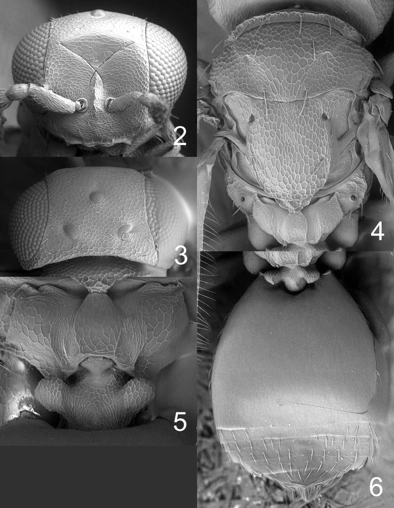
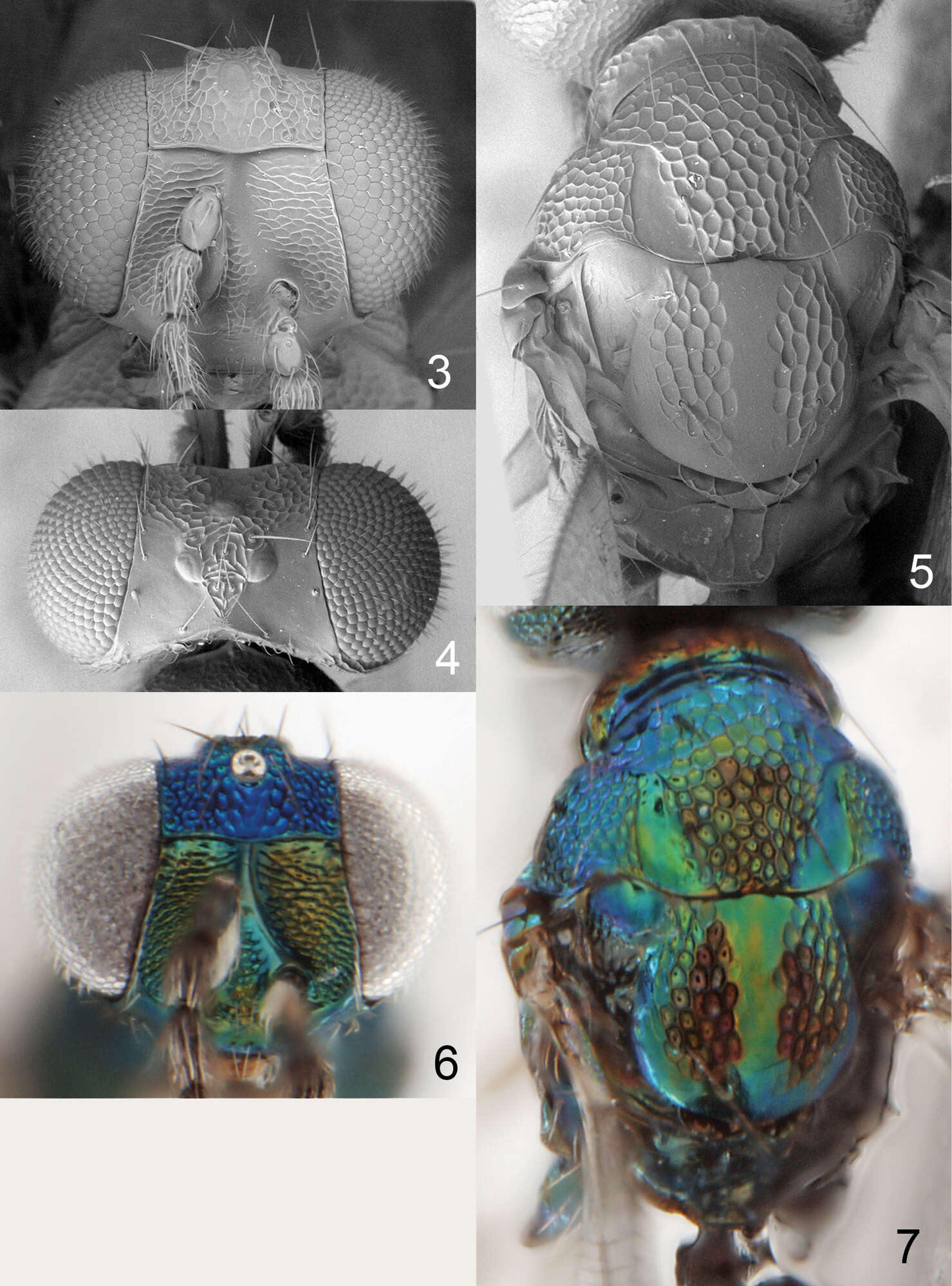
Figures 2–6. Horismenus myrmecophagus female: 2 head in frontal view 3 vertex 4 thoracic dorsum 5 propodeum 6 gaster in dorsal view.
-
Ekaterina Shevtsova, Christer Hansson
Zookeys
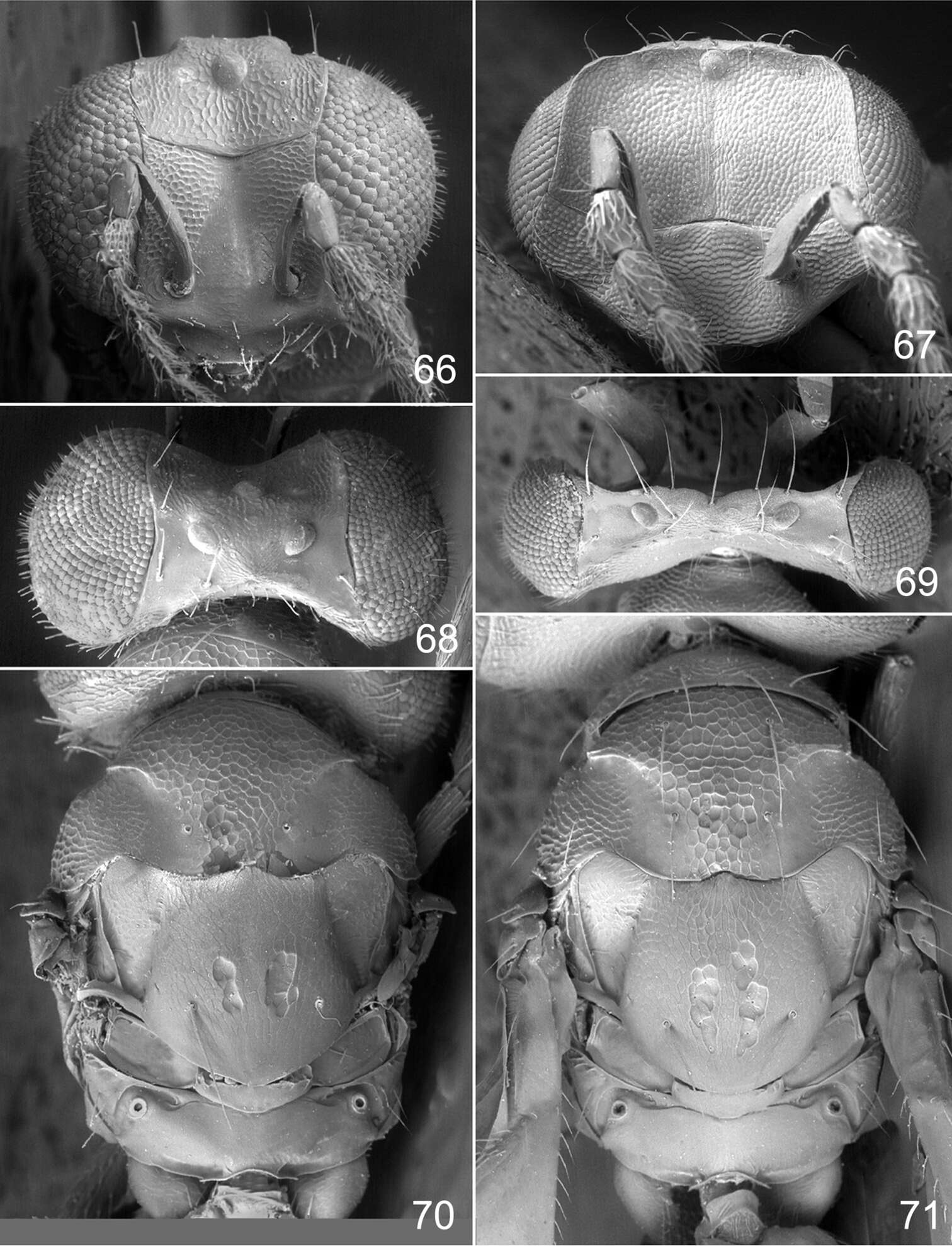
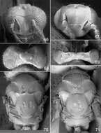
Figures 66–71. Achrysocharoides maieri sp. n.: 66 Head frontal, female 67 Ditto, male 68 Vertex, female 69 Ditto, male 70 Mesosoma dorsal, female. 71 Ditto, male.
-
Christer Hansson, Ekaterina Shevtsova
Zookeys
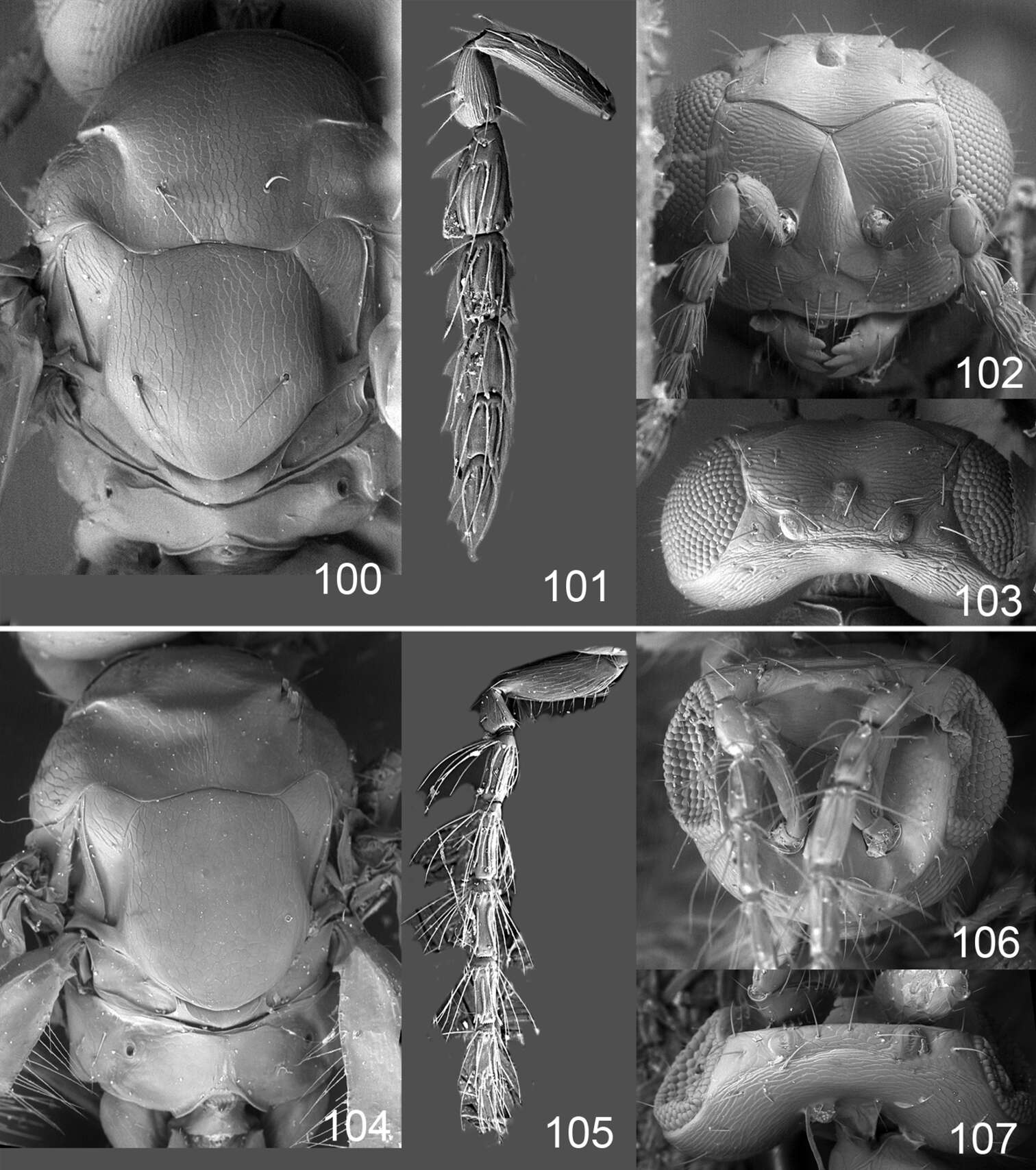
Figures 100–107.Omphale cornula: 100 thoracic dorsum, female 101 antenna, female 102 head in frontal view, female 103 vertex, female 104 thoracic dorsum, male 105 antenna, male 106 head in frontal view, male 107 vertex, male.
-
Zoya Yefremova, Graciela González-Santarosa, J. Refugio Lomeli-Flores, Néstor Bautista-Martínez
Zookeys
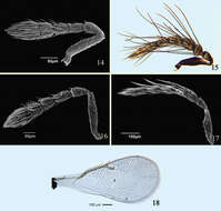
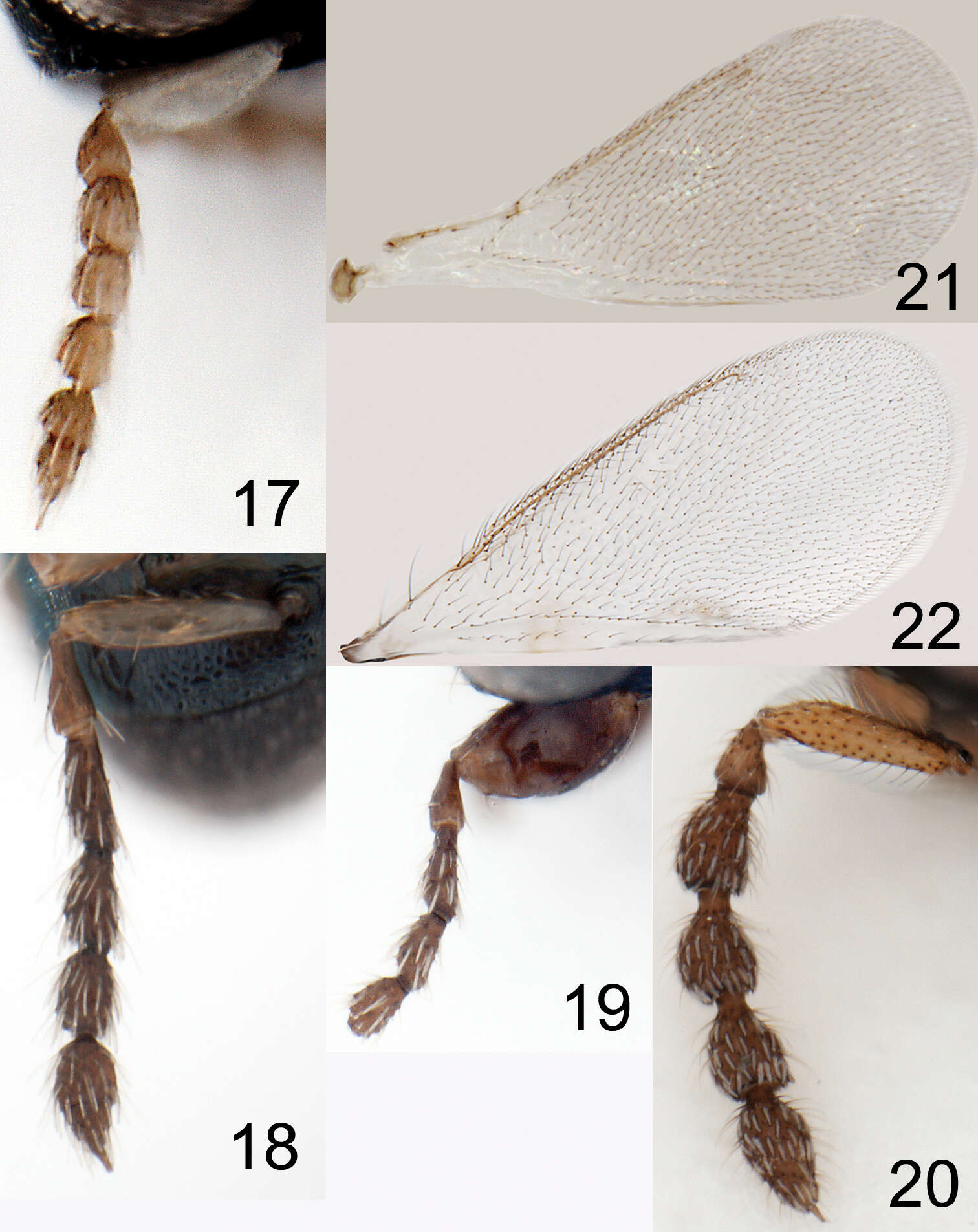
Figures 14–20.Tamarixia schina: 14 Female antenna 15 Male antenna. Tamarixia triozae: 16 Female antenna 17 Male antenna 18 Female fore wing.
-
Huan-Xi Cao, John La Salle, Chao-Dong Zhu
Zookeys
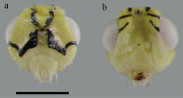
Figure 2.Head (♀): a head in posterior view b head in anterior view. Scale bar: 0.2 mm.
-
Christer Hansson, Jean-Paul Lachaud, Gabriela Pérez-Lachaud
Zookeys
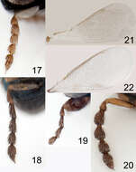
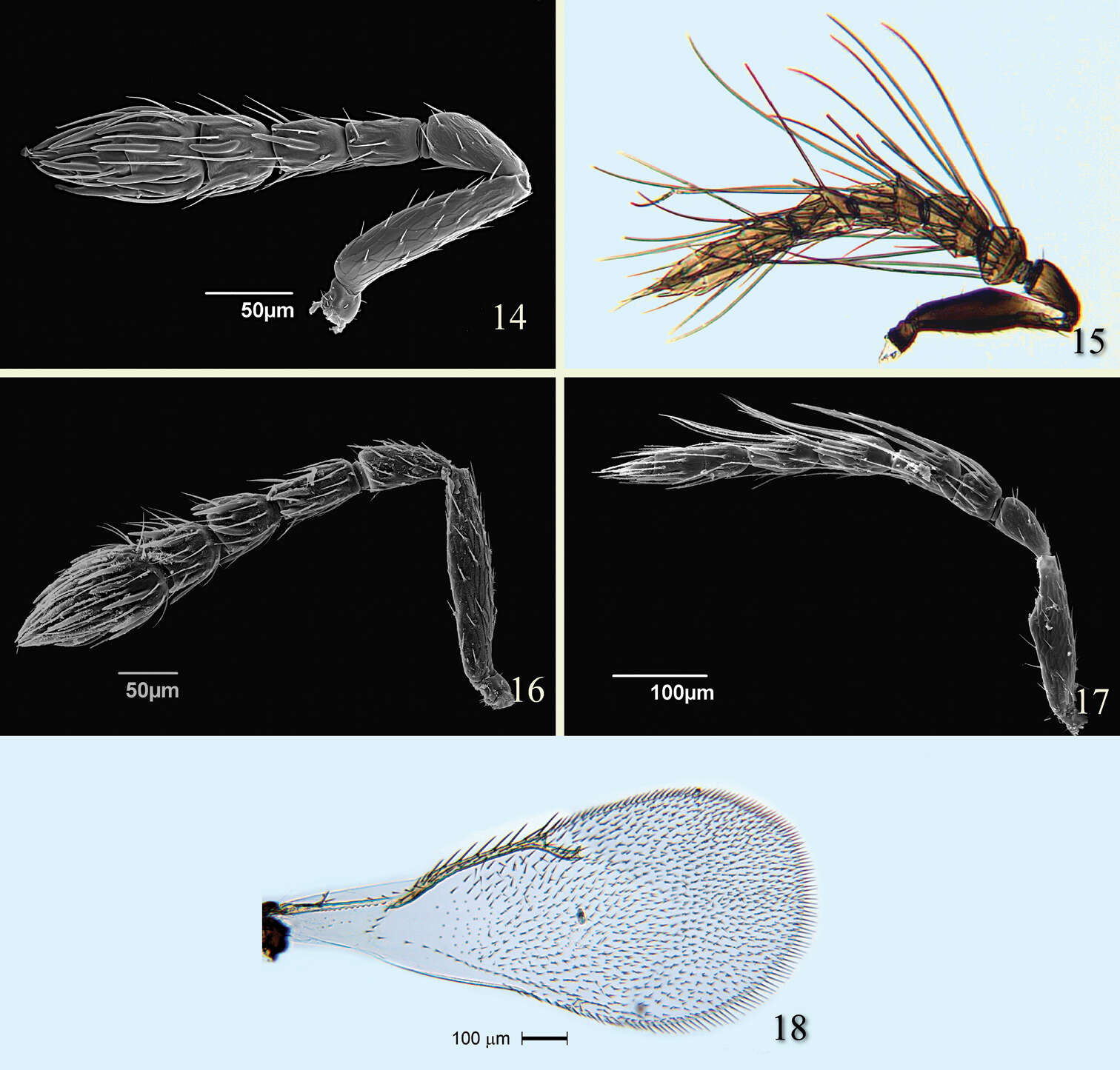
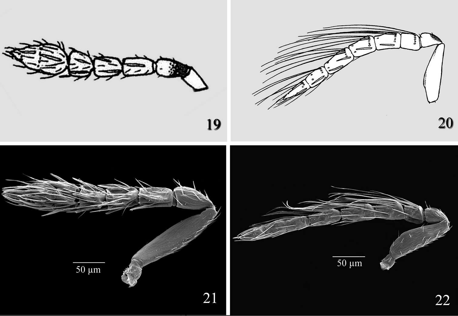
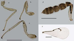
Figures 17–22. 17–20 antenna in lateral view: 17 Horismenus myrmecophagus female 18 H. microdonophagus female 19 H. microdonophagus male (apical 2 flagellomeres missing) 20 Microdonophagus tertius female. 21–22 right fore wing female: 21 H. myrmecophagus 22 H. microdonophagus.
-
Ekaterina Shevtsova, Christer Hansson
Zookeys
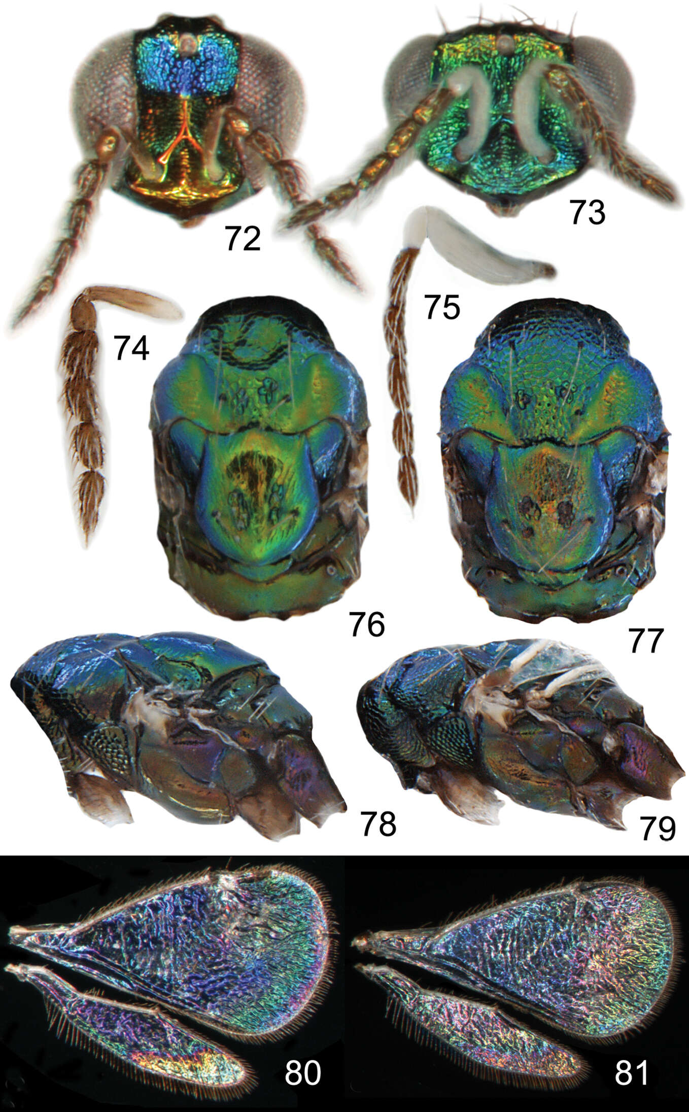
Figures 72–81. Achrysocharoides serotinae sp. n.: 72 Head frontal, female 73 Ditto, male 74 Antenna lateral, female 75 Ditto, male 76 Mesosoma dorsal, female 77 Ditto, male 78 Mesosoma lateral, female 79 Ditto, male 80 Wing interference pattern (WIP), female 81 Ditto, male.
-
Christer Hansson, Ekaterina Shevtsova
Zookeys
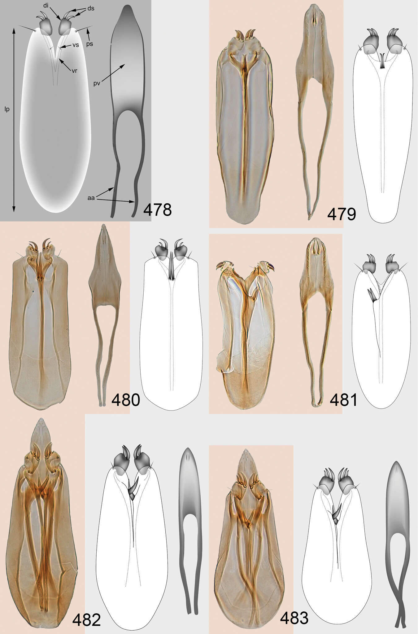
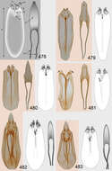
Figures 478–483.Male genitalia (phallobase+aedeagus): 478 Entedon fufius (Walker) (Hymenoptera: Eulophidae: Entedoninae), phallobase to the left, aedeagus to the right, abbreviations: aa = aedeagal apo-demes, di = digitus, ds = digital spines, lp = length of phallobase, ps = parameral setae, pv = penis valve, vr = volsellar ridge, vs = volsellar setae 479–483 Omphale spp.: 479 Omphale admirabilis, length of phallobase 0.30 mm 480 Omphale telephe, length of phallobase 0.32 mm 481 Omphale versicolor, length of phallobase 0.31 mm 482 Omphale chryseis, length of phallobase 0.25 mm 483 Omphale cornula, length of phallobase 0.24 mm.
-
Zoya Yefremova, Graciela González-Santarosa, J. Refugio Lomeli-Flores, Néstor Bautista-Martínez
Zookeys
Figures 19–22.Tamarixia leucaenae: 19 Female antenna 20 Male antenna 21 Tamarixia radiata: 21 Female antenna 22 Male antenna.
-
Huan-Xi Cao, John La Salle, Chao-Dong Zhu
Zookeys
Figure 3.Appendages (♀): a mid leg b fore leg c hind leg d antenna e forewing. Scale bar: 0.5 mm.
-
Christer Hansson, Jean-Paul Lachaud, Gabriela Pérez-Lachaud
Zookeys
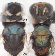
Figures 23–26. 23–24 Horismenus myrmecophagus female: 23 head in frontal view 24 thoracic dorsum. 25–26 Horismenus microdonophagus female: 25 head in frontal view 26 thoracic dorsum.
-
Ekaterina Shevtsova, Christer Hansson
Zookeys
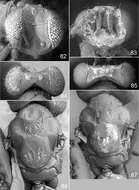
Figures 82–87. Achrysocharoides serotinae sp. n.: 82 Head frontal, female 83 Ditto, male 84 Vertex, female 85 Ditto, male 86 Mesosoma dorsal, female 87 Ditto, male.
-
Christer Hansson, Ekaterina Shevtsova
Zookeys
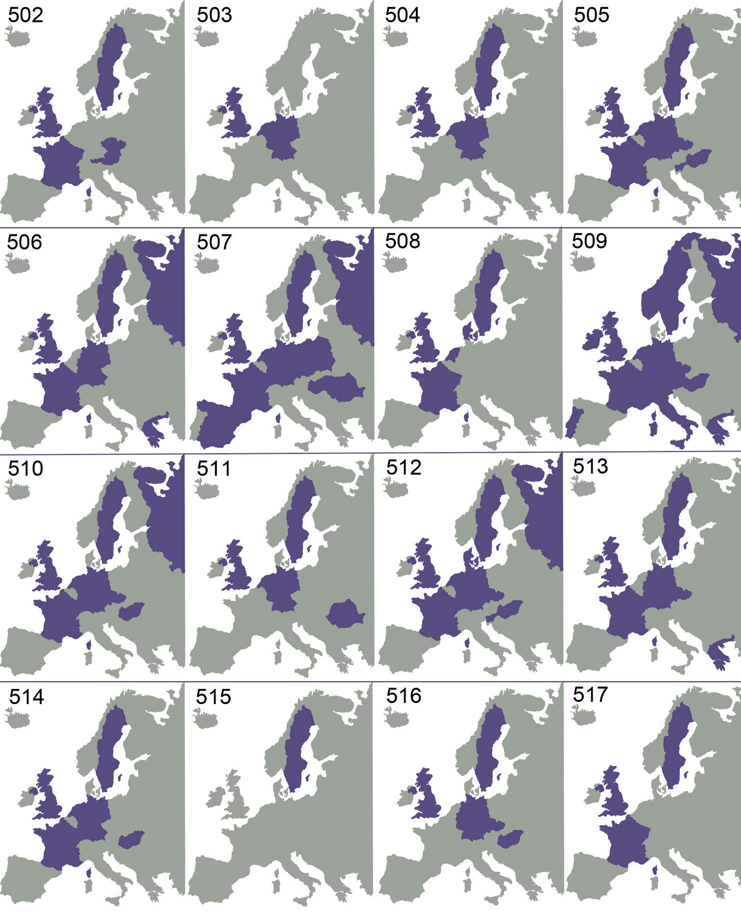
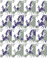
Figures 502–517.Omphale spp. distribution in Europe: 502 Omphale admirabilis 503 Omphale breviventris 504 Omphale telephe 505 Omphale versicolor 506 Omphale acuminata 507 Omphale chryseis 508 Omphale cornula 509 Omphale salicis 510 Omphale theana 511 Omphale brevis 512 Omphale clymene 513 Omphale euphorbiae 514 Omphale incognita 515 Omphale lydia 516 Omphale matrana 517 Omphale nitens.
-
Zoya Yefremova, Graciela González-Santarosa, J. Refugio Lomeli-Flores, Néstor Bautista-Martínez
Zookeys
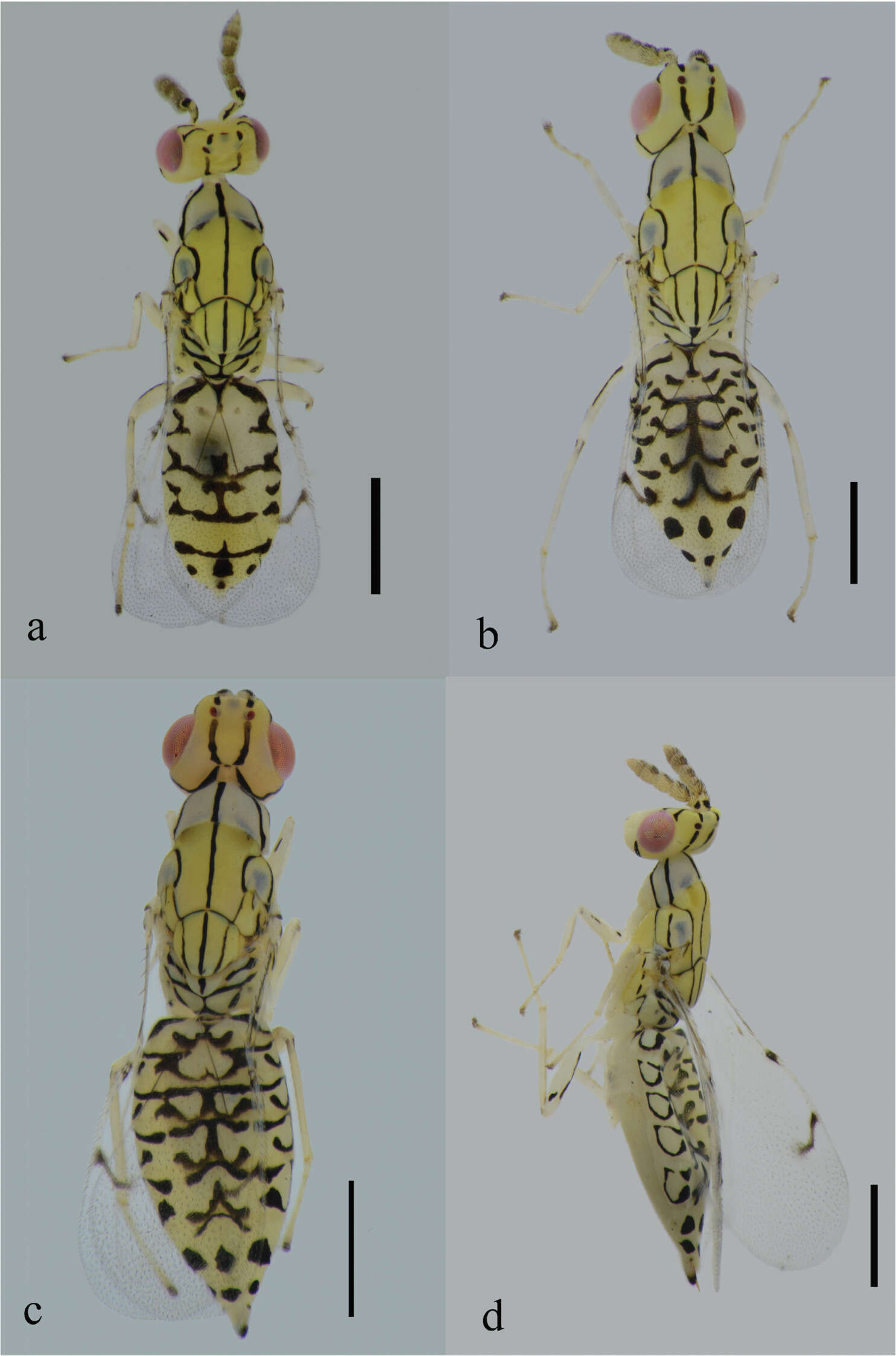
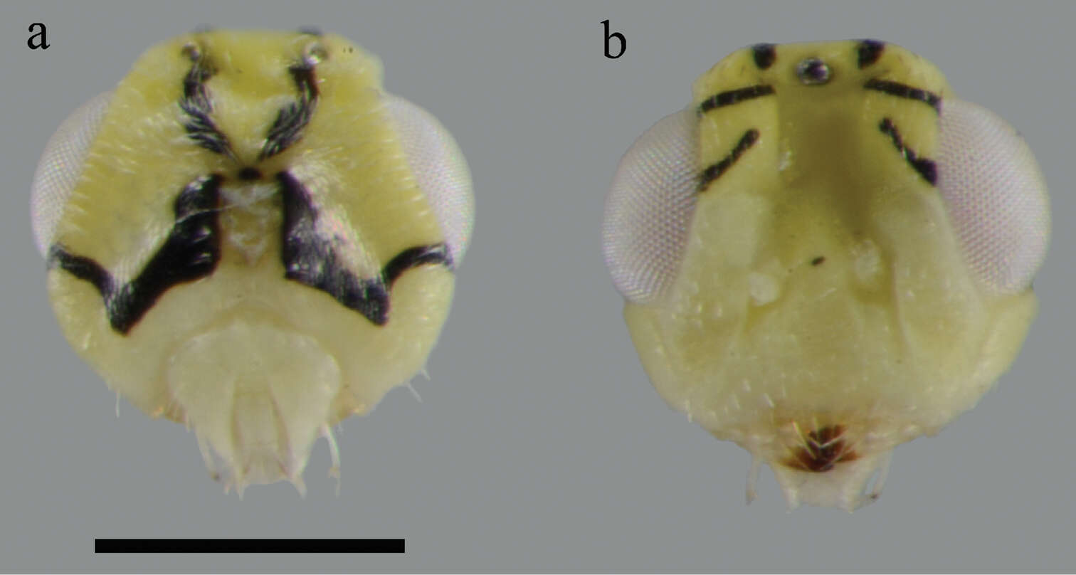
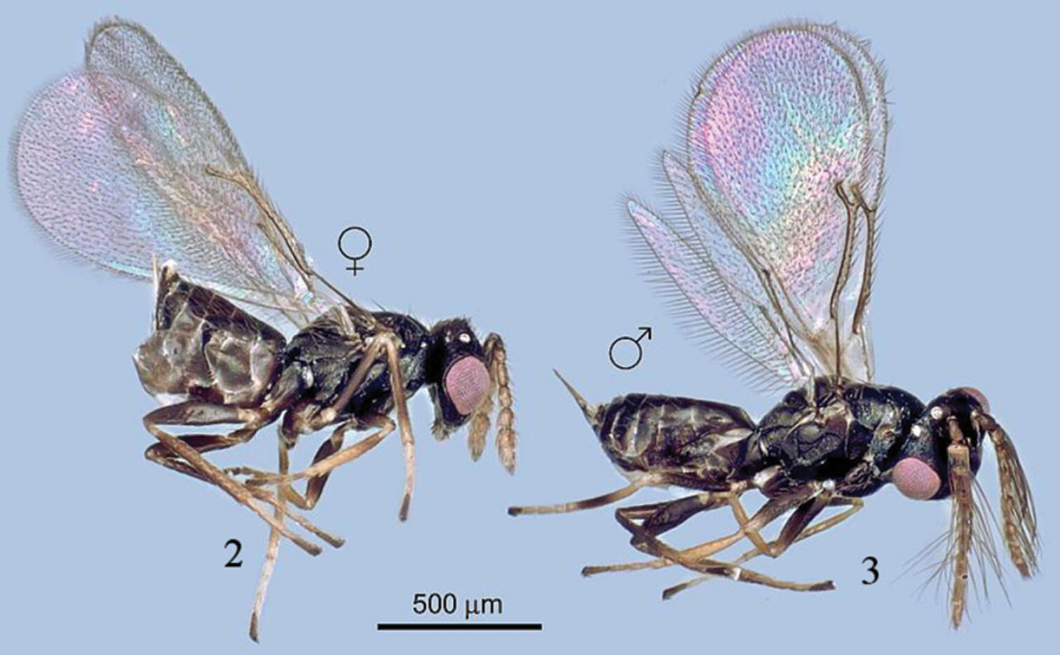
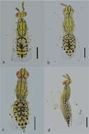
Figures 2–3.Tamarixia aguacatensis, female and male (habitus).
-
Christer Hansson, Jean-Paul Lachaud, Gabriela Pérez-Lachaud
Zookeys
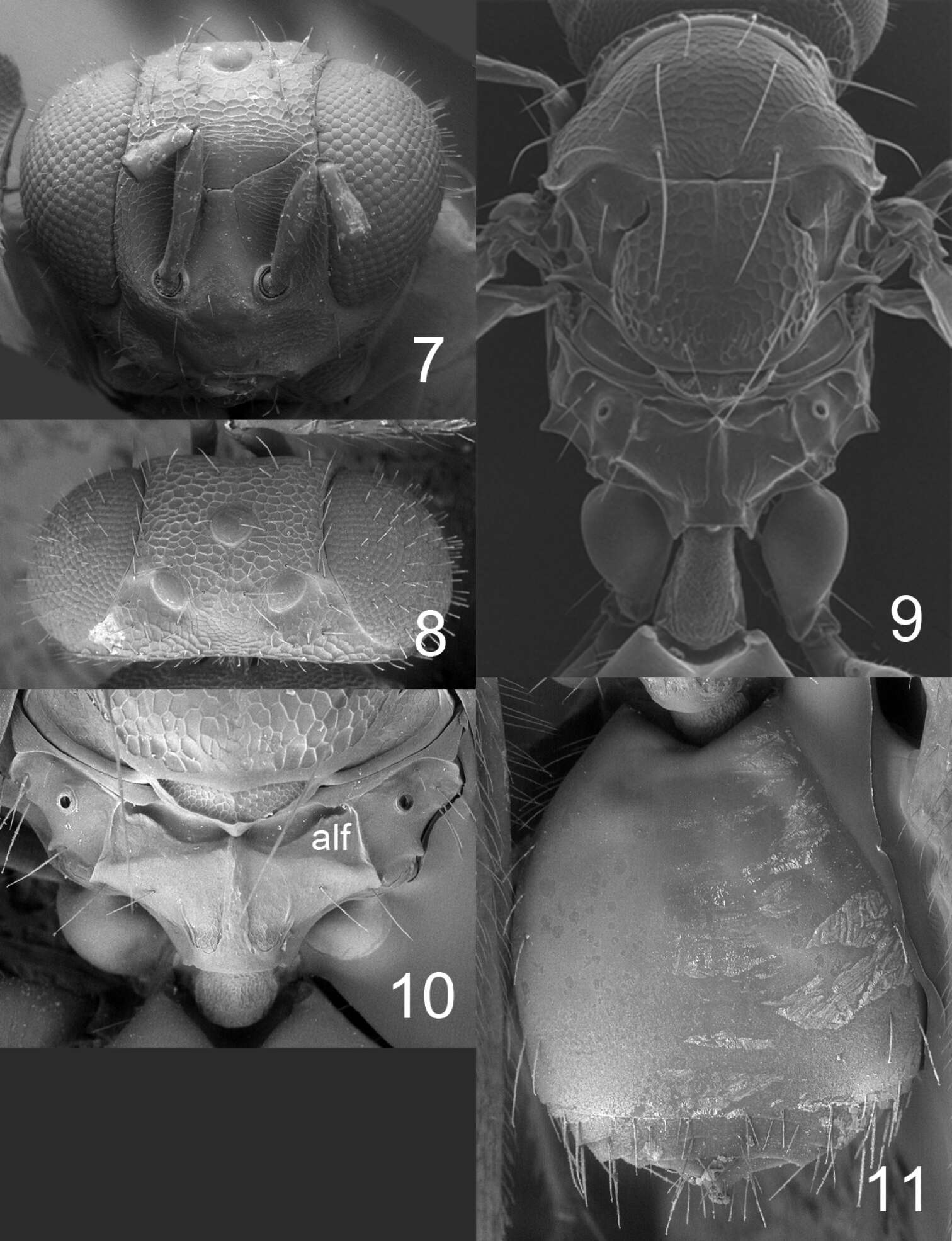
Figures 7–11. Horismenus microdonophagus female: 7 head in frontal view 8 vertex 9 thoracic dorsum and petiole 10 propodeum 11 gaster in dorsal view. Abbreviation alf = anterolateral fovea.
-
Figures 3–7.Achrysocharoides asperulus sp. n., female. 3 Head, frontal 4 Vertex 5 Thoracic dorsum 6 Head, frontal 7 Thoracic dorsum.
-
Christer Hansson, Ekaterina Shevtsova
Zookeys
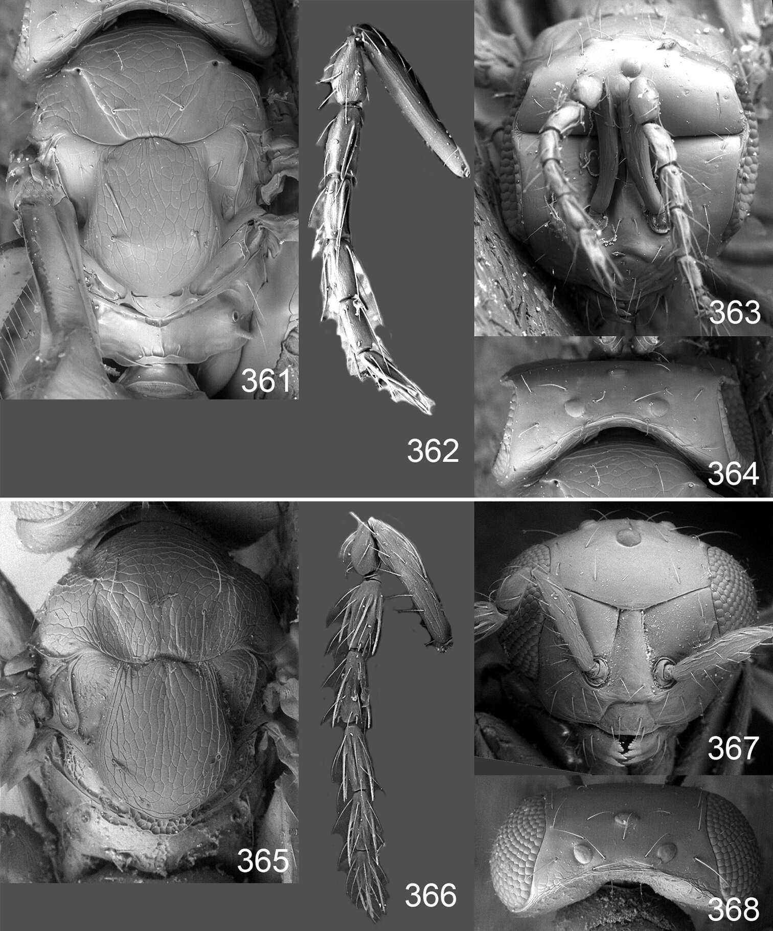
Figures 361–368.Omphale spp., females:361–364. Omphale erugata: 361 thoracic dorsum 362 antenna 363 head in frontal view 364 vertex 365–368. Omphale rossica: 365 thoracic dorsum 366 antenna 367 head in frontal view 368 vertex.
-
Zoya Yefremova, Graciela González-Santarosa, J. Refugio Lomeli-Flores, Néstor Bautista-Martínez
Zookeys
Figures 4–9.Tamarixia aguacatensis. Female: 4 Head, frontal view 5 Mesosoma, lateral view 6 Antenna 7 Mesosoma, dorsal view 9 Propodeum. Male 8 Both antennae on the head.
-
Christer Hansson, Jean-Paul Lachaud, Gabriela Pérez-Lachaud
Zookeys
Figures 17–22. 17–20 antenna in lateral view: 17 Horismenus myrmecophagus female 18 H. microdonophagus female 19 H. microdonophagus male (apical 2 flagellomeres missing) 20 Microdonophagus tertius female. 21–22 right fore wing female: 21 H. myrmecophagus 22 H. microdonophagus.