-
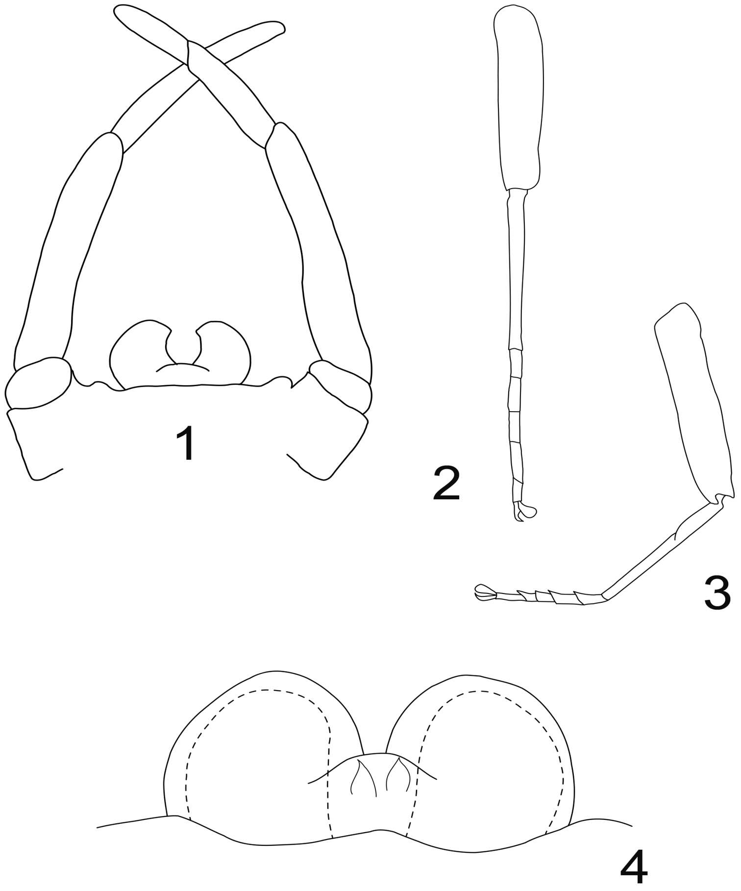
Figures 1–4.Rhithrogeniella ornata Ulmer, 1939. 1 Genitalia of the male imago (holotype) in ventral view 2 Foreleg of a male subimago (paratype) 3 Hindleg of a male subimago (paratype) 4 Penis lobes of a male subimago (paratype): plain line, cuticular structures of the subimago; dotted line, outline of the imago penis lobes.
-
Boonsatien Boonsoong, Dietrich Braasch
Zookeys
Figure 4.A–B Lamella of gills 7 (A) and setae on inner surface of hind tarsi (B) of Asionurus primus Braasch & Soldán, 1986 C bristles on cerci of Rhithrogeniella tonkinensisSoldán & Braasch, 1986 D bristles on cerci of Asionurus namnaoensis Braasch & Boonsoong, 2010 E dorsal view of abdomen of Asionurus rubromaculatus You, Wu, Gui & Hsu, 1981 F lamella of gills 1 of Asionurus gilliesiana Braasch, 1990 G lamella of gills 7 of Asionurus rainulfiana Braasch, 1990.
-
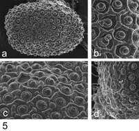
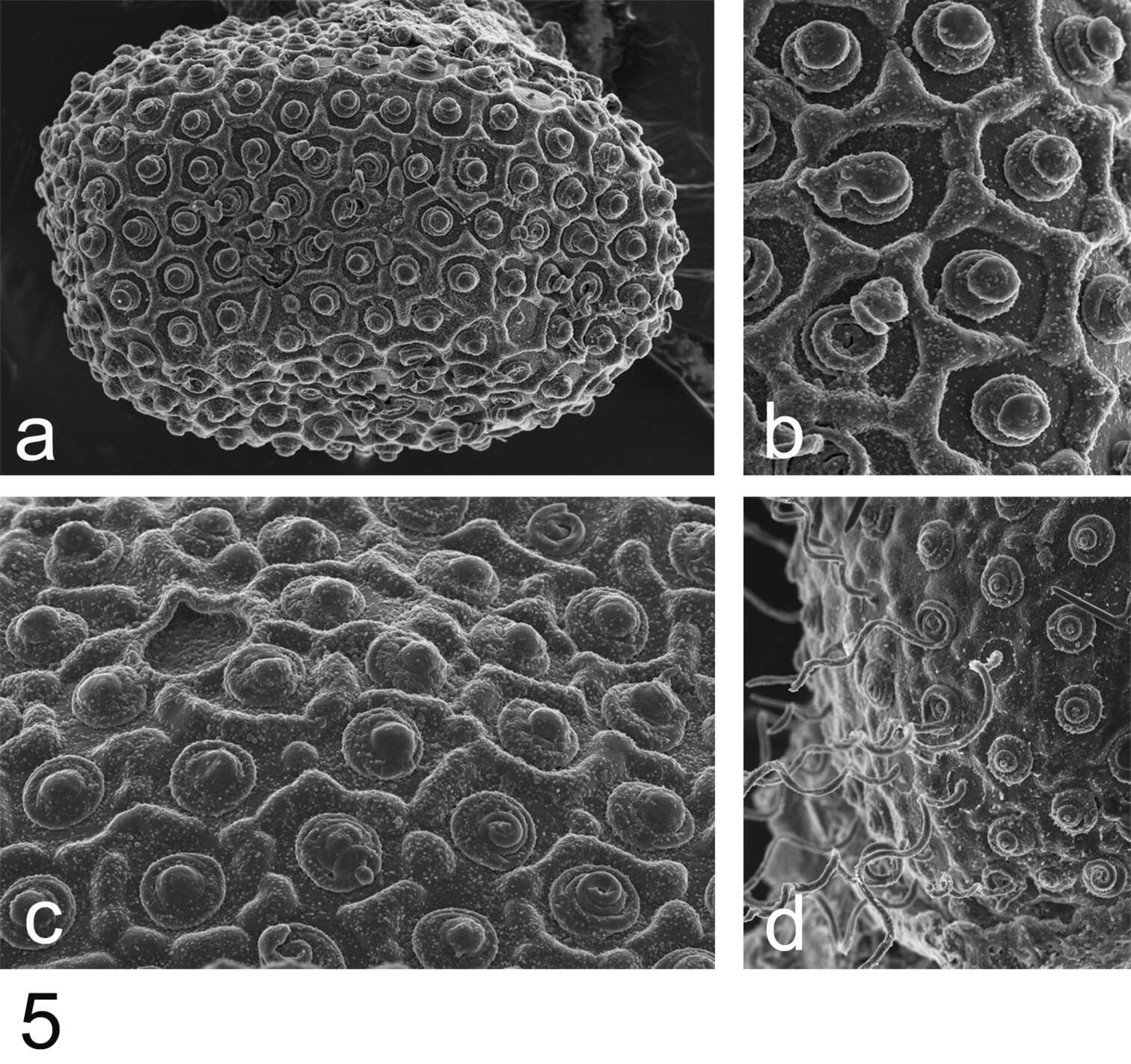
Figure 5.Rhithrogeniella ornata Ulmer, 1939, SEM pictures of egg structures. 5a Egg extracted from a female subimago paratype from Padang, Sumatra 5b Details of the chorionic structure of a female nymph from Ombilin River, Sumatra 5c Details of the chorionic structure and micropyle of a female subimago paratype from Buitenzorg [Bogor], Java 5d chorionic surface of the female allotype from Buitenzorg [Bogor], Java.
-
Boonsatien Boonsoong, Dietrich Braasch
Zookeys
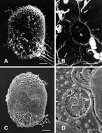
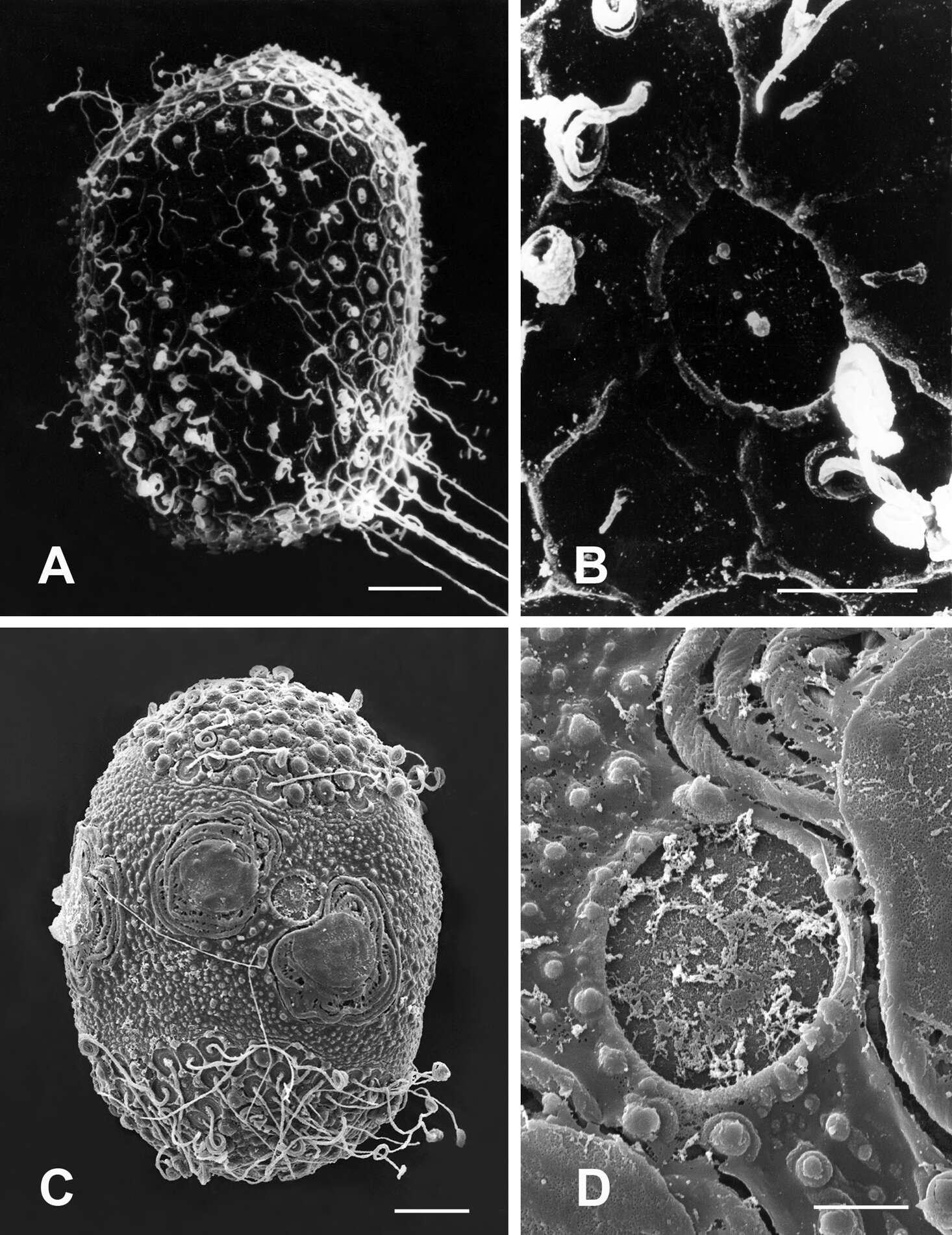
Figure 8.A–B General outline (A) and micropyle (B) of the egg of Rhithrogena tonkinensisSoldán & Braasch, 1986 C–D General outline (C) and micropyle (D) of the egg of Asionurus namnaoensis Braasch & Boonsoong, 2010. Scale bars 20 µm for A and C; 5 µm for B and D.
-
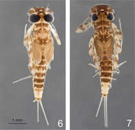
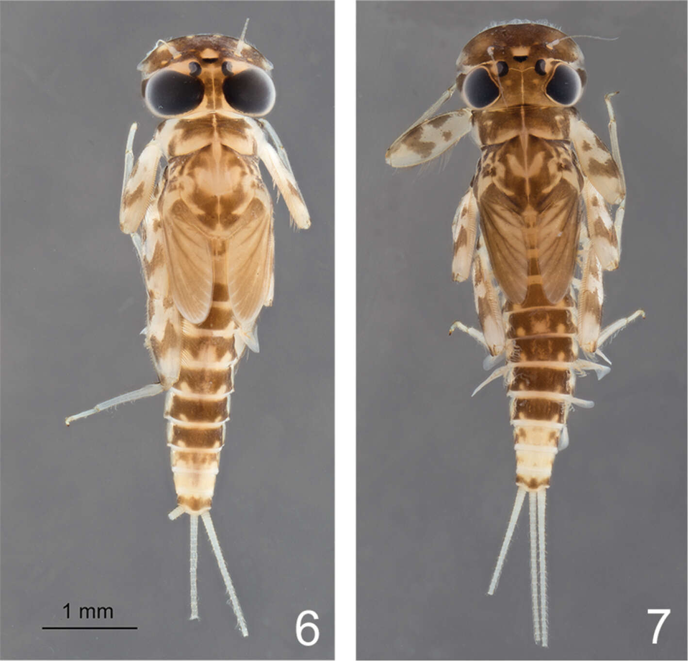
Figures 6–7.Rhithrogeniella ornata Ulmer, 1939. 6 Male nymph 7 Female nymph with slight color variations.
-
Boonsatien Boonsoong, Dietrich Braasch
Zookeys
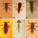
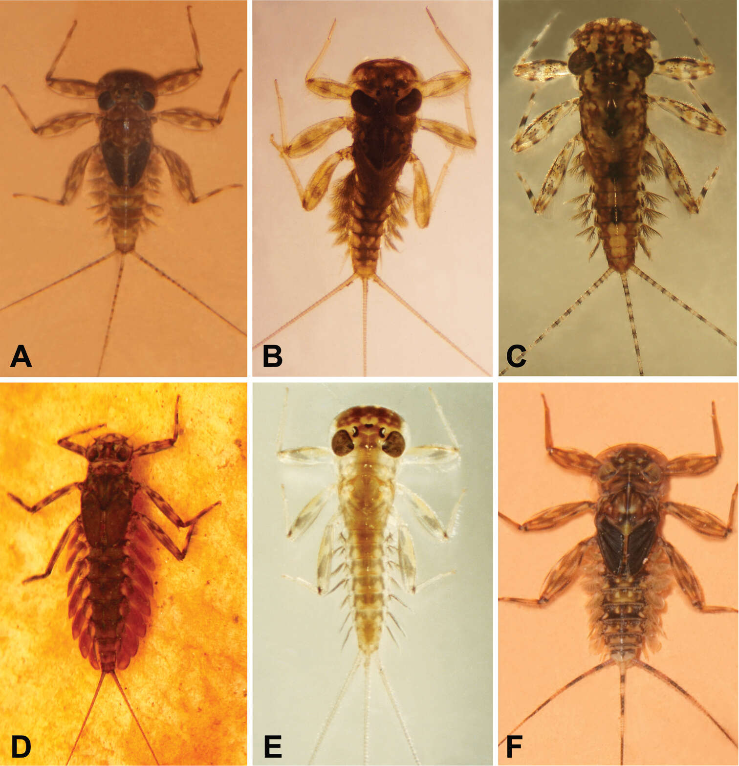
Figure 9.A Habitus of Asionurus namnaoensis Braasch & Boonsoong, 2010 B habitus of Asionurus primus Braasch & Soldán, 1986 Chabitus of Compsoneuria thienemanniUlmer, 1939 Dhabitus of Epeorus khayengensis Boonsoong & Braasch, 2010 E habitus of Rhithrogena tonkinensis Soldán & Braasch, 1986 F habitus of Thalerosphyrus sinuosus Navás, 1933.
-
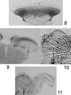
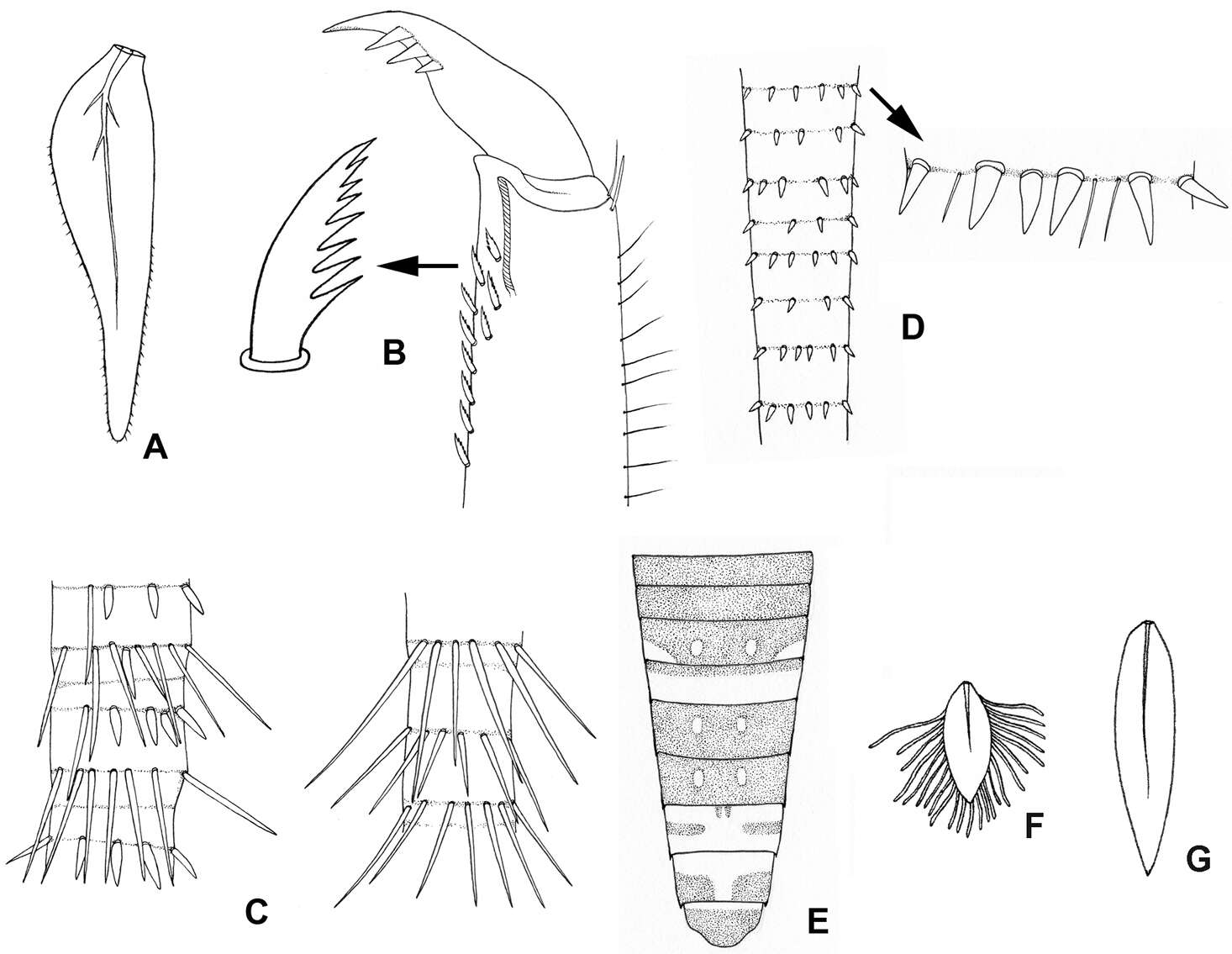
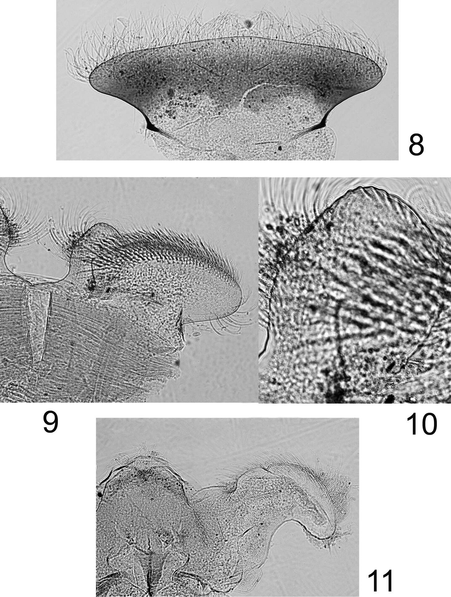
Figures 8–11.Rhithrogeniella ornata Ulmer, 1939, nymphal mouthparts. 8 Labrum in dorsal view 9 Left glossae and paraglossae of the labium 10 Detail of the glossae from 9 11 Hypopharynx, ventral view lingua and left superlingua.
-
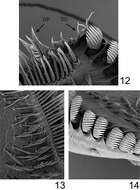
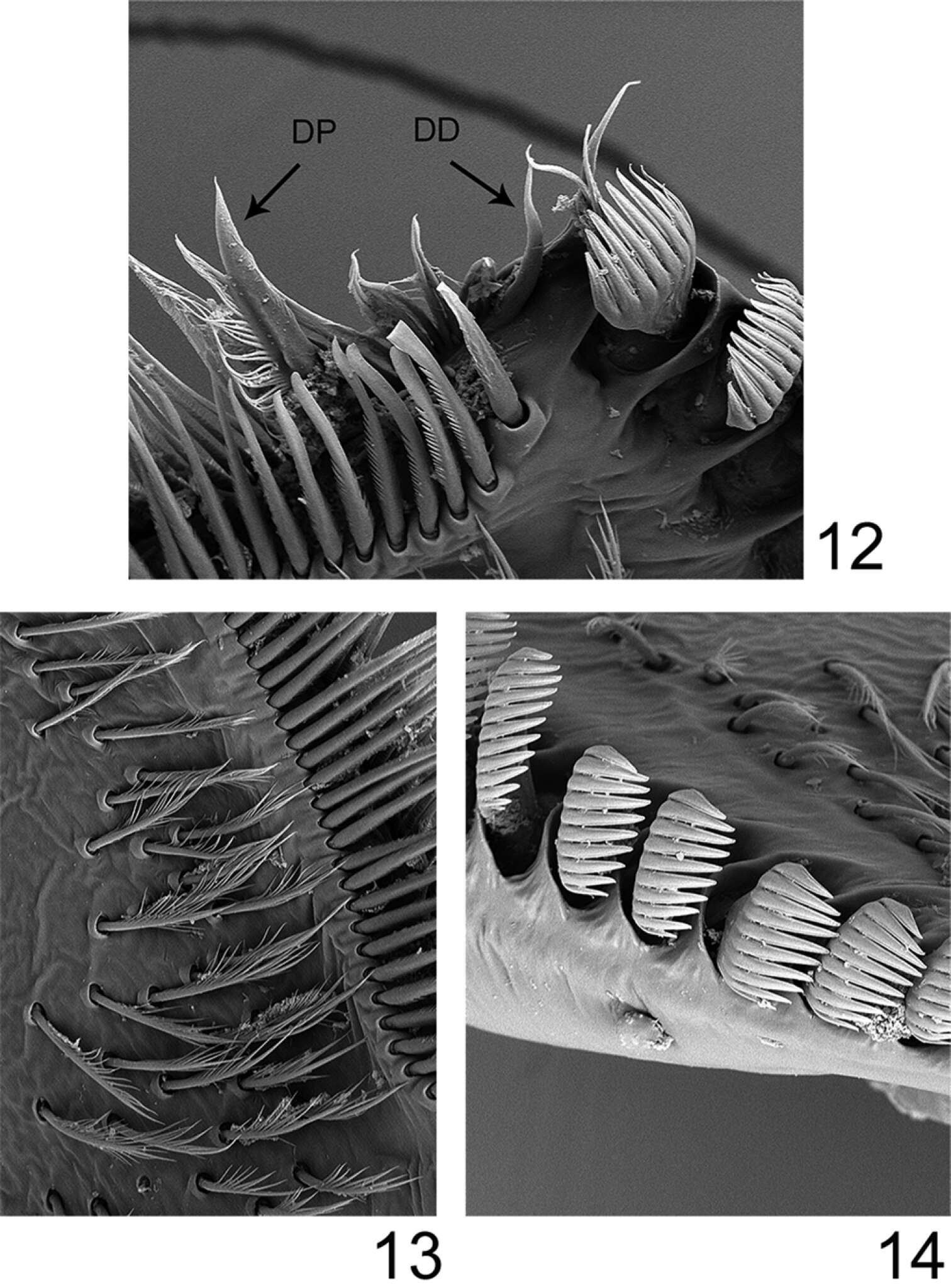
Figures 12–14.Rhithrogeniella ornata Ulmer, 1939, SEM pictures of the maxilla. 12 Dentisetae (DP: proximal dentiseta, DD: distal dentiseta) 13 Fimbriate setae on the ventral surface 14 Comb-shape setae on the crown of the galea-lacinia.
-
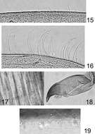
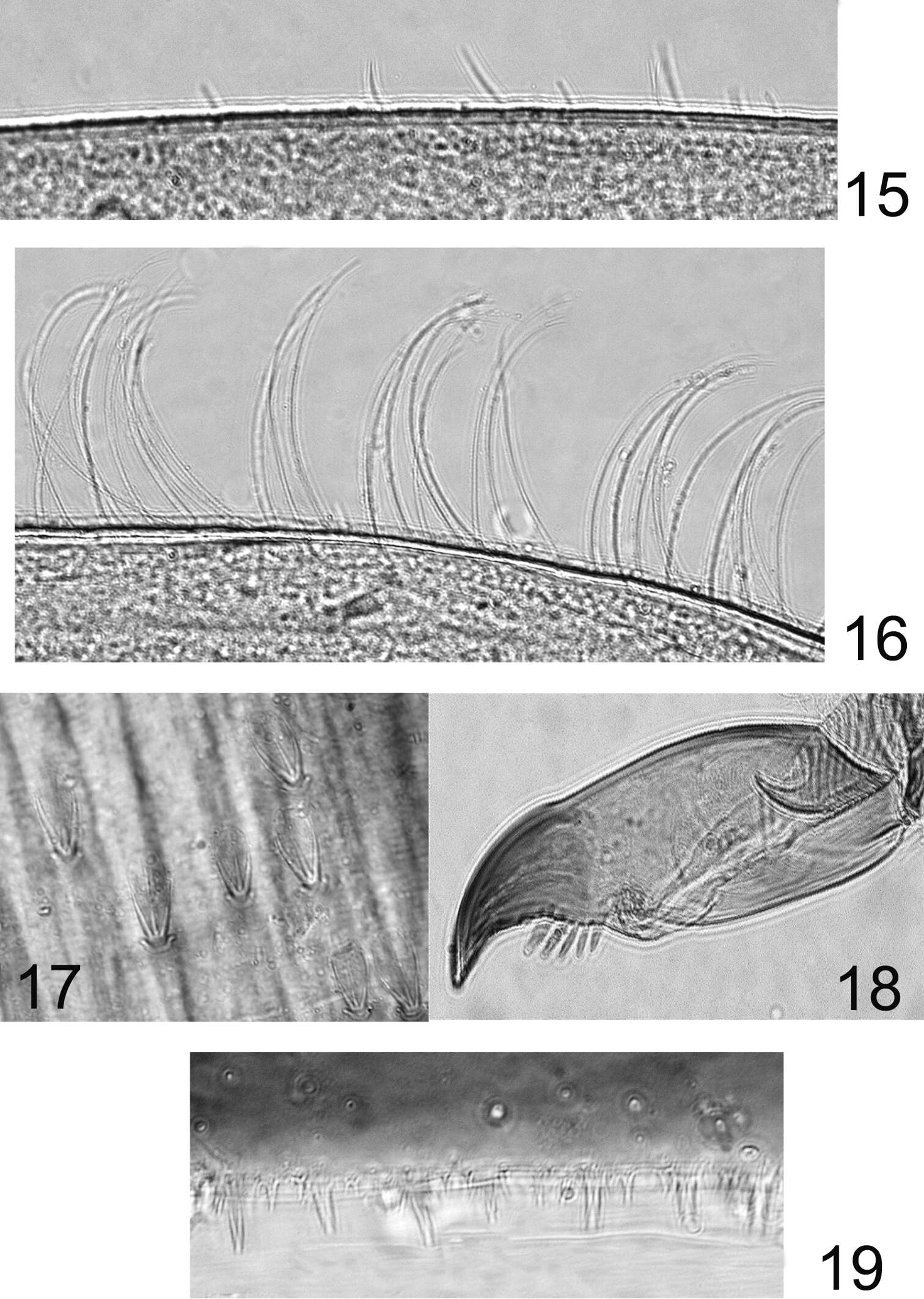
Figures 15–19.Rhithrogeniella ornata Ulmer, 1939. 15 Outer margin of the fore tibia 16 Outer margin of the hind tibia 17 Bristles on the dorsal surface of hind femur 18 Tarsal claw 19 Posterior margin of tergite V.
-
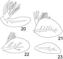
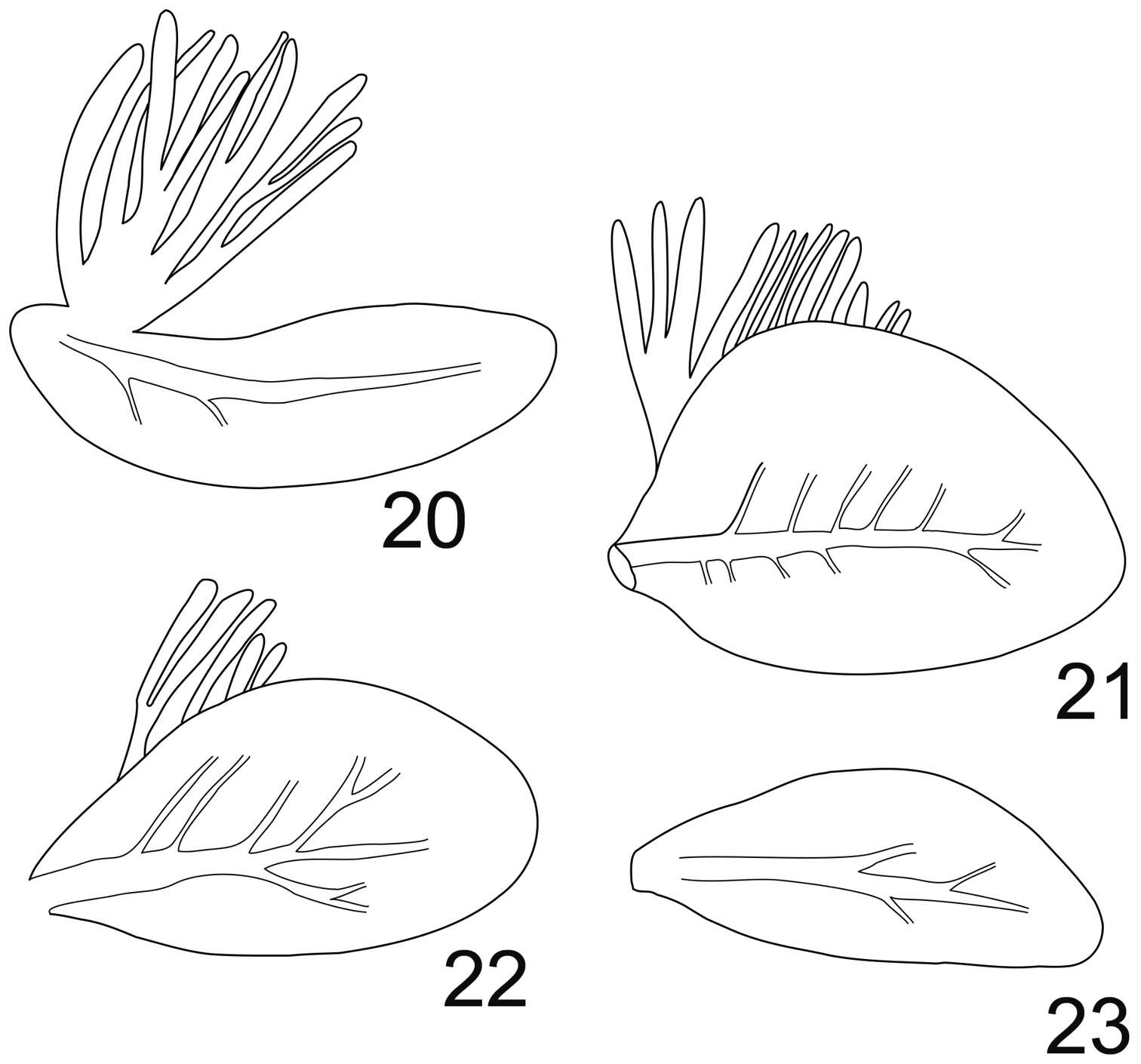
Figures 20–23.Rhithrogeniella ornata Ulmer, 1939. 20 Gill I 21 Gill IV 22 Gill VI 23 Gill VII.
-
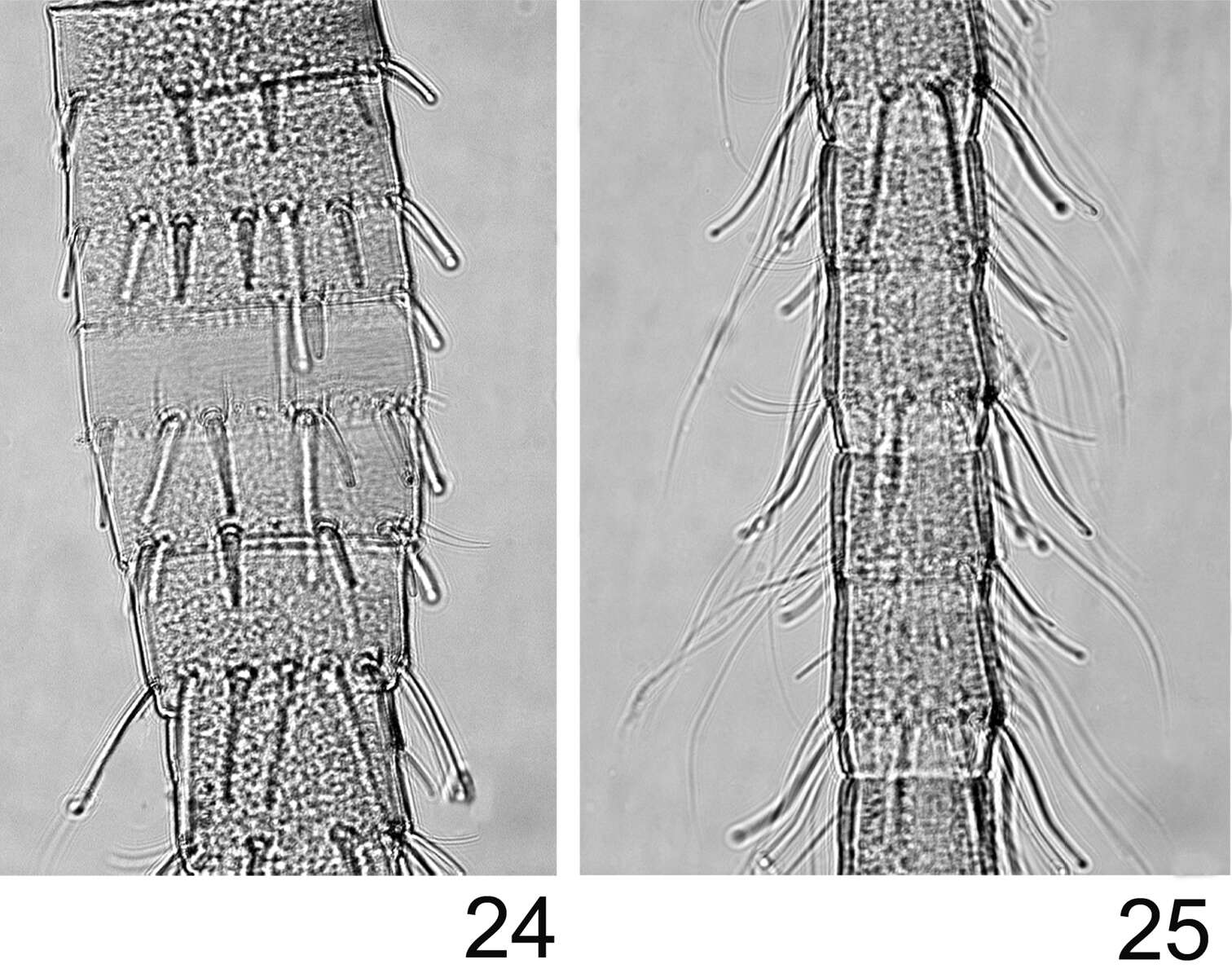
Figures 24–25.Rhithrogeniella ornata Ulmer, 1939. 24 Proximal part of the terminal filament 25 Median part of the terminal filament.