
Les dinofícies (Dinophyceae) són la principal classe de dinoflagel·lats. Inclou les espècies amb un nucli que roman dinocariont durant tot el cicle cel·lular, que és anomenat per l'etapa haploide, i inclou tots els dinoflagel·lats típics, com Peridinium i Gymnodinium. D'altres són més inusuals, incloent-hi alguns colonials, ameboides o paràsits.
Són organismes unicel·lulars, la majoria biflagel·lats, si bé poden aparèixer formes flagel·lat: cocoides, filamentoses, palmeloides o ameboides, relacionades amb la gran varietat de formes de nutrició. Generalment fotosintètics, encara que també hi ha formes heteròtrofes: sapròfites, paràsites, simbiòtiques i holozoiques. Molts autòtrofs marins són auxòtrofs per diverses vitamines.
Com a pigments tenen: clorofil·la a i c, β-caroteno, xantofil·las, peridinina, neoperidinina, dinoxantina, neodinoxantina i diatoxantina. El material de reserva és el midó.
La paret cel·lular o teca, quan es presenta, està composta fonamentalment de cel·lulosa. Presenten dos flagels, situats en solcs o depressions de la superfície de la cèl·lula. Un flagel acronemàtic (llis, acabat en una fibril·la), de disposició posterior, localitzat en un solc longitudinal. Un altre flagel acintat situat en un solc transversal que permet el gir i el desplaçament.
Les dinofícies es classifiquen per la seva morfologia. Les espècies amb teca es divideixen en quatre ordres, basats en la disposició de les plaques de la seva armadura: Peridiniales (per ex. Peridinium), Gonyaulacales (per ex. Ceratium o Gonyaulax), Dinophysiales (per ex. Dinophysis) i Prorocentrales (per ex. Prorocentrum). Els peridinials són probablement parafilètics respecte als altres i amb els arbres d'ARNr es barregen amb espècies que manquen de teca. Els grups sense teca es considera que són polifilètics i es classifiquen en diversos ordres. Exemples de gèneres són Gymnodinium, Amphidinium, Symbiodinium, Dinamoeba i Pfiesteria.
Un grup de dinoflagel·lats paràsits, els blastodinials, a vegades es classifiquen aquí, encara que altres vegades se'ls assigna la seva pròpia classe de les blastodinofícies.
Die Dinoflagellaten (Dinoflagellata; von altgriechisch δῖνος dinos, deutsch ‚wirbelnd‘ und lateinisch flagellum ‚Peitsche‘, ‚Geißel‘), auch als Peridineae und Panzergeißler bezeichnet, sind ein Taxon, das vorwiegend Einzeller umfasst. Zu ihren kennzeichnenden Merkmalen gehören zwei während des mobilen Lebenszyklus vorhandene Flagellen und Chromosomen, die während der Interphase kondensiert sind. Dinoflagellaten haben keine Histone. Weltweit werden rund 2.400 rezente Arten unterschieden (Stand: 2012)[1], die großteils im Meer leben und dabei einen Hauptteil des Phytoplanktons bilden. Der Unterstamm umfasst sowohl autotrophe als auch heterotrophe Arten.
Innerhalb der Dinoflagellaten herrscht eine extrem große Formenvielfalt. Die Größe reicht von 2 µm (Gymnodinium simplex) bis zu 2 mm (Noctiluca miliaris), wobei die meisten Arten zwischen 10 und 100 µm groß werden.
Die Form der freischwimmenden Zelle ist eiförmig bis rundlich, wobei das Anterior meist mehr zugespitzt ist als das Posterior. Die meisten Dinoflagellaten besitzen zwei lange Geißeln. Eine Geißel ist nach hinten gerichtet (longitudinale Geißel), sie liegt im inneren Abschnitt in einer Furche des Zellleibs, ragt aber meist mehr oder weniger lang nach hinten daraus hervor. Die andere Geißel, die in einer Ebene senkrecht dazu schlägt (transversale Geißel), windet sich nach links um den Zellleib, sie liegt meist vollständig innerhalb einer Furche. Die transversale Geißel erlaubt der Zelle Drehungen und trägt am meisten zum Vortrieb bei. Die longitudinale Geißel dient in erster Linie zur Steuerung der Bewegungsrichtung. Diese Anordnung der Geißeln wird als dinokont bezeichnet. Bei den Prorocentrales sitzen, abweichend dazu, beide Geißeln frei am Hinterende der Zelle, dies wird als desmokont bezeichnet. Bei einigen Gattungen treten völlig abweichend gestaltete, zum Teil geißellose Zellen auf.
Bei vielen Arten sind die direkt unterhalb der Zellmembran liegenden Vakuolen mit Zellulose gefüllt und so zu mehr oder weniger massiven Platten verstärkt.[2] Wenn solche intrazellulären Platten vorhanden sind, wird diese Hülle als Theka und die entsprechenden Arten thekat bezeichnet. Wenn die Alveolen nicht oder nur sehr wenig verstärkt sind, werden die Arten athekat oder nackt genannt. Die Theka bildet ein Mosaik aus einzelnen Platten; dieses kann zur Artbestimmung benutzt werden.
Eine Querfurche, das sogenannte Cingulum (Gürtel, en. auch girdle[3]) läuft rund um die Zelle und teilt diese somit in ein Anterior (Episoma) und Posterior (Hyposoma). Ist eine Theka vorhanden, werden die Teile als Epitheka bzw. Hypotheka bezeichnet. Ist keine Theka vorhanden, spricht man von athekaten Dinoflagellaten. Bei morphologischen Beschreibungen dieser Dinoflagellaten werden die Begriffe Epicone und Hypocone anstatt Epi- und Hypotheka verwendet. Nach posterior verläuft ausgehend von der Querfurche eine Längsfurche, der sogenannte Sulcus. Die transversale Geißel schlägt im Cingulum, die longitudinale Geißel im Sulcus.
Die longitudinale Geißel ist meist etwas abgeflacht. Sie trägt gelegentlich einen spärlichen Besatz mit Flimmerhärchen (Mastigonema), der aber auch vollständig fehlen kann. Die transversale Geißel ist innerhalb des furchenartigen Cingulum über eine bandförmige Verbindung längs mit der Zelle verbunden. Sie schlägt mit einer wellenartigen Bewegung. Ihre freie Außenkante ist meist mit Härchen besetzt. Das Cingulum umgibt die Zelle meist nicht kreisförmig, sondern ist etwas spiralig gestaltet, so dass das hintere Ende der transversalen Geißel weiter hinten zu liegen kommt als die Wurzel, die Spirale ist meist relativ flach, kann aber bei einigen Gattungen recht steil sein. Beim Schlag wird die Zelle so in eine Drehbewegung (immer nach links) versetzt.
Der äußere Region des Zellkörpers der Dinoflagellaten weist eine Reihe morphologischer Besonderheiten auf. Unterhalb der Zellmembran sitzt ein System von flachen Vakuolen, die als amphiesmale Vesikel oder Alveolen bezeichnet werden, diese haben die Dinoflagellaten mit einer Reihe anderer Einzeller wie den Wimperntierchen (Ciliaten) gemeinsam, mit denen sie, nach diesem Merkmal, im Taxon der Alveolata vereinigt werden. Die äußere Region, die die Vakuolen enthält, wird als Amphiesma oder auch Cortex (Rinde) bezeichnet. Innerhalb der Vesikel wird bei den gepanzerten (thekaten) Dinoflagellaten, in jeweils einem Vesikel immer eine Platte aus Zellulose abgeschieden, die sich letztlich zu einer geschlossenen Hülle verbinden können. Durch die Bildung und Lage innerhalb einer Vakuole liegt die Hülle allerdings innerhalb der Zelle (intrazellulär) und ist also von der Zellmembran umschlossen. Bei wenigen Dinoflagellaten sind die Vakuolen des Amphiesmas ausschließlich mit Flüssigkeit gefüllt. Bei vielen anderen enthalten sie festes Material, dass sich aber nicht zu einem geschlossenen Panzer versteift, diese werden gemeinsam athekat (also: ohne Theka) genannt. Bei den thekaten Dinoflagellaten wird die Anordnung der Platten zur Bestimmung der Gattungen und Arten verwendet, jede Platte hat dazu in einem ausgefeilten System jeweils einen besonderen Namen erhalten. Unterhalb der Vesikel sitzt bei manchen Arten eine zweite, dünne Lage aus Fasern, die Pellicula genannt wird. Sie enthält neben Zellulose das Polymer Sporopollenin. Bei vielen Dinoflagellaten kann der äußere Panzer abgeworfen werden (Ecdysis genannt), die Pellicula bildet dann die äußere Hülle von Cysten genannten Überdauerungsstadien.
Einige basale athekate Dinoflagellaten, zum Beispiel der Gattung Oxyrrhis, besitzen auf der Oberfläche (also extrazellulär) kleine, oft sternförmige Schüppchen aus Zellulose. Andere, wie Dicroerisma und Actinscus besitzen interne Skelettelemente aus Siliciumdioxid. Bei Achradina und Monaster können diese die Zelle körbchenartig einschließen.
Innerhalb der Eukaryoten besitzt der Zellkern der Dinoflagellaten einzigartige Eigenschaften, er wird deshalb mit dem besonderen Ausdruck Dinokaryon belegt. Die DNA ist bei ihnen nicht in Nukleosomen organisiert, deren charakteristische Proteine, die Histone, fehlen fast vollständig. Insgesamt ist der Proteinanteil des Zellkerns weitaus geringer als bei anderen Eukaryoten, meist nur etwa 10 Prozent. Anstelle der Histone werden nur bei ihnen vorkommende, besondere Proteine nachgewiesen, deren Herkunft durch horizontalen Gentransfer aus Viren nachgewiesen werden konnte (dinoflagellate viral nucleoproteins; DVNPs). Während früher angenommen wurde, dass Histone völlig fehlen, wurden inzwischen alle Histonfamilien, wenn auch in geringerem Gehalt und in teilweise stark abweichender Struktur, bei den Dinoflagellaten nachgewiesen, sie haben vermutlich bei ihnen eine besondere Rolle bei der Transkription beibehalten.[4]
Sowohl der DNA-Gehalt der Dinoflagellaten gehört zu den höchsten bei allen Eukaryoten, auch ihr Genom ist ungewöhnlich umfangreich. Die Chromosomen sind auch während der Interphase kondensiert und im Elektronenmikroskop sichtbar. Die Chromosomen bilden eine Girlandenstruktur, wobei die einzelnen Fibrillen nur 2,5 nm im Durchmesser haben. Die übrigen Eukaryoten besitzen Fibrillen mit zehnfachem Durchmesser mit einem zentralen Nucleohistonstrang. Die Struktur der Chromosomen wurde mit Flüssigkristallen verglichen. Der Gehalt an nicht-kodierender DNA der Dinoflagellaten ist außergewöhnlich hoch. Es wird angenommen, dass nur die äußeren, schleifenförmigen Enden der Chromosomen, die aus dem Zellkern nach außen vorragen, kodierende Abschnitte enthalten. Auch die Mitose ist bei ihnen äußerst ungewöhnlich. Die den Nukleus umgebende Membran bleibt während des gesamten Mitosezyklus erhalten. Bei der Teilung bilden sich fingerförmige Einstülpungen, die letztlich den Kern ganz durchdringen und so Torus-artige Strukturen hervorbringen. Die Mitosespindel wird innerhalb des Torus ausgebildet, wobei seine Anheftungsstellen (die Kinetochoren) in der inneren Membran des Torus sitzen. Je nach Verwandtschaftsgruppe werden zwischen einem und fünf (oder sechs) solcher Tunnel durch den Zellkern ausgebildet. Auch während der Interphasen ist der Zellkern, neben der üblichen Kernhülle, durch ein Netzwerk aus Membranen durchzogen, aus denen ie Tunnelstrukturen gebildet werden.[5]
Weiterhin ist nur innerhalb der Dinoflagellaten die modifizierte Base Hydroxymethyluracil (HOMeU) in der DNA nachgewiesen. Mit einem Gesamtanteil von 4–19 % ersetzt sie 12–70 % der Thymin-Basen.[6] Die Chromosomenzahl schwankt zwischen 5 bei Syndinium turbo und 274 bei Ceratium hirundinella.[7]
Der Chloroplast hat wie der der Euglenida drei Membranen. Im Unterschied zu diesen gleicht er aber nicht dem einer Grünalge, sondern dem einer Rotalge, was auf die Herkunft von einer symbiontischen Rotalge hindeutet.[8]
Viele Dinoflagellaten besitzen komplexe, der Verteidigung oder dem Beutererwerb dienende Organellen, die Extrusomen, Trichozysten, Mucocysten oder Nematocysten genannt werden (genauso benannt, aber nicht homolog zu den Nematocysten der Nesseltiere). Bei Polykrikos kofoidii wurden die Nematocysten im Detail untersucht. Dinoflagellaten der Gymnodiniales besitzt harpunen-artige Nematocysten, die der räuberischen Ernährung dienen. Sie arbeiten quasi im Tandem mit einem weiteren, Taeniocyste genannten, Organell, mit dem sie einen morphologisch verbundenen Komplex bilden. Die Taeniocysten bilden dabei eine weitere Art von Extrusomen. Die Nematocysten bestehen aus einer quergestreiften Kapsel, die im Zellinneren liegt und durch eine deckelartige Struktur (Operculum) außen aus der Zellhülle hervorragt. Im Inneren der quer gestreiften Kapsel liegt ein aufgerollter Faden, an dessen Vorderende ein verstärkte, stilett-artige Spitze sitzt. Die Nematocysten feuern, indem der Deckel abgeworfen wird und der Faden mit der Spitze, unter hohem Druck, herausgeschleudert wird. Auslösend dafür könnte entweder, wie bei den Nematocysten der Nesseltiere, erhöhter osmotischer Druck sein, oder die Kapselwand wird muskelartig kontrahiert. Bei anderen Dinoflagellaten-Arten sitzt anstelle eines Stiletts eine hohle nadelartige Spitze an, die in Art einen Injektionsnadel ein Toxin appliziert. Bei Nematodinium sitzt innerhalb der Kapsel noch ein Ring von Unterkapseln, die den Vortrieb des Stiletts weiter verstärken. Die Nematocysten der Dinoflagellaten gehören zu den komplexesten Organellen überhaupt.[9]
Einige Arten sind zur Biolumineszenz fähig, wobei dieses Leuchten eine Reaktion auf mechanische Stimulation ist. In der Natur sind dies Deformationen der Zellmembran, die durch Scherkräfte hervorgerufen werden. Stark aufgewühltes Wasser, wie brechende Wellen oder schnell schwimmende Fische können solche Stimulationen auslösen. Im Labor kann auch mittels Chemikalien eine Reaktion induziert werden. Zu den Dinoflagellaten gehören die einzigen biolumineszenten autotrophen Lebewesen wie etwa Vertreter der Gattungen Gonyaulax, Protogonyaulax, Pyrodinium und Pyrocystis. Auch bei heterotrophen Arten wie Noctiluca miliaris oder einigen Vertretern der Gattungen Ceratium kann Biolumineszenz beobachtet werden.
Das emittierte Licht ist blau-grün und hat ein Maximum bei 474–476 nm. Da diese Wellenlänge nahe dem maximalen Transmissionsgrad des Meerwassers liegt, wird angenommen, dass die Sichtbarkeit des Lichtes den selektiven Vorteil verursacht. In Experimenten mit leuchtenden und nicht-leuchtenden Spezies konnte gezeigt werden, dass im Falle von Biolumineszenz die Prädation vermindert wurde. Vermutlich werden Feinde durch den Lichtblitz abgeschreckt. Wie bei fast allen Arten der Biolumineszenz ist dies auf eine Reaktion von Luciferasen und Luciferinen zurückzuführen.
Einige Arten produzieren äußerst starke Gifte. Das Saxitoxin beispielsweise wird von Vertretern der Gattung Alexandrium (Gonyaulax) produziert. Wenn die giftigen Dinoflagellaten von Muscheln gefressen werden, reichert sich das Gift in den Muscheln an und kann dann auch für Menschen gefährlich werden. Bei einer Massenvermehrung von giftigen Arten wird soviel Gift produziert, dass auch Fische und andere Meereslebewesen getötet werden.[10] Karenia brevis produziert die Brevetoxine und kann bei den von ihnen erzeugten „Roten Tiden“ zu Massensterben bei Fischen, Vögeln und Säugern führen.
Die Krankheit Ciguatera,[11] eine Art Fischvergiftung, wird durch Stoffwechselprodukte der Art Gambierdiscus toxicus hervorgerufen. Über die Nahrungskette gelangen die Dinoflagellaten-Toxine Ciguatoxin und Maitotoxin in Fische, die dadurch ebenfalls stark giftig werden. Die Vergiftung kann unter Umständen beim Menschen tödlich verlaufen.
Das Toxin von Pfiesteria piscicida dagegen wird nicht über die Nahrungskette angereichert, sondern ist direkt giftig für Fische und Menschen.[12]
Dinoflagellaten sind kosmopolitisch im Salz- wie auch im Süßwasser verbreitet und können dort aufgrund ihres Formenreichtums viele Habitate besiedeln. Rund 75 % aller Arten werden dem marinen Plankton zugerechnet,[1] mit der größten Artenvielfalt in tropischen Gewässern. Sie sind aber auch benthische Lebewesen und dringen auch in die Sedimente ein. Darüber hinaus sind sie ebenfalls in der Polarregion und in Meereis anzutreffen.
Im Süßwasser sind weniger Arten verbreitet. Weltweit sind 420 Arten aus Binnengewässern bekannt (etwa 17 Prozent der Artenzahl),[1] die Seen, Tümpel und Moore besiedeln. Das Verbreitungsgebiet reicht etwa vom Äquator bis 78° nördlicher Breite (Insel Spitzbergen). Die Höhenunterschiede reichen von −209 Meter in Israel bis auf 4150 Meter in Hochgebirgsseen von Mexiko.
Da einige Arten Symbiosen eingehen oder als Parasiten leben, werden auch Lebewesen als Habitate genutzt. Beispielsweise leben Dinoflagellaten als Endosymbionten in vielen Korallen und werden dann als Zooxanthellen bezeichnet. Autotrophe Arten sind auf lichtdurchflutete Wasserschichten angewiesen, heterotrophe Arten können auch in vollkommen dunkle Tiefen vordringen.
Etwa die Hälfte der Dinoflagellaten ist autotroph und kann mit Hilfe der Assimilation der Chloroplasten anorganischen Kohlenstoff nutzen. Jedoch sind fast sämtliche photosynthetisch aktive Arten auxotroph und benötigen Vitamine (Cobalamine, Biotin, Thiamin) für katalytische Zwecke. Diese werden über Phagocytose aufgenommen. Autotrophe Arten gehen auch eine Symbiose mit Nesseltieren (Cnidaria), insbesondere Korallen, Weichtieren (Mollusca) aber auch Foraminiferen (Foraminifera) und Wimpertierchen (Ciliata), ein.
Heterotrophe Dinoflagellaten ernähren sich von einem vielfältigen Spektrum von Planktonorganismen, das von Nanoplankton bis zu großen Kieselalgen reicht.[13][14] Darunter fallen auch Dinoflagellaten der eigenen wie auch anderer Arten, Detritus und selbst Eier und Larven von Ruderfußkrebsen. Im einfachsten Fall wird die Nahrung durch Phagocytose aufgenommen (beispielsweise Noctiluca miliaris). Durch spezielle Zellstrukturen wie Pedunkel oder Pallium können sich heterotrophe Dinoflagellaten aber auch von Organismen ernähren, die um ein Vielfaches größer als sie selbst sind (beispielsweise Pfiesteria[15] oder Protoperidinium).[13][14]
Die autotrophen Arten enthalten Plastiden mit Chlorophyll a bzw. einige Arten auch Chlorophyll c. Als Haupt-Carotinoid enthalten sie meist Peridinin anstatt von Fucoxanthin. Ihre Färbung reicht von gelbbraun bis rötlich, da das Chlorophyll von braunen und gelben Carotinoiden und roten Xanthophyllen überdeckt wird. Stärke ist das Hauptassimilationsprodukt, das in Körnchen außerhalb der Chloroplasten gespeichert wird. Es wurden aber auch fettartige Stoffe nachgewiesen. Die Plastiden sind meist mit drei Membranen umgeben, von denen eine mit dem endoplasmatischen Retikulum verbunden ist.
Grundsätzlich können Dinoflagellaten sehr verschiedene Plastiden beherbergen, die vom Grundtyp abweichen. Dies ist auf Phagotrophie zurückzuführen, die auch bei autotrophen Arten aufrechterhalten wird. Dies führte in der Stammesgeschichte zu einer weiteren, tertiären Endocytobiose. Die aufgenommenen Organismen können hierbei aus unterschiedlichen Gruppen, wie Haptophyta, Cryptophyceae, Heterokontophyta oder eines Chlorophyten zurückgehen. Der ursprünglich von den Rotalgen stammende Chloroplast ist hierbei völlig oder weitgehend zurückgebildet und erscheint im letzteren Fall als inaktiver Augenfleck (Stigma). Gelegentlich ist in den Chloroplasten auch ein Nucleomorph enthalten.
Die Fraßmechanismen heterotropher Dinoflagellaten lassen sich mit drei Grundtypen beschreiben.
Die Grundtypen Myzozytose und Pallium kommen hauptsächlich bei thekaten (gepanzerten) Arten vor und werden gelegentlich als extrazelluläre Verdauung bezeichnet. Dies stimmt genau betrachtet nicht, denn in jedem Fall wird die Beute in einer Fraßvakuole innerhalb des Zellplasmas verdaut, jedoch können sich diese Fraßvakuolen außerhalb der Theka befinden. Dies kann als Überwindung der Beschränkungen durch die Theka interpretiert werden und eröffnete den heterotrophen Arten ein erweitertes Beutespektrum.
Der untypische Dinoflagellat Noctiluca miliaris besitzt einen kurzen Tentakel, der wie eine Leimrute eingesetzt wird. Nahrungspartikel wie Kieselalgen und Detritus bleiben daran hängen und werden dann vom Tentakel zum Cytostom befördert.
Der thekate Dinoflagellat Stoeckeria algicida dagegen nutzt ein schlagartig ausgestoßenes Proteinfilament (englisch: tow filament), um die Beute über eine vergleichsweise große Entfernung einzufangen.[18] Ein vergleichbares Filament wird von Protoperidinium benutzt, um sich an Kieselalgen-Ketten zu verankern.[13]
Die Fortpflanzung erfolgt überwiegend vegetativ. Bei bepanzerten Arten werden die Platten in der Regel schräg zum Gürtel gesprengt, wobei die fehlende Hälfte später nachwächst. Es besteht aber auch die Möglichkeit, dass der Panzer abgeworfen und von den Tochterzellen völlig neu gebildet wird. Unter ungünstigen Lebensbedingungen entstehen dickwandige, überdauerungsfähige Zysten.
Geschlechtliche Fortpflanzung wurde nur bei wenigen Arten nachgewiesen. Hierbei wurden Anisogamie mit zygotischem Kernphasenwechsel als auch Isogameten, die in Gametangien entstehen, freigesetzt werden und miteinander verschmelzen, beschrieben.
Zusammen mit den Kieselalgen sind die Dinoflagellaten die Hauptprimärproduzenten organischer Stoffe im Meer, bilden dort also zusammen mit den Kieselalgen den Hauptteil der Basis der Nahrungspyramide. In Hochgebirgsseen können sie bis zu 50 % der Biomasse ausmachen.
Die heterotrophen Dinoflagellaten können mit ihren spezialisierten Fraßmechanismen ein weites Spektrum von Beuteorganismen fressen, das von Nanoplankton kleiner als 10 µm bis zu großen kettenbildenden Kieselalgen reicht. Dadurch stellen die heterotrophen Dinoflagellaten einen wichtigen Teil der mikrobiellen Schleife[19] dar (englisch microbial loop[20]).
Unter für sie günstigen Bedingungen vermehren sich in tropischen und subtropischen Gewässern bestimmte Arten in extremem Ausmaß, so dass sich die oberen Schichten des Meeres rot bis braun färben. Man nennt diese Algenblüte auch Rote Flut oder Rote Tide (englisch red tide).
Durch ihre sehr widerstandsfähige, organische Zellwand werden Dinoflagellatenzysten nicht durch Kalklösung zerstört, sondern bleiben auch nach langen Zeiträumen erhalten. Außerdem haben viele Zysten eine charakteristische Form. Das spielt für die Altersdatierung (Biostratigraphie) von Sedimenten eine entscheidende Rolle.
Andere Fossilgruppen wie Foraminiferen besitzen eine zu geringe Artenvielfalt und Dinoflagellatenzysten treten nahezu in allen Gewässern auf, wo sie heute als Klimaindikatoren verwendet werden. Erst 1988 begann man in Deutschland mit der Aufstellung von „Dinoflagellaten-Zonen“, die nun regelmäßig verbessert werden.
Durch die teilweise sehr komplexen Lebenszyklen der Dinoflagellaten ist die systematische Gliederung Gegenstand wissenschaftlicher Diskussion. Das Taxon gilt als polyphyletisch. Die hier angeführte Gliederung (Gattungen exemplarisch) folgt im Wesentlichen Adl et al. 2012:[21]
Soweit nicht unter Einzelnachweisen angegeben, basiert der Artikel auf folgenden Unterlagen:
Die Dinoflagellaten (Dinoflagellata; von altgriechisch δῖνος dinos, deutsch ‚wirbelnd‘ und lateinisch flagellum ‚Peitsche‘, ‚Geißel‘), auch als Peridineae und Panzergeißler bezeichnet, sind ein Taxon, das vorwiegend Einzeller umfasst. Zu ihren kennzeichnenden Merkmalen gehören zwei während des mobilen Lebenszyklus vorhandene Flagellen und Chromosomen, die während der Interphase kondensiert sind. Dinoflagellaten haben keine Histone. Weltweit werden rund 2.400 rezente Arten unterschieden (Stand: 2012), die großteils im Meer leben und dabei einen Hauptteil des Phytoplanktons bilden. Der Unterstamm umfasst sowohl autotrophe als auch heterotrophe Arten.
Dinophyceae je razred dinoflagelata.[1][2][3] Obuhvata bentoske dinoflagelate koji se nalaze u sedimentima.
Dinophyceae su grupa protista, jednocelijskih organizama iz koljrna Dinoflagellata koja uključuje one vrste dinoflagelata čije jedro ostaje dinokariontno tokom cijelog ciklusa, kojim dominira haploidni stadij.[4] Uključuje sve tipske dinoflagelate, poput rodova Peridinium i Gymnodinium, pored drugih neobičnijih, uključujući neke kolonijalne, ameboidne ili parazitske oblike.
Dinofícea su jednoćelijski organizmi, većina sa dva biča, iako se mogu pojaviti aflagelirani oblici: kokoidi, vlaknasti, palmeloidi ili ameboidi, koji su povezani sa velikom raznolikošću oblika prehrane. Općenito su fotosintetski, mada postoje i heterotrofni oblici: saprofitski, paraziti, simbiotski i holozoici. Mnogi morski autotrofi su auksotrofi za nekoliko vitamina.
Ćelijski zid ili teke, kada ih ima, sastavljene su uglavnom od celuloze. Imaju dvije flagele, smještene u utorima ili udubljenjima čekijske površine. Bič je akronematski (glatka, završna fibrila), stražnji, smješten u uzdužnom utoru ili brazdi. Još jedan aktinski bič smješten je u poprečnom utoru, cingulumu, ekvatorski, koji omogućava rotaciju i pomicanje. Kao pigment imaju hlorofil a i c, β-karoten, ksantofile, peridinin, neoperidinin , dinoksantin, neodinoksantin i diatoksantin. Supstanca za skladištenje je skrob.
Dinophyceae su grupa protista, jednoćelijskih organizama iz koljena Dinoflagellata koja uključuje one vrste dinoflagelata čije jedro ostaje dinokariontno tokom cijelog ciklusa, kojim dominira haploidni stadij.[4] Uključuje sve tipske dinoflagelate, poput rodova Peridinium i Gymnodinium, pored drugih neobičnijih, uključujući neke kolonijalne, ameboidne ili parazitske oblike.
Dinofícea su jednoćelijski organizmi, većina sa dva biča, iako se mogu pojaviti aflagelirani oblici: kokoidi, vlaknasti, palmeloidi ili ameboidi, koji su povezani sa velikom raznolikošću oblika prehrane. Općenito su fotosintetski, mada postoje i heterotrofni oblici: saprofitski, paraziti, simbiotski i holozoici. Mnogi morski autotrofi su auksotrofi za nekoliko vitamina.
Ćelijski zid ili teke, kada ih ima, sastavljene su uglavnom od celuloze. Imaju dvije flagele, smještene u utorima ili udubljenjima čekijske površine. Bič je akronematski (glatka, završna fibrila), stražnji, smješten u uzdužnom utoru ili brazdi. Još jedan aktinski bič smješten je u poprečnom utoru, cingulumu, ekvatorski, koji omogućava rotaciju i pomicanje. Kao pigment imaju hlorofil a i c, β-karoten, ksantofile, peridinin, neoperidinin , dinoksantin, neodinoksantin i diatoksantin. Supstanca za skladištenje je skrob.
Dinofícea su klasificirane po svojoj morfologiji. Vrste sa tekama podijeljene su u četiri reda na temelju rasporeda njihovih ploča u oklopu: Peridiniales (npr. Peridinium), Gonyaulacales (npr. Ceratium, Gonyaulax ), Dinophysiales (ex. Dinophysis ) i Prorocentrales (ex. Prorocentrum). Peridinijales su vjerovatno parafiletski u odnosu na ostale, a na rRNK filogenetskim stablima pomiješani su s vrstama kojima nedostaje teka. Skupine bez teka smatraju se polifilitskim i svrstavaju se u nekoliko redova. Primjeri rodova su Gymnodinium, Amphidinium, Symbiodinium i Dinamoeba.[5][6] Skupina dinoflagelatnih parazita s dinokarionom, Blastodiniophyceae, je nevažeća. Uključivala je, između ostalog, i poznati rod Pfiesteria, pored Oodinijum i Haplozoon, koji su sada podijeljeni u nekoliko redova dinoficea.
Proces simbiogeneze uključio bi se u porijeklo dinoficea, dan biološkom fuzijom dinoflagelatog heterotrofnog predatora koji je postao domaćin endosimbiontske alge Haptophyte. Zbog toga su njihovi tipiski plastidi naslijedili prisustvo hlorofile a, c1, c2, c3, β-karoten i raznolike ksantofile. Obje grupe većinom imaju peridinin, kao i Brachidiniales koji poseduju fukoksantin i povezane su s tim monofiletskim porijeklom.[7]
Dio podskupina filogenetski je povezan kako slijedi:[8]
Dinophyceae TekaPojava teke tipska za dinoficea bila bi jedinstvena pojava, pa bi dinoflagelatni oblici sa tekama bili monofiletska skupina. Teke sastoje se od celuloznih ploča unutar subkutikularnih alveola.[8]
Dinophyceae es un classe de Dinoflagellata.
Dinophyceae je razred dinoflagelata. Obuhvata bentoske dinoflagelate koji se nalaze u sedimentima.
The dinoflagellates (Greek δῖνος dinos "whirling" and Latin flagellum "whip, scourge") are a monophyletic group of single-celled eukaryotes constituting the phylum Dinoflagellata[5] and are usually considered algae. Dinoflagellates are mostly marine plankton, but they also are common in freshwater habitats. Their populations vary with sea surface temperature, salinity, and depth. Many dinoflagellates are photosynthetic, but a large fraction of these are in fact mixotrophic, combining photosynthesis with ingestion of prey (phagotrophy and myzocytosis).[6][7]
In terms of number of species, dinoflagellates are one of the largest groups of marine eukaryotes, although substantially smaller than diatoms.[8] Some species are endosymbionts of marine animals and play an important part in the biology of coral reefs. Other dinoflagellates are unpigmented predators on other protozoa, and a few forms are parasitic (for example, Oodinium and Pfiesteria). Some dinoflagellates produce resting stages, called dinoflagellate cysts or dinocysts, as part of their lifecycles, and are known from 84 of the 350 described freshwater species, and form a little more than 10% of the known marine species.[9][10] Dinoflagellates are alveolates possessing two flagella, the ancestral condition of bikonts.
About 1,555 species of free-living marine dinoflagellates are currently described.[11] Another estimate suggests about 2,000 living species, of which more than 1,700 are marine (free-living, as well as benthic) and about 220 are from fresh water.[12] The latest estimates suggest a total of 2,294 living dinoflagellate species, which includes marine, freshwater, and parasitic dinoflagellates.[2]
A rapid accumulation of certain dinoflagellates can result in a visible coloration of the water, colloquially known as red tide (a harmful algal bloom), which can cause shellfish poisoning if humans eat contaminated shellfish. Some dinoflagellates also exhibit bioluminescence—primarily emitting blue-green light. Thus, some parts of the ocean light up at night giving blue-green light.
The term "dinoflagellate" is a combination of the Greek dinos and the Latin flagellum. Dinos means "whirling" and signifies the distinctive way in which dinoflagellates were observed to swim. Flagellum means "whip" and this refers to their flagella.[13]
In 1753, the first modern dinoflagellates were described by Henry Baker as "Animalcules which cause the Sparkling Light in Sea Water",[14] and named by Otto Friedrich Müller in 1773.[15] The term derives from the Greek word δῖνος (dînos), meaning whirling, and Latin flagellum, a diminutive term for a whip or scourge.
In the 1830s, the German microscopist Christian Gottfried Ehrenberg examined many water and plankton samples and proposed several dinoflagellate genera that are still used today including Peridinium, Prorocentrum, and Dinophysis.[16]
These same dinoflagellates were first defined by Otto Bütschli in 1885 as the flagellate order Dinoflagellida.[17] Botanists treated them as a division of algae, named Pyrrophyta or Pyrrhophyta ("fire algae"; Greek pyrr(h)os, fire) after the bioluminescent forms, or Dinophyta. At various times, the cryptomonads, ebriids, and ellobiopsids have been included here, but only the last are now considered close relatives. Dinoflagellates have a known ability to transform from noncyst to cyst-forming strategies, which makes recreating their evolutionary history extremely difficult.
Dinoflagellates are unicellular and possess two dissimilar flagella arising from the ventral cell side (dinokont flagellation). They have a ribbon-like transverse flagellum with multiple waves that beats to the cell's left, and a more conventional one, the longitudinal flagellum, that beats posteriorly.[18][19][20] The transverse flagellum is a wavy ribbon in which only the outer edge undulates from base to tip, due to the action of the axoneme which runs along it. The axonemal edge has simple hairs that can be of varying lengths. The flagellar movement produces forward propulsion and also a turning force. The longitudinal flagellum is relatively conventional in appearance, with few or no hairs. It beats with only one or two periods to its wave. The flagella lie in surface grooves: the transverse one in the cingulum and the longitudinal one in the sulcus, although its distal portion projects freely behind the cell. In dinoflagellate species with desmokont flagellation (e.g., Prorocentrum), the two flagella are differentiated as in dinokonts, but they are not associated with grooves.
Dinoflagellates have a complex cell covering called an amphiesma or cortex, composed of a series of membranes, flattened vesicles called alveoli (= amphiesmal vesicles) and related structures.[21][22] In thecate ("armoured") dinoflagellates, these support overlapping cellulose plates to create a sort of armor called the theca or lorica, as opposed to athecate ("nude") dinoflagellates. These occur in various shapes and arrangements, depending on the species and sometimes on the stage of the dinoflagellate. Conventionally, the term tabulation has been used to refer to this arrangement of thecal plates. The plate configuration can be denoted with the plate formula or tabulation formula. Fibrous extrusomes are also found in many forms.[23][24]
A transverse groove, the so-called cingulum (or cigulum) runs around the cell, thus dividing it into an anterior (episoma) and posterior (hyposoma). If and only if a theca is present, the parts are called epitheca and hypotheca, respectively. Posteriorly, starting from the transverse groove, there is a longitudinal furrow called the sulcus. The transverse flagellum strikes in the cingulum, the longitudinal flagellum in the sulcus.[25][24]
Together with various other structural and genetic details, this organization indicates a close relationship between the dinoflagellates, the Apicomplexa, and ciliates, collectively referred to as the alveolates.[23]
Dinoflagellate tabulations can be grouped into six "tabulation types": gymnodinoid, suessoid, gonyaulacoid–peridinioid, nannoceratopsioid, dinophysioid, and prorocentroid.
The chloroplasts in most photosynthetic dinoflagellates are bound by three membranes, suggesting they were probably derived from some ingested algae. Most photosynthetic species contain chlorophylls a and c2, the carotenoid beta-carotene, and a group of xanthophylls that appears to be unique to dinoflagellates, typically peridinin, dinoxanthin, and diadinoxanthin. These pigments give many dinoflagellates their typical golden brown color. However, the dinoflagellates Karenia brevis, Karenia mikimotoi, and Karlodinium micrum have acquired other pigments through endosymbiosis, including fucoxanthin.[26] This suggests their chloroplasts were incorporated by several endosymbiotic events involving already colored or secondarily colorless forms. The discovery of plastids in the Apicomplexa has led some to suggest they were inherited from an ancestor common to the two groups, but none of the more basal lines has them. All the same, the dinoflagellate cell consists of the more common organelles such as rough and smooth endoplasmic reticulum, Golgi apparatus, mitochondria, lipid and starch grains, and food vacuoles. Some have even been found with a light-sensitive organelle, the eyespot or stigma, or a larger nucleus containing a prominent nucleolus. The dinoflagellate Erythropsidinium has the smallest known eye.[27]
Some athecate species have an internal skeleton consisting of two star-like siliceous elements that has an unknown function, and can be found as microfossils. Tappan[28] gave a survey of dinoflagellates with internal skeletons. This included the first detailed description of the pentasters in Actiniscus pentasterias, based on scanning electron microscopy. They are placed within the order Gymnodiniales, suborder Actiniscineae.[5]
The formation of thecal plates has been studied in detail through ultrastructural studies.[22]
'Core dinoflagellates' (dinokaryotes) have a peculiar form of nucleus, called a dinokaryon, in which the chromosomes are attached to the nuclear membrane. These carry reduced number of histones. In place of histones, dinoflagellate nuclei contain a novel, dominant family of nuclear proteins that appear to be of viral origin, thus are called Dinoflagellate viral nucleoproteins (DVNPs) which are highly basic, bind DNA with similar affinity to histones, and occur in multiple posttranslationally modified forms.[29] Dinoflagellate nuclei remain condensed throughout interphase rather than just during mitosis, which is closed and involves a uniquely extranuclear mitotic spindle.[30] This sort of nucleus was once considered to be an intermediate between the nucleoid region of prokaryotes and the true nuclei of eukaryotes, so were termed "mesokaryotic", but now are considered derived rather than primitive traits (i. e. ancestors of dinoflagellates had typical eukaryotic nuclei). In addition to dinokaryotes, DVNPs can be found in a group of basal dinoflagellates (known as Marine Alveolates, "MALVs") that branch as sister to dinokaryotes (Syndiniales).[31]

Dinoflagellates are protists and have been classified using both the International Code of Botanical Nomenclature (ICBN, now renamed as ICN) and the International Code of Zoological Nomenclature (ICZN). About half of living dinoflagellate species are autotrophs possessing chloroplasts and half are nonphotosynthesising heterotrophs.
The peridinin dinoflagellates, named after their peridinin plastids, appear to be ancestral for the dinoflagellate lineage. Almost half of all known species have chloroplasts, which are either the original peridinin plastids or new plastids acquired from other lineages of unicellular algae through endosymbiosis. The remaining species have lost their photosynthetic abilities and have adapted to a heterotrophic, parasitic or kleptoplastic lifestyle.[32][33]
Most (but not all) dinoflagellates have a dinokaryon, described below (see: Life cycle, below). Dinoflagellates with a dinokaryon are classified under Dinokaryota, while dinoflagellates without a dinokaryon are classified under Syndiniales.
Although classified as eukaryotes, the dinoflagellate nuclei are not characteristically eukaryotic, as some of them lack histones and nucleosomes, and maintain continually condensed chromosomes during mitosis. The dinoflagellate nucleus was termed 'mesokaryotic' by Dodge (1966),[34] due to its possession of intermediate characteristics between the coiled DNA areas of prokaryotic bacteria and the well-defined eukaryotic nucleus. This group, however, does contain typically eukaryotic organelles, such as Golgi bodies, mitochondria, and chloroplasts.[35]

Jakob Schiller (1931–1937) provided a description of all the species, both marine and freshwater, known at that time.[36] Later, Alain Sournia (1973, 1978, 1982, 1990, 1993) listed the new taxonomic entries published after Schiller (1931–1937).[37][38][39][40][41] Sournia (1986) gave descriptions and illustrations of the marine genera of dinoflagellates, excluding information at the species level.[42] The latest index is written by Gómez.[2]
English-language taxonomic monographs covering large numbers of species are published for the Gulf of Mexico,[43] the Indian Ocean,[44] the British Isles,[45] the Mediterranean[46] and the North Sea.[47]
The main source for identification of freshwater dinoflagellates is the Süsswasser Flora.[48]
Calcofluor-white can be used to stain thecal plates in armoured dinoflagellates.[49]
Dinoflagellates are found in all aquatic environments: marine, brackish, and fresh water, including in snow or ice. They are also common in benthic environments and sea ice.
All Zooxanthellae are dinoflagellates and most of them are members within Symbiodiniaceae (e.g. the genus Symbiodinium).[50] The association between Symbiodinium and reef-building corals is widely known. However, endosymbiontic Zooxanthellae inhabit a great number of other invertebrates and protists, for example many sea anemones, jellyfish, nudibranchs, the giant clam Tridacna, and several species of radiolarians and foraminiferans.[51] Many extant dinoflagellates are parasites (here defined as organisms that eat their prey from the inside, i.e. endoparasites, or that remain attached to their prey for longer periods of time, i.e. ectoparasites). They can parasitize animal or protist hosts. Protoodinium, Crepidoodinium, Piscinoodinium, and Blastodinium retain their plastids while feeding on their zooplanktonic or fish hosts. In most parasitic dinoflagellates, the infective stage resembles a typical motile dinoflagellate cell.
Three nutritional strategies are seen in dinoflagellates: phototrophy, mixotrophy, and heterotrophy. Phototrophs can be photoautotrophs or auxotrophs. Mixotrophic dinoflagellates are photosynthetically active, but are also heterotrophic. Facultative mixotrophs, in which autotrophy or heterotrophy is sufficient for nutrition, are classified as amphitrophic. If both forms are required, the organisms are mixotrophic sensu stricto. Some free-living dinoflagellates do not have chloroplasts, but host a phototrophic endosymbiont. A few dinoflagellates may use alien chloroplasts (cleptochloroplasts), obtained from food (kleptoplasty). Some dinoflagellates may feed on other organisms as predators or parasites.[52]
Food inclusions contain bacteria, bluegreen algae, small dinoflagellates, diatoms, ciliates, and other dinoflagellates.[53][54][55][56][57][58][59]
Mechanisms of capture and ingestion in dinoflagellates are quite diverse. Several dinoflagellates, both thecate (e.g. Ceratium hirundinella,[58] Peridinium globulus[56]) and nonthecate (e.g. Oxyrrhis marina,[54] Gymnodinium sp.[60] and Kofoidinium spp.[61]), draw prey to the sulcal region of the cell (either via water currents set up by the flagella or via pseudopodial extensions) and ingest the prey through the sulcus. In several Protoperidinium spp., e.g. P. conicum, a large feeding veil—a pseudopod called the pallium—is extruded to capture prey which is subsequently digested extracellularly (= pallium-feeding).[62][63] Oblea, Zygabikodinium, and Diplopsalis are the only other dinoflagellate genera known to use this particular feeding mechanism.[63][64][65] Katodinium (Gymnodinium) fungiforme, commonly found as a contaminant in algal or ciliate cultures, feeds by attaching to its prey and ingesting prey cytoplasm through an extensible peduncle.[66] Two related species, polykrikos kofoidii and neatodinium, shoots out a harpoon-like organelle to capture prey.[67]
Some mixotrophic dinoflagellates are able to produce neurotoxins that have anti-grazing effects on larger copepods and enhance the ability of the dinoflagellate to prey upon larger copepods. Toxic strains of K. veneficum produce karlotoxin that kills predators who ingest them, thus reducing predatory populations and allowing blooms of both toxic and non-toxic strains of K. veneficum. Further, the production of karlotoxin enhances the predatory ability of K. veneficum by immobilizing its larger prey.[68] K. arminger are more inclined to prey upon copepods by releasing a potent neurotoxin that immobilizes its prey upon contact. When K. arminger are present in large enough, they are able to cull whole populations of its copepods prey.[69]
The feeding mechanisms of the oceanic dinoflagellates remain unknown, although pseudopodial extensions were observed in Podolampas bipes.[70]
Dinoflagellate blooms are generally unpredictable, short, with low species diversity, and with little species succession.[71] The low species diversity can be due to multiple factors. One way a lack of diversity may occur in a bloom is through a reduction in predation and a decreased competition. The first may be achieved by having predators reject the dinoflagellate, by, for example, decreasing the amount of food it can eat. This additionally helps prevent a future increase in predation pressure by cause predators that reject it to lack the energy to breed. A species can then inhibit the growth of its competitors, thus achieving dominance.[72]
Dinoflagellates sometimes bloom in concentrations of more than a million cells per millilitre. Under such circumstances, they can produce toxins (generally called dinotoxins) in quantities capable of killing fish and accumulating in filter feeders such as shellfish, which in turn may be passed on to people who eat them. This phenomenon is called a red tide, from the color the bloom imparts to the water. Some colorless dinoflagellates may also form toxic blooms, such as Pfiesteria. Some dinoflagellate blooms are not dangerous. Bluish flickers visible in ocean water at night often come from blooms of bioluminescent dinoflagellates, which emit short flashes of light when disturbed.
A red tide occurs because dinoflagellates are able to reproduce rapidly and copiously as a result of the abundant nutrients in the water. Although the resulting red waves are an interesting visual phenomenon, they contain toxins that not only affect all marine life in the ocean, but the people who consume them, as well.[73] A specific carrier is shellfish. This can introduce both nonfatal and fatal illnesses. One such poison is saxitoxin, a powerful paralytic neurotoxin.[74][75][76]
Human inputs of phosphate further encourage these red tides, so strong interest exists in learning more about dinoflagellates, from both medical and economic perspectives. Dinoflagellates are known to be particularly capable of scavenging dissolved organic phosphorus for P-nutrient, several HAS species have been found to be highly versatile and mechanistically diversified in utilizing different types of DOPs.[74][75][76] The ecology of harmful algal blooms is extensively studied.[77]

At night, water can have an appearance of sparkling light due to the bioluminescence of dinoflagellates.[78][79] More than 18 genera of dinoflagellates are bioluminescent,[80] and the majority of them emit a blue-green light.[81] These species contain scintillons, individual cytoplasmic bodies (about 0.5 µm in diameter) distributed mainly in the cortical region of the cell, outpockets of the main cell vacuole. They contain dinoflagellate luciferase, the main enzyme involved in dinoflagellate bioluminescence, and luciferin, a chlorophyll-derived tetrapyrrole ring that acts as the substrate to the light-producing reaction. The luminescence occurs as a brief (0.1 sec) blue flash (max 476 nm) when stimulated, usually by mechanical disturbance. Therefore, when mechanically stimulated—by boat, swimming, or waves, for example—a blue sparkling light can be seen emanating from the sea surface.[82]
Dinoflagellate bioluminescence is controlled by a circadian clock and only occurs at night.[83] Luminescent and nonluminescent strains can occur in the same species. The number of scintillons is higher during night than during day, and breaks down during the end of the night, at the time of maximal bioluminescence.[84]
The luciferin-luciferase reaction responsible for the bioluminescence is pH sensitive.[82] When the pH drops, luciferase changes its shape, allowing luciferin, more specifically tetrapyrrole, to bind.[82] Dinoflagellates can use bioluminescence as a defense mechanism. They can startle their predators by their flashing light or they can ward off potential predators by an indirect effect such as the "burglar alarm". The bioluminescence attracts attention to the dinoflagellate and its attacker, making the predator more vulnerable to predation from higher trophic levels.[82]
Bioluminescent dinoflagellate ecosystem bays are among the rarest and most fragile,[85] with the most famous ones being the Bioluminescent Bay in La Parguera, Lajas, Puerto Rico; Mosquito Bay in Vieques, Puerto Rico; and Las Cabezas de San Juan Reserva Natural Fajardo, Puerto Rico. Also, a bioluminescent lagoon is near Montego Bay, Jamaica, and bioluminescent harbors surround Castine, Maine.[86] Within the United States, Central Florida is home to the Indian River Lagoon which is abundant with dinoflagellates in the summer and bioluminescent ctenophore in the winter.[87]
Dinoflagellates produce characteristic lipids and sterols.[88] One of these sterols is typical of dinoflagellates and is called dinosterol.
Dinoflagellate theca can sink rapidly to the seafloor in marine snow.[89]
Dinoflagellates have a haplontic life cycle, with the possible exception of Noctiluca and its relatives.[5] The life cycle usually involves asexual reproduction by means of mitosis, either through desmoschisis or eleuteroschisis. More complex life cycles occur, more particularly with parasitic dinoflagellates. Sexual reproduction also occurs,[90] though this mode of reproduction is only known in a small percentage of dinoflagellates.[91] This takes place by fusion of two individuals to form a zygote, which may remain mobile in typical dinoflagellate fashion and is then called a planozygote. This zygote may later form a resting stage or hypnozygote, which is called a dinoflagellate cyst or dinocyst. After (or before) germination of the cyst, the hatchling undergoes meiosis to produce new haploid cells. Dinoflagellates appear to be capable of carrying out several DNA repair processes that can deal with different types of DNA damage.[92]

The life cycle of many dinoflagellates includes at least one nonflagellated benthic stage as a cyst. Different types of dinoflagellate cysts are mainly defined based on morphological (number and type of layers in the cell wall) and functional (long- or short-term endurance) differences. These characteristics were initially thought to clearly distinguish pellicle (thin-walled) cysts from resting (double-walled) dinoflagellate cysts. The former were considered short-term (temporal) and the latter long-term (resting) cysts. However, during the last two decades further knowledge has highlighted the great intricacy of dinoflagellate life histories.[93]

More than 10% of the approximately 2000 known marine dinoflagellate species produce cysts as part of their life cycle (see diagram on the right). These benthic phases play an important role in the ecology of the species, as part of a planktonic-benthic link in which the cysts remain in the sediment layer during conditions unfavorable for vegetative growth and, from there, reinoculate the water column when favorable conditions are restored.[93]
Indeed, during dinoflagellate evolution the need to adapt to fluctuating environments and/or to seasonality is thought to have driven the development of this life cycle stage. Most protists form dormant cysts in order to withstand starvation and UV damage.[94] However, there are enormous differences in the main phenotypic, physiological and resistance properties of each dinoflagellate species cysts. Unlike in higher plants most of this variability, for example in dormancy periods, has not been proven yet to be attributed to latitude adaptation or to depend on other life cycle traits.[95][96] Thus, despite recent advances in the understanding of the life histories of many dinoflagellate species, including the role of cyst stages, many gaps remain in knowledge about their origin and functionality.[93]
Recognition of the capacity of dinoflagellates to encyst dates back to the early 20th century, in biostratigraphic studies of fossil dinoflagellate cysts. Paul Reinsch was the first to identify cysts as the fossilized remains of dinoflagellates.[97] Later, cyst formation from gamete fusion was reported, which led to the conclusion that encystment is associated with sexual reproduction.[90] These observations also gave credence to the idea that microalgal encystment is essentially a process whereby zygotes prepare themselves for a dormant period.[98] Because the resting cysts studied until that time came from sexual processes, dormancy was associated with sexuality, a presumption that was maintained for many years. This attribution was coincident with evolutionary theories about the origin of eukaryotic cell fusion and sexuality, which postulated advantages for species with diploid resting stages, in their ability to withstand nutrient stress and mutational UV radiation through recombinational repair, and for those with haploid vegetative stages, as asexual division doubles the number of cells.[94] Nonetheless, certain environmental conditions may limit the advantages of recombination and sexuality,[99] such that in fungi, for example, complex combinations of haploid and diploid cycles have evolved that include asexual and sexual resting stages.[100][93]
However, in the general life cycle of cyst-producing dinoflagellates as outlined in the 1960s and 1970s, resting cysts were assumed to be the fate of sexuality,[90][101] which itself was regarded as a response to stress or unfavorable conditions. Sexuality involves the fusion of haploid gametes from motile planktonic vegetative stages to produce diploid planozygotes that eventually form cysts, or hypnozygotes, whose germination is subject to both endogenous and exogenous controls. Endogenously, a species-specific physiological maturation minimum period (dormancy) is mandatory before germination can occur. Thus, hypnozygotes were also referred to as "resting" or "resistant" cysts, in reference to this physiological trait and their capacity following dormancy to remain viable in the sediments for long periods of time. Exogenously, germination is only possible within a window of favorable environmental conditions.[93]
Yet, with the discovery that planozygotes were also able to divide it became apparent that the complexity of dinoflagellate life cycles was greater than originally thought.[102][103] Following corroboration of this behavior in several species, the capacity of dinoflagellate sexual phases to restore the vegetative phase, bypassing cyst formation, became well accepted.[104][105] Further, in 2006 Kremp and Parrow showed the dormant resting cysts of the Baltic cold water dinoflagellates Scrippsiella hangoei and Gymnodinium sp. were formed by the direct encystment of haploid vegetative cells, i.e., asexually.[106] In addition, for the zygotic cysts of Pfiesteria piscicida dormancy was not essential.[107][93]
One of the most striking features of dinoflagellates is the large amount of cellular DNA that they contain. Most eukaryotic algae contain on average about 0.54 pg DNA/cell, whereas estimates of dinoflagellate DNA content range from 3–250 pg/cell,[30] corresponding to roughly 3000–215 000 Mb (in comparison, the haploid human genome is 3180 Mb and hexaploid Triticum wheat is 16 000 Mb). Polyploidy or polyteny may account for this large cellular DNA content,[108] but earlier studies of DNA reassociation kinetics and recent genome analyses do not support this hypothesis.[109] Rather, this has been attributed, hypothetically, to the rampant retroposition found in dinoflagellate genomes.[110][111]
In addition to their disproportionately large genomes, dinoflagellate nuclei are unique in their morphology, regulation, and composition. Their DNA is so tightly packed that exactly how many chromosomes they have is still uncertain.[112]
The dinoflagellates share an unusual mitochondrial genome organisation with their relatives, the Apicomplexa.[113] Both groups have very reduced mitochondrial genomes (around 6 kilobases (kb) in the Apicomplexa vs ~16kb for human mitochondria). One species, Amoebophrya ceratii, has lost its mitochondrial genome completely, yet still has functional mitochondria.[114] The genes on the dinoflagellate genomes have undergone a number of reorganisations, including massive genome amplification and recombination which have resulted in multiple copies of each gene and gene fragments linked in numerous combinations. Loss of the standard stop codons, trans-splicing of mRNAs for the mRNA of cox3, and extensive RNA editing recoding of most genes has occurred.[115][116] The reasons for this transformation are unknown. In a small group of dinoflagellates, called 'dinotoms' (Durinskia and Kryptoperidinium), the endosymbionts (diatoms) still have mitochondria, making them the only organisms with two evolutionarily distinct mitochondria.[117]
In most of the species, the plastid genome consist of just 14 genes.[118]
The DNA of the plastid in the peridinin-containing dinoflagellates is contained in a series of small circles called minicircles.[119] Each circle contains one or two polypeptide genes. The genes for these polypeptides are chloroplast-specific because their homologs from other photosynthetic eukaryotes are exclusively encoded in the chloroplast genome. Within each circle is a distinguishable 'core' region. Genes are always in the same orientation with respect to this core region.
In terms of DNA barcoding, ITS sequences can be used to identify species,[120] where a genetic distance of p≥0.04 can be used to delimit species,[121] which has been successfully applied to resolve long-standing taxonomic confusion as in the case of resolving the Alexandrium tamarense complex into five species.[122] A recent study[123] revealed a substantial proportion of dinoflagellate genes encode for unknown functions, and that these genes could be conserved and lineage-specific.
Dinoflagellates are mainly represented as fossils by dinocysts, which have a long geological record with lowest occurrences during the mid-Triassic,[124] whilst geochemical markers suggest a presence to the Early Cambrian.[125] Some evidence indicates dinosteroids in many Paleozoic and Precambrian rocks might be the product of ancestral dinoflagellates (protodinoflagellates).[126][127] Dinoflagellates show a classic radiation of morphologies during the Late Triassic through the Middle Jurassic.[128][129][130] More modern-looking forms proliferate during the later Jurassic and Cretaceous.[128] This trend continues into the Cenozoic, albeit with some loss of diversity.[128][124]
Molecular phylogenetics show that dinoflagellates are grouped with ciliates and apicomplexans (=Sporozoa) in a well-supported clade, the alveolates. The closest relatives to dinokaryotic dinoflagellates appear to be apicomplexans, Perkinsus, Parvilucifera, syndinians, and Oxyrrhis.[131] Molecular phylogenies are similar to phylogenies based on morphology.[132][133]
The earliest stages of dinoflagellate evolution appear to be dominated by parasitic lineages, such as perkinsids and syndinians (e.g. Amoebophrya and Hematodinium).[134][135][136][137]
All dinoflagellates contain red algal plastids or remnant (nonphotosynthetic) organelles of red algal origin.[138] The parasitic dinoflagellate Hematodinium however lacks a plastid entirely.[139] Some groups that have lost the photosynthetic properties of their original red algae plastids has obtained new photosynthetic plastids (chloroplasts) through so-called serial endosymbiosis, both secondary and tertiary. Like their original plastids, the new chloroplasts in these groups can be traced back to red algae, except from those in the members of the genus Lepidodinium, which possess plastids derived from green algae, possibly Trebouxiophyceae or Ulvophyceae.[140][141] Lineages with tertiary endosymbiosis are Dinophysis, with plastids from a cryptomonad,[142] the Karenia, Karlodinium, and Takayama, which possess plastids of haptophyte origin, and the Kryptoperidiniaceae, Durinskia and Kryptoperidinium, which have plastids derived from diatoms[143][144] Some species also perform kleptoplasty.[145]
Dinoflagellate evolution has been summarized into five principal organizational types: prorocentroid, dinophysoid, gonyaulacoid, peridinioid, and gymnodinoid.[146] The transitions of marine species into fresh water have been frequent events during the diversification of dinoflagellates and have occurred recently.[147]
Many dinoflagellates also have a symbiotic relationship with cyanobacteria, called cyanobionts, which have a reduced genome and has not been found outside their hosts. The Dinophysoid dinoflagellates have two genera, Amphisolenia and Triposolenia, that contain intracellular cyanobionts, and four genera; Citharistes, Histioneis, Parahistioneis, and Ornithocercus, that contain extracellular cyanobionts.[148] Most of the cyanobionts are used for nitrogen fixation, not for photosynthesis, but some don't have the ability to fix nitrogen. The dinoflagellate Ornithocercus magnificus is host for symbionts which resides in an extracellular chamber. While it is not fully known how the dinoflagellate benefit from it, it has been suggested it is farming the cyanobacteria in specialized chambers and regularly digest some of them.[149]
Recently, the living fossil Dapsilidinium pastielsii was found inhabiting the Indo-Pacific Warm Pool, which served as a refugium for thermophilic dinoflagellates,[150] and others such as Calciodinellum operosum and Posoniella tricarinelloides were also described from fossils before later being found alive.[151][152]
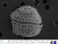
Unknown dinoflagellate under SEM (Dinophyceae)

Symbiodinium sp. (Dinophyceae): zooxanthella, a coral endosymbiont
The dinoflagellates (Greek δῖνος dinos "whirling" and Latin flagellum "whip, scourge") are a monophyletic group of single-celled eukaryotes constituting the phylum Dinoflagellata and are usually considered algae. Dinoflagellates are mostly marine plankton, but they also are common in freshwater habitats. Their populations vary with sea surface temperature, salinity, and depth. Many dinoflagellates are photosynthetic, but a large fraction of these are in fact mixotrophic, combining photosynthesis with ingestion of prey (phagotrophy and myzocytosis).
In terms of number of species, dinoflagellates are one of the largest groups of marine eukaryotes, although substantially smaller than diatoms. Some species are endosymbionts of marine animals and play an important part in the biology of coral reefs. Other dinoflagellates are unpigmented predators on other protozoa, and a few forms are parasitic (for example, Oodinium and Pfiesteria). Some dinoflagellates produce resting stages, called dinoflagellate cysts or dinocysts, as part of their lifecycles, and are known from 84 of the 350 described freshwater species, and form a little more than 10% of the known marine species. Dinoflagellates are alveolates possessing two flagella, the ancestral condition of bikonts.
About 1,555 species of free-living marine dinoflagellates are currently described. Another estimate suggests about 2,000 living species, of which more than 1,700 are marine (free-living, as well as benthic) and about 220 are from fresh water. The latest estimates suggest a total of 2,294 living dinoflagellate species, which includes marine, freshwater, and parasitic dinoflagellates.
A rapid accumulation of certain dinoflagellates can result in a visible coloration of the water, colloquially known as red tide (a harmful algal bloom), which can cause shellfish poisoning if humans eat contaminated shellfish. Some dinoflagellates also exhibit bioluminescence—primarily emitting blue-green light. Thus, some parts of the ocean light up at night giving blue-green light.
Los dinoflagelados (Dinoflagellata, Dinophyta o Pyrrhophyta) son un extenso grupo de protistas flagelados, con unas 2400 especies conocidas.[2][3][4] El nombre proviene del griego dinos, girar y del latín, flagellum, látigo, describiendo el movimiento rotatorio propio de estos organismos.[5] Estos microorganismos son unicelulares (aunque pueden formar colonias) y forman parte del fitoplancton de agua dulce (unas 220 especies) y marino (el resto).[6] Aproximadamente la mitad son fotosintéticos y poseen pigmentos con clorofila a y c2 y carotenoides. Al ser su nutrición principalmente autótrofa son productores primarios por lo que, junto a las diatomeas y otros grupos de fitoplancton, constituyen el nivel trófico primario en la cadena alimentaria acuática. Ciertas especies fotosintéticas como las zooxantelas son endosimbiontes de animales invertebrados como los corales, anémonas y almejas y protozoos marinos desarrollando una relación mutualista con los arrecifes coralinos. Otros son heterótrofos o mixótrofos y se alimentan de otros dinoflagelados, protozoos y diatomeas, además, algunas formas son parásitas (véase por ejemplo, Oodinium y Pfiesteria).[7] Sus poblaciones se distribuyen en función de la temperatura, salinidad y profundidad del agua. Algunos dinoflagelados pueden emitir luz a través de la bioluminiscencia, otros son responsables de las mareas rojas y floraciones algales nocivas (FAN o bloom de algas).
La mayoría de los dinoflagelados tienen un tamaño entre 50 y 500 µm, por lo que se los considera parte del fitoplancton, si bien Noctiluca puede llegar hasta los 2 mm de diámetro. Son unicelulares, aunque como excepción, algunas especies pueden formar colonias o seudocolonias. El rasgo más característico de los dinoflagelados es la presencia de dos flagelos disimilares que les proporcionan movimientos característicos. Uno de ellos es ondulado y rodea la célula transversalmente, se denomina flagelo transversal y le permite un movimiento giratorio distintivo del cual proviene el nombre dinoflagelado (del griego dinos, girando). El otro está localizado en el lado posterior de forma longitudinal, funciona como timón y es responsable de su movimiento vertical, este se denomina flagelo longitudinal.
En las especies dinocontas los flagelos se alojan en dos ranuras, denominadas cíngulo, la transversal, y sulcus, la longitudinal. Los flagelos emergen de la intersección de los dos surcos. Los dinoflagelados basales tienen células desmocontas, no presentan cíngulo ni sulcos y los flagelos emergen de un poro localizado en la parte apical. En este caso, uno de los flagelos es anterior y recuerda al flagelo transversal ondulado de los dinoflagelados típicos.
Los dinoflagelados tienen una cubierta celular compleja llamada anfiesma, integrada por vesículas planas denominadas alvéolos corticales. Morfológicamente, se distinguen dos tipos de dinoflagelados: tecados y desnudos. En las formas tecadas, los alvéolos se apoyan en placas de celulosa entrelazadas que componen una especie de armadura llamada teca. La teca o cobertura de la pared celular exhibe diversas formas en la morfología externa dependiendo de la especie y a veces de la etapa del dinoflagelado. Las formas sin armadura, "atecadas" o "desnudas" tienden a ser frágiles y a deformarse fácilmente, mientras que la pared celular de los dinoflagelados armados es más rígida e inflexible. En muchas especies también se encuentran extrusomas fibrosas.
Aproximadamente la mitad de los dinoflagelados presentan cloroplastos y los demás son heterótrofos (fagotrofos o parásitos osmotrofos). Aunque algunas especies con cloroplastos son totalmente autótrofas, la mayoría son mixótrofas, combinando la nutrición autótrofa y heterótrofa. El grupo de los dinoflagelados incluye cloroplastos procedentes de al menos de seis fuentes diferentes. Los cloroplastos de los dinoflagelados ancestrales derivan probablemente de la endosimbiosis secundaria de un alga roja. Posteriormente algunos grupos de dinoflagelados los reemplazaron por cloroplastos procedentes de otros grupos de algas mediante endosimbiosis secundarias o terciarias posteriores, incluyendo cloroplastos procedentes de Chlorophyta, Heterokontophyta, Cryptophyta y Haptophyta.
Los dinoflagelados fotosintéticos típicos (del tipo peridiniales) presentan cloroplastos en forma de discos o varillas, tilacoides usualmente en grupos de tres, varios tipos de pirenoides y algunas xantofilas específicas. Los pigmentos incluyen clorofilas a y c2, peridinina (un tipo de pirenoide exclusivo de los dinoflagelados), β-caroteno y pequeñas cantidades de dinoxantina y diadinoxantina. Las distintas combinaciones de pigmentos les proporcionan una coloración amarilla, pardo amarillenta, parda, verde azul, etc. Los pirenoides se encuentran junto al cloroplasto y como productos de reserva utilizan almidón, producido en el exterior del plasto, y aceites. Los cloroplastos están rodeados por tres membranas (en algunos casos de dos), lo que sugiere que proceden probablemente de la endosimbiosis secundaria de algún alga, que por los tipos de clorofila que contienen, se supone un alga roja.
Sin embargo, algunas especies presentan cloroplastos con diferente pigmentación y estructura, algunos de los cuales conservan un nucleomorfo. Ello sugiere que estos cloroplastos fueron incorporados por varios acontecimientos endosimbióticos que implicaban formas ya coloreadas o secundariamente descoloridas. Es decir, estos dinoflagelados reemplazaron sus cloroplastos procedentes de la endosimbiosis secundaria de un alga roja por otros procedentes de endosimbiosis secundarias o terciarias de otros tipos de algas. Estos cloroplastos presentan cuatro membranas y clorofilas a y b cuando proceden de endosimbiosis secundarias de algas verdes, y clorofilas a y c cuando proceden de endosimbiosis terciarias de otros tipos de algas. En concreto, existen cloroplastos de procedencia de los siguientes grupos:[8][9]
El descubrimiento de apicoplastos en los apicomplejos sugiere que los plastos de estos dos grupos se originaron de un antepasado común que realizó una endosimbiosis secundaria con un alga roja.
Los dinoflagelados típicos presentan un núcleo de características únicas denominado dinocarión. En este tipo de núcleo, los cromosomas se fijan a la envoltura nuclear, contienen una enorme cantidad de ADN, están muy organizados, carecen de histonas al contrario que los demás eucariotas y no presentan una verdadera interfase. Esta clase de núcleo fue una vez considerado una forma intermedia entre el nucleoide de los procariontes y los núcleos verdaderos de los eucariontes y fue llamado mesocarión, pero ahora se considera una forma avanzada más que primitiva. Los dinoflagelados basales, sin embargo, presentan núcleos similares al resto de los eucariotas, mientras que en Noctilucales el dicarión está presente solo en las etapas juveniles.
La célula de los dinoflagelados contiene los orgánulos más comunes tales como el retículo endoplasmático, aparato de Golgi, mitocondrias, gránulos de lípidos y almidón y vacuolas endoplasmáticas. Además, algunos dinoflagelados, la mayoría de agua dulce, presentan una mancha ocular, un orgánulo sensible a la luz que les permiten determinan la dirección e intensidad de la luz. Dependiendo de la especie, la mancha ocular presenta diferentes tipos de organización, que va desde la más sencilla de un glóbulo libre en el citoplasma hasta un orgánulo complejo u ocelo compuesto de una lente con retinoide. Muchos dinoflagelados disponen de tricocistos que disparan filamentos mucilaginosos.
Algunas de estas características morfológicas y genéticas indican una relación cercana entre Dinoflagellata, Apicomplexa y Ciliophora, que son agrupados en Alveolata.
La reproducción de los dinoflagelados es principalmente asexual, que en condiciones favorables puede ser muy rápida originando poblaciones que pueden a llegar a 60 millones de individuos por litro de agua. También se da reproducción sexual. En los dinoflagelados típicos el núcleo es dinocarión durante todo el ciclo vital y son generalmente haploides. La reproducción sexual tiene lugar por fusión de dos individuos para formar un zigoto, que puede seguir siendo móvil o formar un quiste inmóvil, que más adelante experimentará una meiosis para producir nuevas células haploides.
En un ciclo de vida típico, cuando las condiciones llegan a ser críticas, generalmente por falta de alimento o por inexistencia de luz, dos dinoflagelados se fusionarán formando un planozigoto. Este continúa su movilidad hasta que después de unos días pierde sus flagelos. A continuación tiene lugar una etapa no muy diferente de la hibernación llamada hipnozigoto. La membrana se expande abriendo la teca, el protoplasma se contrae y se forma una nueva teca más dura en el cual algunas veces se forman espinas. El quiste recién formado se deposita en el fondo marino. Cuando las condiciones vuelven a ser favorables, rompe su teca, pasa por una etapa temporal denominada planomeiocito y después retorna rápidamente a la forma dinoflagelada a principio del ciclo.
La proliferación de los dinoflagelados junto con otras bacterias y ciliados puede llegar a ser tóxica, fenómeno que se conoce como "floraciones algales nocivas" (FAN), o puede producir cambios de color en el agua tornándola roja por la biomasa y pigmentación de estos organismos; este otro fenómeno es conocido como mareas rojas y no es tóxico.[10][11][12] La causa de éstas puede ser natural, por factores como la salinidad, cantidad de luz, turbulencia y temperatura, pero de igual forma la actividad humana es un elemento importante en la proliferación de estos organismos en su hábitat. Uno de los procesos naturales en los que el ser humano ha intervenido y a la vez perjudicado ha sido el ciclo de nitrógeno. A causa de este se afecta la acidificación, eutrofización y proliferación de algas nocivas dentro de distintos cuerpos de agua.
Algunas fuentes principales de nitrógeno inorgánico lo son aguas residuales sin tratamiento, infiltración en basureros, campos de cultivo, bosques quemados, emisiones a la atmósfera de combustibles fósiles y residuos de granjas de animales, estos desechos provienen de diferentes fuentes pero terminan en un mismo lugar, en lagos, ríos y océanos. La acidificación se presenta cuando hay desbalances en el valor del pH del agua de los ríos y lagos. Entre sus efectos negativos se encuentra la disminución de la diversidad, fotosíntesis y productividad del fitoplancton, disminución en la actividad alimentaria y diversidad en animales acuáticos. Al ser afectados estos productores primarios y consumidores de la cadena alimentaria en el ecosistema acuático se afectan los eslabones, se desequilibran los niveles tróficos de esta cadena y se pone en riesgo todo el ecosistema acuático.
Una gran acumulación de fósforo y nitrógeno en los cuerpos de agua, los cambios climáticos, el aumento en la radiación ultravioleta son otros factores por los que se producen estas floraciones, ya que estos elementos promueven su desarrollo y mantenimiento. Las toxinas de los dinoflagelados podrían ser producidas a través de simbiosis con bacterias, selección natural, reservas de nitrógeno, metabolitos secundarios, como mecanismos de defensa o competencia. Las FAN tienen una gran importancia ecológica ya que los animales que se alimentan de estos microorganismos tóxicos, se intoxican, enferman, mueren y transmiten el tóxico a través de la cadena alimentaria que a su vez afecta a los humanos que ingieran estos organismos contaminados, reducen el oxígeno disuelto en el agua y causan la muerte de cientos de peces y corales. De igual manera, tienen una gran importancia social ya que constituyen una amenaza a la salud de las personas, la economía, el turismo, la pesca y la acuicultura.
En términos de salud las personas pueden sufrir diferentes intoxicaciones como la intoxicación paralizante por moluscos, intoxicación diarreica por moluscos, intoxicación neurotóxica por moluscos y intoxicación amnésica por moluscos o ciguatera o veneno ciguatérico de pescado, y a consecuencia de éstos, algunos efectos y síntomas que pueden presentar son cuadros neuróticos, parestesia bucal, dolor abdominal, cefalea, alteración del pulso, insuficiencia respiratoria, paros cardiorrespiratorios y hasta la muerte. La pesca se ve afectada ya que los peces y otros animales están contaminados, el turismo disminuye ya que las facilidades recreacionales no están en condiciones saludables y la economía se ve afectada por ambos elementos y muchos más. Debido a la intensidad de ocurrencias de las floraciones algales nocivas mundialmente, se han creado distintas organizaciones nacionales e internacionales dedicadas a la investigación, manejo y prevención de dicho fenómeno. Su estudio es de gran importancia y aportación al conocimiento científico, al desarrollo de métodos, herramientas, modelos, pronósticos y tecnologías en los programas de investigación.
Debe tenerse en cuenta que no todas las floraciones de dinoflagelados son peligrosas. Los parpadeos azulados visibles en el agua del océano por la noche son producidos a menudo por las floraciones de dinoflagelados bioluminescentes, que emiten ráfagas cortas de luz como mecanismo de defensa.
Los fósiles más antiguos de dinoflagelados corresponde a acritarcos que datan del Mesoproterozoico y se conocen como Shuiyousphaeridium y Dictyosphaera.[13] Posteriormente aparecen quistes de dinoflagelados que se encuentran como microfósiles a partir del período Triásico hace 245 a 208 millones de años, aumentando su número y diversidad y formando una parte importante de la microflora marina del Jurásico medio, aunque se encuentran restos químicos de dinosporina (sustancia que compone a los dinoflagelados) en rocas del Silúrico. La presencia de dinosteranes, un esterol mayormente asociado con los dinoflagelados, sustenta una radiación Mesozoica de estos microorganismos, lo cual muestra que hubo un aumento dramático entre el Periodo Pérmico y el Cretáceo (Hackett et al., 2004) hasta el día de hoy. Puesto que ciertas especies se adaptan a distintas condiciones del agua superficial, estos fósiles se pueden utilizar para reconstruir las condiciones superficiales oceánicas.[14]
La clasificación de los dinoflagelados es difícil y comprende cinco clases, de las cuales, las tres primeras son basales o constituyen linajes altamente divergentes y a veces se clasifican aparte.[1] Los grupos basales presentan un núcleo celular similar al resto de los eucariotas. Los dinoflagelados típicos con dinocarión pertenecen a la clase Dinophyceae y en menor medida a Noctiluciphyceae.
Las relaciones son las siguientes:[15][19]
DinoflagellataLos dinoflagelados (Dinoflagellata, Dinophyta o Pyrrhophyta) son un extenso grupo de protistas flagelados, con unas 2400 especies conocidas. El nombre proviene del griego dinos, girar y del latín, flagellum, látigo, describiendo el movimiento rotatorio propio de estos organismos. Estos microorganismos son unicelulares (aunque pueden formar colonias) y forman parte del fitoplancton de agua dulce (unas 220 especies) y marino (el resto). Aproximadamente la mitad son fotosintéticos y poseen pigmentos con clorofila a y c2 y carotenoides. Al ser su nutrición principalmente autótrofa son productores primarios por lo que, junto a las diatomeas y otros grupos de fitoplancton, constituyen el nivel trófico primario en la cadena alimentaria acuática. Ciertas especies fotosintéticas como las zooxantelas son endosimbiontes de animales invertebrados como los corales, anémonas y almejas y protozoos marinos desarrollando una relación mutualista con los arrecifes coralinos. Otros son heterótrofos o mixótrofos y se alimentan de otros dinoflagelados, protozoos y diatomeas, además, algunas formas son parásitas (véase por ejemplo, Oodinium y Pfiesteria). Sus poblaciones se distribuyen en función de la temperatura, salinidad y profundidad del agua. Algunos dinoflagelados pueden emitir luz a través de la bioluminiscencia, otros son responsables de las mareas rojas y floraciones algales nocivas (FAN o bloom de algas).
Dinoflagellata • Dinophytes, Dinoflagellés, Péridiniens
Les Dinophytes (Dinophyta), encore appelées Dinoflagellés ou Péridiniens, sont un embranchement d'organismes eucaryotes unicellulaires. Ce sont des microorganismes aquatiques (marins ou dulçaquicoles). Ils sont très diversifiés, en particulier par leurs comportements alimentaires. Certains sont hétérotrophes, d'autres sont mixotrophes et d'autres encore sont des organismes photosynthétiques qui sont donc assimilés à des algues unicellulaires, en majorité biflagellées. Environ 50 % des dinoflagellés sont hétérotrophes. Ils sont comme leur nom l'indique dotés de flagelles, mais certains sont « benthiques » et vivent en épiphytes sur des macroalgues ou encore dans les interstices des substrats sableux (tout en pouvant migrer verticalement pour se reproduire).
Ces microorganismes jouent un rôle trophique majeur, en particulier les protistes hétérotrophes herbivores qui régulent les populations de microalgues, et qui en mangeant ce phytoplancton en transfèrent les nutriments (mais aussi certains polluants qu'ils peuvent éventuellement bioconcentrer) vers les niveaux trophiques supérieurs (métazoaires, puis poissons, mammifères marins..).
Les études moléculaires montrent que les dinoflagellés ont un ancêtre commun dont on tente encore de savoir s'il était photosynthétique ou non[1]. Pour cela on étudie les gènes plastidiaux importés dans le génome nucléaire à la suite d'endosymbioses. On sait aussi grâce à ces études génétiques que plusieurs lignées hétérotrophes sont apparues très tôt au cours de l’évolution [1].
Ce groupe est encore assez mal connu. On a récemment encore en Bretagne — lors d'une étude des dinophyta du sédiment — trouvé plusieurs espèces jusqu'alors non décrites de Prorocentrum, Sinophysis ou Cabra[2].
Le mot Dinoflagellata, viendrait du grec ancien δεινός / deinόs « qui inspire la crainte » ou δῖνος / dinos, « tournoiement », et du latin flagellum, « fouet », littéralement « flagelle tournoyant ».
Chez beaucoup d'espèces, la cellule est protégée par une thèque constituée de plaques rigides de cellulose incrustée de silice.
Les flagelles reposent dans deux sillons superficiels : le transversal ou équatorial, appelé « cingulum », le longitudinal, appelé « sulcus » (du latin cingulum, ceinture, et sulcus, sillon) .
Le sillon équatorial sépare la cellule en une partie supérieure (l’épicone) et une partie inférieure (l’hypocone). postérieur (hyposome), et, si une thèque est présente, ces deux parties sont appelées respectivement « épithèque » et « hypothèque ».
Certaines espèces ont un « appareil photosensible », et d’autres un véritable ocelle, qui servirait vraisemblablement à détecter les proies.
Nombreuses sont les espèces photosynthétiques. Mais la grande variété de chloroplastes témoigne de plusieurs endosymbioses secondaires. Il y a eu en effet, chez un ancêtre du groupe, une endosymbiose secondaire avec un eucaryote de la lignée rouge. Certains Dinoflagellés ont perdu cet endosymbiote, et parmi ceux-ci beaucoup sont restés hétérotrophes. Mais d’autres ont effectué une endosymbiose dite tertiaire avec des Straménopiles, des Cryptophytes ou d’autres Dinoflagellés.
Les Dinoflagellés font partie du plancton d’eaux douces et surtout des eaux marines. Certaines espèces sont aussi retrouvées dans la neige. D’autres espèces, les Zooxanthelles, vivent en symbiose avec des protistes ou des Invertébrés marins (éponges, Coraux, Vers plats, Mollusques, Crustacés). Les espèces chlorophylliennes effectuent la photosynthèse grâce à des plastes. Les espèces non pigmentées se nourrissent de plancton. Il existe des formes bioluminescentes comme Noctiluca qui utilise une enzyme, la luciférase, et un substrat, la luciférine. L’accumulation de Noctiluca miliaris et Noctiluca scintillans entraîne des efflorescences et des phénomènes de marées rouges dans certaines conditions.
Certaines espèces d'algues planctoniques, essentiellement des Dinoflagellés et des Diatomées, peuvent sécréter des phycotoxines (des toxines algales) dans certaines circonstances mal connues, en particulier à l'occasion des multiplications soudaines (on parle alors d'efflorescences localisées, bloom en anglais). Ces toxines seraient un moyen de protection et de limitation de la prédation par les phytophages (zooplancton, coquillages filtreurs).
Ces toxines provoquent différents troubles, base de la classification :
La production et la pêche des coquillages comestibles sont, dans la plupart des pays, placées sous le contrôle d'une autorité chargée de la surveillance des eaux marines et des coquillages produits[6], afin de garantir l'innocuité des denrées.
Cependant, la plupart des espèces de Dinoflagellés ne sont pas toxiques et sont à la base de la nutrition de nombreuses espèces : zooplancton, poissons.
Les études génétiques ont essentiellement concerné des dinoflagellés photosynthétiques. Crypthecodinium cohnii est le premier dinoflagellé hétérotrophe ayant fait l'objet d'une étude génomique[7]
Étant donné la taille de leur génome le séquençage s'avère particulièrement coûteux. Seul les génomes de Symbiodinium microadriaticum, Symbiodinium minutum et Symbiodinium kawagutii, des symbiotes des coraux, ont été séquencés en 2016 et leur taille est entre 1 et 5 Gb [8].
Les plus anciens restes fossiles incontestables de Dinoflagellés datent du Trias(~250 Ma). Une forme présumée, datée du Silurien (420 Ma), Arpylorus sp., serait plutôt une structure de stockage produite par un arthropode[9]. Certaines thèques fossiles pouvant avoir appartenu à des Dinoflagellés primitifs existent dans le Cambrien inférieur (-540 Ma).
Liste des ordres de Dinoflagellata selon World Register of Marine Species (5 janvier 2014)[10] : ...
Liste de Dinoflagellés communs (2 200 espèces connues) :
Dinoflagellata • Dinophytes, Dinoflagellés, Péridiniens
Les Dinophytes (Dinophyta), encore appelées Dinoflagellés ou Péridiniens, sont un embranchement d'organismes eucaryotes unicellulaires. Ce sont des microorganismes aquatiques (marins ou dulçaquicoles). Ils sont très diversifiés, en particulier par leurs comportements alimentaires. Certains sont hétérotrophes, d'autres sont mixotrophes et d'autres encore sont des organismes photosynthétiques qui sont donc assimilés à des algues unicellulaires, en majorité biflagellées. Environ 50 % des dinoflagellés sont hétérotrophes. Ils sont comme leur nom l'indique dotés de flagelles, mais certains sont « benthiques » et vivent en épiphytes sur des macroalgues ou encore dans les interstices des substrats sableux (tout en pouvant migrer verticalement pour se reproduire).
Ces microorganismes jouent un rôle trophique majeur, en particulier les protistes hétérotrophes herbivores qui régulent les populations de microalgues, et qui en mangeant ce phytoplancton en transfèrent les nutriments (mais aussi certains polluants qu'ils peuvent éventuellement bioconcentrer) vers les niveaux trophiques supérieurs (métazoaires, puis poissons, mammifères marins..).
Les études moléculaires montrent que les dinoflagellés ont un ancêtre commun dont on tente encore de savoir s'il était photosynthétique ou non. Pour cela on étudie les gènes plastidiaux importés dans le génome nucléaire à la suite d'endosymbioses. On sait aussi grâce à ces études génétiques que plusieurs lignées hétérotrophes sont apparues très tôt au cours de l’évolution .
Ce groupe est encore assez mal connu. On a récemment encore en Bretagne — lors d'une étude des dinophyta du sédiment — trouvé plusieurs espèces jusqu'alors non décrites de Prorocentrum, Sinophysis ou Cabra.
Skoruþörungar (fræðiheiti: Dinophyceae) eru stærsti flokkur svipuþörunga. Þessir þörungar mynda þörungablóma við strendur þegar líður á sumar og sumar tegundir þeirra eru helsta ástæðan fyrir hættulegum eiturefnum í skelfiski eins og kræklingi.
Einkenni í útliti þessara einfrumu þörunga eru tvær skorur sem liggja í kross þvert yfir frumuna. Önnur skoran, þverskoran, nær umhverfis alla frumuna, en hin, langskoran, er misjöfn eftir tegundum. Skoruþörungar hafa tvær mis langar svipur og liggja þær hvor í sinni skorunni. Svipan í langskorunni er að jafnaði meira áberandi. [1] Nokkurskonar brynja umlykur margar tegundir skoruþörunga. Hún getur verið hornótt og er samsett úr nokkuð þykkum plötum. Þeir þörungar sem hafa þetta útlit eru oft kallaðir hornþörungar eða brynþörungar (Dinophyceae). Aðrar tegundir eru nánast berar og getur lögun þeirra verið breytileg. [2] Í flestum tilvikum er kjarni skoruþörunga áberandi. Frumurnar sjálfar eru í ýmsum litum og sumar jafnvel rauðar. Í þörungablóma er oft talað um blóðsjó vegna litarins. Grænukornin í þeim eru yfirleitt gulbrún, en sumar tegundir hafa engin grænukorn (Dinoflagellata). Þær geta neytt fastrar fæðu og flokkast því oft með dýrum. Sumir skoruþörungar hafa rauðan augndíl.[3]
Skoruþörungar eru flokkaðir eftir sköpulagi en taldir vera af samsíða þróunarlínum. Þeir hópar sem eru með hulu eru flokkaðir í fjóra ættbálka eftir því hvernig brynplötur þeirra raðast.
Ættbálkar (auk ýmissa ættkvísla) skoruþörunga sem eru án hulu eru taldir vera fjölstofna (þ.e. tilheyra ólíkum þróunarlínum).
Skourþörunga má finna bæði í sjó og vötnum, þó meira sé um þá í sjó. Þeir búa oft í samlífi eða jafnvel í samhjálp með öðrum sjávardýrum, þar á meðal kóraldýrum og svömpum.[4]
Skoruþörungar eru meðal þeirra svifþörunga sem mynda svokallaðan þörungablóma við strendur og í fjörðum á vorin, sumrin og svo aftur á haustin. [5] Mjög mikill þörungablómi getur litað vötn og sjó í rauðum eða brúnum lit. Þetta er oft kallað mor eða bljóstjór og skapast vegna gríðarlegs fjölda þörunga á viðkomandi svæði. [6]
Vissar tegundir skoruþörunga geta valdið matareitrun í mönnum vegna neyslu á skelfiski. Meðal þeirra eitrana sem menn geta veikst af er svokölluð DSP (Diarrhetic shellfish poisoning) eitrun. Eitrunin á sér stað þegar menn neyta skelfiskt sem nærst hefur á þörungunum með þeim afleiðingum að eitur safnast upp í þeim. Þetta gerist aðeins ef mikið magn þörunga er í sjó á svæðinu. DSP eitrun veldur ógleði og uppköstum, niðurgangi og verkjum í kviði. Veikindin ganga yfirleitt yfir á þrem sólarhringum, en þó er talið að í um 15% tilvika valdi hún dauða. [7]
Skoruþörungar (fræðiheiti: Dinophyceae) eru stærsti flokkur svipuþörunga. Þessir þörungar mynda þörungablóma við strendur þegar líður á sumar og sumar tegundir þeirra eru helsta ástæðan fyrir hættulegum eiturefnum í skelfiski eins og kræklingi.
Le dinoflagellate (dal greco δῖνος dînos, "vortice", e il latino flagellum, "flagello", "frusta"), conosciute anche come pirrofite, peridinee o dinoficee, sono alghe microscopiche per lo più unicellulari e flagellate, che rappresentano uno dei più importanti gruppi del fitoplancton sia marino che d'acqua dolce con oltre 2000 specie viventi.
Le dinoflagellate, assieme ai Ciliati e agli Apicomplexa, appartengono al supergruppo degli Alveolata ovvero quegli organismi unicellulari che possiedono un sistema di alveoli corticali al di sotto del plasmalemma.
La cellula è dotata di una struttura peculiare, l'anfiesma e consiste di un periplasto, nella regione sottostante a questo può essere presente un velo di cellulosa. L'anfiesma è formato da delle vescicole, gli alveoli, che possono essere vuote o contenere glucani, in questo caso formano delle placche che rivestono la cellula, si parla in questo caso di teca.
Sono presenti due flagelli, entrambi provvisti di peli laterali, differenti l'uno dall'altro per struttura e orientamento.
Esistono due principali morfotipi: le Dinoconte e le Desmoconte. Nelle Dinoconte la cellula presenta due scanalature, una equatoriale (cingolo) e una longitudinale (solco). Quest'ultimo divide la cellula in due parti, dette epicono (o epiteca) e ipocono (o ipoteca). I due flagelli emergono in posizione ventrale all'intersezione tra il cingolo e il solco. Nelle Desmoconte le placche tecali sono organizzate a formare due valve distinte e i due flagelli fuoriescono in posizione apicale.
Il cloroplasto nelle dinoflagellate è stato originariamente acquisito mediante un evento di endosimbiosi secondaria con un'alga rossa. Nella maggior parte delle specie i cloroplasti sono circondati da tre membrane, i tilacoidi sono in gruppi di tre e contengono clorofilla di tipo a e c2, caroteni, e xantofille tra cui la peridinina. In alcuni casi questo cloroplasto è stato perso e talvolta un altro è stato acquisito mediante altri eventi di endosimbiosi (secondaria o terziaria) con altre alghe eucariote (diatomea, criptoficea, haptofita, alga verde) presentando pertanto un corredo pigmentario differente. Molte dinoflagellate (circa la metà delle specie) sono prive di cloroplasti e sono pertanto eterotrofe obbligate.
Le sostanze di riserva sono granuli di amido che si accumulano all'esterno del cloroplasto e sostanze lipidiche.
Hanno un nucleo voluminoso e primitivo, che viene detto dinocarion: il DNA non è associato a istoni, e durante la mitosi i cromosomi rimangono attaccati alla membrana nucleare, persistente, e non al fuso.
Sulla superficie sono presenti le tricocisti, organuli bastoncellari che sono sparati all'esterno attraverso i pori presenti sulla parete e che hanno funzione di difesa.
Si riproducono abitualmente per via vegetativa: la cellula si divide longitudinalmente, trasversalmente od obliquamente. A parte poche eccezioni, le dinoflagellate sono aploidi e presentano un ciclo vitale aplonte, con meiosi zigotica. La riproduzione sessuata avviene per produzione di gameti che non sono distinguibili dalle cellule vegetative. Dopo la fusione si origina un planozigote che poi perde i flagelli, sviluppa una spessa parete, e spesso diviene una cellula di resistenza (cisti) che resta dormiente nei sedimenti per periodi anche molto lunghi. Alla germinazione si riforma il planozigote che compie la meiosi riformando le cellule vegetative aploidi.
Le cisti sono caratterizzate da una teca molto resistente agli agenti chimici. Possono avere un aspetto simile o molto diverso da quello della cellula madre e presentano un'apertura detta archeopilo, attraverso cui avverrà la germinazione. Si distinguono cisti di resistenza e cisti temporanee, prodotte come strategia di sopravvivenza a lungo e a breve termine. Entrambe possono essere prodotte sia per via sessuata che per via vegetativa. La produzione di cisti ha un ampio significato ecologico, in quanto consente la sopravvivenza delle specie in condizioni avverse, ed ha un importante ruolo nella dispersione delle specie.
Le dinoflagellate sono molto abbondanti in tutti gli oceani, particolarmente nelle regioni tropicali. Nelle aree temperate hanno il loro massimo sviluppo tipicamente in estate, in condizioni di stabilità della colonna d'acqua. In alcuni casi proliferano intensamente raggiungendo abbondanze molto elevate, dell'ordine di milioni di cellule per litro, dando origine al fenomeno delle "maree rosse".
Alcune specie producono biotossine, composti che hanno attività tossica per l'uomo e per altri vertebrati. Comunemente le biotossine vengono trasferite tramite la catena alimentare e vengono accumulate in organismi vettori (per lo più molluschi bivalvi, ma anche pesci). Il consumo di molluschi o pesci contaminati causa biointossicazioni, tra cui le più importanti sono la PSP (Paralytic Shellfish Poisoning), la DSP (Diarrhetic Shellfish Poisoning), NSP (Neurotoxic Shellfish Poisoning) e la ciguatera.
Pfiesteria piscicida è una specie eterotrofa che produce una ittiotossina neurotossica ed è responsabile di morie di pesce lungo le coste Atlantiche degli USA.
Alcune (es. Symbiodinium spp.) sono simbionti di invertebrati marini, come i coralli, e prendono il nome di zooxantelle. Tali simbiosi sono molto importanti per la formazione e lo stato di salute delle barriere coralline tropicali.
Le dinoflagellate (dal greco δῖνος dînos, "vortice", e il latino flagellum, "flagello", "frusta"), conosciute anche come pirrofite, peridinee o dinoficee, sono alghe microscopiche per lo più unicellulari e flagellate, che rappresentano uno dei più importanti gruppi del fitoplancton sia marino che d'acqua dolce con oltre 2000 specie viventi.
Le dinoflagellate, assieme ai Ciliati e agli Apicomplexa, appartengono al supergruppo degli Alveolata ovvero quegli organismi unicellulari che possiedono un sistema di alveoli corticali al di sotto del plasmalemma.
Šarvadumbliai (lot. Pyrrophyta) – eukariotų (Eukaryota) domeno pirmuonių (Protista) karalystės skyriaus dumbliai.
Šarvadumbliams priklauso judrūs, rečiau nejudrūs, vienaląsčiai arba kolonijiniai žiuželiniai dumbliai. Ląstelės ovališkos, kiaušiniškos, elipsiškos arba pupelės formos su išgaubta nugarine ir plokščia arba įgaubta pilveline pusėmis. Spalva nuo gelsvai rudos ir rausvos.
Iš viso yra apie 1000 rūšių, gyvenančių daugiausia jūrose ir sudarančių didžiąją dalį fitoplanktono. Lietuvoje yra 2 klasės:
Fureflagellater (Dinophyceae) er en gruppe av mikroskopiske alger som lever i hav og innsjøer. Visse arter lever også i symbioseforhold f.eks. i koralldyr. Dinoflagellatene er en stor gruppe alger som fins i alle deler av verden, fra polene til tropiske farvann.
Kjennetegn for gruppen er at de har to flageller, den ene ligger i en fure som går rundt cellen den andre går bakover. Flagellene som ligger i furen får cellen til å snurre om sin egen akse mens flagellen som ligger bakover får den til å bevege seg framover. Cellene er asymmetriske og den karakteristiske måten å svømme på, at den skrur seg gjennom vannet, er gunstig pga det lave reynoldstallet (lav masse og hastighet).
Dinoflagellater vokser relativt sakte sammenlignet med mange andre algegrupper, men klarer å konkurrere under næringsfattige forhold. Derfor er denne gruppen vanligst om sommeren i norske farvann etter at termoklinen, dvs. den horisontale lagdelingen av vannmassen, har blitt bygget opp.
En rekke dinoflagellater er giftige, og gruppen står bak en rekke av de HAB (=harmful algal blooms) som blir rapportert rundt om i verden.
Os dinoflagelados (divisão Dinophyta, segundo os botânicos, ou filo Dinoflagellata (Dinoflagellata - do grego "dino", rodopiantes) para os protozoologistas são um grande grupo de protistas flagelados. A maior parte das espécies pertencem ao plâncton marinho (mais especificamente do fitoplâncton), mas são também comuns em água doce. Estão intimamente relacionados com os protozoários ciliados, tais como Paramecium e Vorticella, e com os Apicomplexa (o filo do qual o parasita da malária faz parte).
São organismos que em sua maioria apresentam formas unicelulares, ocorrendo algumas raras formas filamentosas; em sua grande maioria, são flageladas (dois flagelos eucarióticos diferentes), mas existem formas imóveis, amebóides, cocóides e coloniais palmelóides.
São conhecidas por volta de 2.000 a 4.000 espécies, incluindo formas protistas flagelados. A maior parte das espécies pertencem ao plâncton marinho (marinhas e de mais especificamente do fitoplâncton), mas são também comuns em água doce sendo que, metade delas é fóssil e estão distribuídas em 550 gêneros. Seu corpo possuem aspecto semelhante a uma medusa ou a um verme.
São, geralmente, divididos entre as classes Dinophyceae, Noctiluciphyceae e Syndiniophyceae.
Muitas destas espécies têm capacidade fotossintética e formam o maior componente do fitoplâncton depois das diatomáceas. Algumas, as zooxantelas são endosimbiontes de animais ou protistas marinhos e têm um importante papel na biologia dos corais. São heterotróficas (saprófitos, parasitas e holozóicas), bem como formas que vivem em simbiose com animais.
Todos os dinoflagelados são unicelulares com dois flagelos diferentes: um flagelo longitudinal, orientado segundo o eixo da célula, e um flagelo transversal que rodeia a célula. Em muitas espécies, estes flagelos encontram-se em sulcos: o longitudinal chamado sulcus e o transversal, cingulum. É o flagelo transversal que provoca a maior parte do movimento da célula, geralmente em forma de hélice, donde provém o nome destes seres (dinos, em grego, significa, rodar).
Têm sido encontrados com frequência quistos fossilizados de dinoflagelados desde o período Triássico (de há 200 milhões de anos), mas já se encontraram do Siluriano (400 milhões de anos) e pensa-se que alguns dos antigos acritarcas com uma idade de 1,8 biliões de anos, também representem dinoflagelados.Mas ainda não se comprovou se os cistos fósseis mais antigos sejam mesmo de Dinophyta.
Existem espécies marinhas, espécies dulciaquícolas e planctônicas. Ainda ocorrem espécies que são: endozóoicas, simbióticas e parasitas.
A maioria são monadais isto é, se locomovem por meio de dois flagelos eucarióticos. Além disso, podem apresentar placas rígidas de celulose formando uma estrutura denominada teca. Esta estrutura em mares abertos se apresenta em tamanho grande, semelhantes a velas, e auxiliam na flutuação.
Há ainda formas imóveis, amebóides, cocóides e coloniais.
Em torno de 50% das espécies não possuem aparato fotossintetizante (plastídios) e são heterótrofas. Por isso, muitas vezes a nutrição ocorre por ingestão de partículas sólidas ou pela absorção de compostos orgânicos dissolvidos. A alimentação de alguns dinoflagelados ocorre por meio da projeção de uma estrutura tubular (conhecida como pedúnculo) que suga a matéria orgânica para dentro da célula. Ao termino da alimentação este pedúnculo é recolhido para dentro da célula.
Muitas das espécies fotossintéticas formam o maior componente do fitoplâncton depois das diatomáceas. Algumas, as zooxantelas são endosimbiontes de animais ou protistas marinhos e têm um importante papel na biologia dos corais. Algumas espécies não têm aquela capacidade e são predadores de outros protistas, havendo ainda espécies parasitas.
Em torno de 20% dos dinoflagelados produzem um ou mais compostos tóxicos. O Pfiesteria piscicida usa essa toxina como meio de capturar alimento. Com a presença de peixes, seus cistos bentônicos são estimulados a germinar o que darão origem às células natantes de Pfiesteria.
Essas células se produzem e liberam uma toxina que paralisa o sistema respiratório do peixe, causando sua morte por asfixia. Com a decomposição deste animal, os dinoflagelados estendem seus pedúnculos e se alimentam da musculatura do peixe. Após a alimentação eles rapidamente retornam ao estágio de cistos bentônicos.
Devido à presença ou não de um núcleo diferenciado, denominado dinocarion (possui cromossomos sempre condensados, mesmo na interfase, e com pouca quantidade de histonas), essas algas foram divididas entre as classes:
Dinophyceae: constituído por organismos fotossintéticos ou não, possuindo um dinocarion em todas as fases de seu ciclo de vida.
Blastodiniphyceae: formado pelos organismos parasitas não fotossintéticas de peixes e que apresentam um dinocarion em apenas uma parte da vida.
Noctiluciphyceae: composto por organismos flagelados fagotróficos não fotossintéticos que contam com a presença de um dinocarion em apenas um estagio de seu ciclo biológico.
Syndiniophyceae: formado pelos organismos que são caracterizados pela ausência de dinocarion.
São parasitas marinhos não fotossintéticos de outros dinoflagelados, invertebrados, ovos de peixes e mamiferos.
Os dinoflagelados possuem uma complexa parede celular chamada anfiesma, e situa-se sobre o plasmalema. A anfiesma é composta de vesículas achatadas, os alvéolos. Em algumas formas, estes alvéolos contêm placas sobrepostas de celulose que formam o que se chama a teca, que pode ter formas muito variadas.
Estas características, em conjunto com recentes estudos genéticos, levaram a juntar este filo com os Apicomplexa e ciliados num clado denominado Alveolata.
Os cloroplastos, nas espécies que os possuem, são rodeados por três membranas e podem ainda possuir um núcleo celular, sugerindo que eles possam ser derivados de uma "alga" endosimbionte. Os pigmentos são tipicamente as clorofilas a e c2 Já os pigmentos acessórios são principalmente β-caroteno, peridinina e outros carotenoides como a giroxantina diéster, que é característico de dinoflagelados que causam marés vermelhas. O carboidrato de reserva é constituído de amido, o qual é sintetizado fora do plastídio e presente no citoplasma na forma de grãos.
Os dinoflagelados têm uma forma peculiar de núcleo celular, denominado dinocarion, em que os cromossomos se encontram condensados, mesmo na interfase, e com pouca quantidade de histonas e mantêm se na fase condensada durante a interfase. Devido a essa escassez de histonas ocorre a presença de uma dupla hélice de DNA de 2,5 nm de diâmetro. Os dinoflagelados têm de 5 a 10 vezes mais DNA no se núcleo que quaisquer outros eucariontes.
A mitose que ocorre nesses organismos é muito particular: a membrana nuclear permanece intacta (mitose fechada) e, na prófase, feixes de microtúbulos penetram no núcleo e situam-se em canais citoplasmáticos. Na anáfase, os núcleos-filhos são formados por constrição da membrana nuclear, os cromossomos-filhos separam-se e migram-se ao longo do feixe em direção aos polos.
Muitos dinoflagelados são haploides, e se reproduzem principalmente por fissão binária, mas a reprodução sexuada também ocorre, através a fusão de dois indivíduos que formam um zigoto, que pode manter-se na sua forma típica, ou enquistar e, mais tarde, sofrer meiose e produzir novas células haploides.
Muitos dinoflagelados possuem células vegetativas haploides, (com exceção do Noctiluca, que parece ter células vegetativas diplóides) e se reproduzem principalmente por divisão celular longitudinal.
A Reprodução Assexuada cada célula receberá um dos flagelos e uma porção da teca. Em seguida cada célula filha reconstitui as partes que faltam em sua sequência intrincada.
Já a Reprodução Sexuada, ocorre através da fusão de duas células haploides que formará uma célula diploide, e a partir daí a célula com sua teca celulósica crescem. Na sequência, haverá a transformação das células diploides em cistos fossilizáveis (encistamento), que encerram o material celular e constituem a fase imóvel do ciclo. Após o encistamento, a parede celulósica se desagrega e o cisto é sedimentado. Em condições ambientais favoráveis, o protoplasma sai do cisto (excistamento) através de uma abertura chamada arqueopilo e reinicia-se o ciclo com a formação de novas células móveis.
Os dinoflagelados são um dos grupos mais abundantes no plâncton marinho e, como na sua maioria são autotróficos, têm um importante papel na produção primária do globo. No entanto, algumas espécies podem causar impacto econômico negativo ao contaminar os organismos aquáticos. O gênero Alexandrium, por exemplo, produz toxinas que tem forte poder paralisante e ao entrar em contato com organismos filtradores vem provocando mortes de pessoas que os consumirem. O dinoflagelado bentônico Gambierdiscus toxicus é responsável pela Ciguatera (envenenamento resultante do consumo da carne contaminada de determinados peixes tropicais marinhos).
Algumas espécies são responsáveis pelas marés vermelhas que em condições favoráveis de temperatura e nutrientes, elas podem reproduzir-se rapidamente em enormes quantidades e as toxinas que produzidas por elas em muitas vezes causam a morte em massa de outras espécies marinhas. Além disso, estas toxinas podem acumular-se no corpo de animais que se alimentam por filtração, como os bivalves, e podem prejudicar a saúde dos seres humanos que os consumirem.
Alguns dinoflagelados sem aquela pigmentação, como Pfiesteria, podem também produzir florescimentos tóxicos que provocam perdas neurológicas de percepção próximas do mal de Alzheimer.
No entanto, nem todos os florescimentos de dinoflagelados são desagradáveis: nos mares tropicais, podem por vezes apreciar-se, em noites escuras, lindas cenas de luzes à superfície da água e, por vezes, mesmo na rebentação, causadas por dinoflagelados bioluminescentes, de entre os quais o mais famoso é o Noctiluca, que se pode ver a olho nu.
Também servem como alimento para larvas de certos peixes (aquacultura) e com indicadores bioestratigráficos.
Os dinoflagelados foram, pela primeira vez descritos por Otto Bütschli em 1885 como ordem Dinoflagellida, da classe dos flagelados. Os botânicos tratam-nos como uma divisão das algas, denominada Pyrrhophyta, por causa das formas bioluminescentes (do grego pyrrhos = fogo); também já foram classificados como Dinophyta ou Dinoflagellata. Vários outros protistas, como as criptomonadinas, ebriídeos e elobiopsídeos foram por vezes incluídos no grupo dos dinoflagelados, mas aparentemente não têm afinidade evolutiva com eles.
Devido à sua complexa parede celular, que serve para compostar de vesículas achatadas, os alvéolos, em conjunto com recentes estudos genéticos, levaram a juntar este filo com os Apicomplexa e ciliados num clade denominado Alveolata.
|título= (ajuda)
|título= (ajuda)
Os dinoflagelados (divisão Dinophyta, segundo os botânicos, ou filo Dinoflagellata (Dinoflagellata - do grego "dino", rodopiantes) para os protozoologistas são um grande grupo de protistas flagelados. A maior parte das espécies pertencem ao plâncton marinho (mais especificamente do fitoplâncton), mas são também comuns em água doce. Estão intimamente relacionados com os protozoários ciliados, tais como Paramecium e Vorticella, e com os Apicomplexa (o filo do qual o parasita da malária faz parte).
São organismos que em sua maioria apresentam formas unicelulares, ocorrendo algumas raras formas filamentosas; em sua grande maioria, são flageladas (dois flagelos eucarióticos diferentes), mas existem formas imóveis, amebóides, cocóides e coloniais palmelóides.
São conhecidas por volta de 2.000 a 4.000 espécies, incluindo formas protistas flagelados. A maior parte das espécies pertencem ao plâncton marinho (marinhas e de mais especificamente do fitoplâncton), mas são também comuns em água doce sendo que, metade delas é fóssil e estão distribuídas em 550 gêneros. Seu corpo possuem aspecto semelhante a uma medusa ou a um verme.
São, geralmente, divididos entre as classes Dinophyceae, Noctiluciphyceae e Syndiniophyceae.
Muitas destas espécies têm capacidade fotossintética e formam o maior componente do fitoplâncton depois das diatomáceas. Algumas, as zooxantelas são endosimbiontes de animais ou protistas marinhos e têm um importante papel na biologia dos corais. São heterotróficas (saprófitos, parasitas e holozóicas), bem como formas que vivem em simbiose com animais.
Todos os dinoflagelados são unicelulares com dois flagelos diferentes: um flagelo longitudinal, orientado segundo o eixo da célula, e um flagelo transversal que rodeia a célula. Em muitas espécies, estes flagelos encontram-se em sulcos: o longitudinal chamado sulcus e o transversal, cingulum. É o flagelo transversal que provoca a maior parte do movimento da célula, geralmente em forma de hélice, donde provém o nome destes seres (dinos, em grego, significa, rodar).
Têm sido encontrados com frequência quistos fossilizados de dinoflagelados desde o período Triássico (de há 200 milhões de anos), mas já se encontraram do Siluriano (400 milhões de anos) e pensa-se que alguns dos antigos acritarcas com uma idade de 1,8 biliões de anos, também representem dinoflagelados.Mas ainda não se comprovou se os cistos fósseis mais antigos sejam mesmo de Dinophyta.
Dinophyceae — клас джгутикових найпростіших типу Динофлагелляти (Dinoflagellata). Масове розмноження представників класу призводить до так званих червоних припливів.
Характерним для класу є присутність двох джгутиків: один в центрі в канавці, призначений для обертання навколо власної осі; інший на кінці тіла — для поступального руху вперед. Вони мають твердий корпус з целюлози.
Більшість з них живуть вільно як прісноводний або морський планктон. Види роду зооксантелл мають симбіотичні відносини з губками, медузами, коралами, актиніями. Корали отримують гліцерин як побічний продукт фотосинтезу, який є важливим у формуванні коралових рифів.
Водорості цього класу характеризуються наявністю хлорофілу і бета-каротину, ці пігменти завжди знаходиться в хроматофорах, хоча деякі види сапрофітів можуть бути без пігментів. Деякі види є біолюмінесцентними, випромінюють світло, коли їх потривожать, ймовірно, щоб налякати хижаків.
Масове розмноження окремих видів може викликати загибель риби.
Dinophyceae — клас джгутикових найпростіших типу Динофлагелляти (Dinoflagellata). Масове розмноження представників класу призводить до так званих червоних припливів.
橫裂甲藻綱(Dinophyceae)為藻類植物之一植物綱。該植物於植物分類表上,歸於甲藻門 (Pyrrophyta),該植物綱轄下有多甲藻目(Peridi-niales) 、變形甲藻目(Dinamoebidiales)等等植物目。
橫裂甲藻綱(Dinophyceae)為藻類植物之一植物綱。該植物於植物分類表上,歸於甲藻門 (Pyrrophyta),該植物綱轄下有多甲藻目(Peridi-niales) 、變形甲藻目(Dinamoebidiales)等等植物目。
와편모충류(渦鞭毛蟲類, dinoflagellates)는 대형 편모충류 원생생물 분류군의 하나이다. 대부분 해양 플랑크톤이지만, 담수 환경에서도 발견된다. 분포하는 개체수는 해수면 온도와 염도 또는 수심 등에 의존한다. 모든 와편모충류의 약 절반은 광합성을 하며, 이들은 규조류를 제외하고 해양 진핵생물 조류 중에서 가장 큰 분류군을 형성한다.
현재, 약 1,555종의 자유 생활하는 와편모충류를 기술하고 있다.[1] 또한 1,700종 이상의 해양 종과 220여 종의 담수 종을 포함하여 약 2,000여 종으로 추산하기도 한다.[2] 가장 크게 추산하는 경우는 현존하는 와편모충류를 2,294종으로 집계하며, 해수와 담수 그리고 기생하는 와편모충류를 포함하고 있다.[3]
"dinoflagellate"라는 용어는 그리스어 dinos와 라틴어 flagellum의 합성어이다. Dinos는 "소용돌이치는"을 의미하며 와편모충류가 헤엄치는 것이 관찰된 독특한 방식을 의미한다. Flagellum은 "whip"을 의미하며 이것은 편모를 나타낸다.
1753년 Henry Baker는 최초의 현대 와편모충류를 "바다에서 반짝이는 빛을 일으키는 동물들"로 묘사했고, 1773년 Otto Friedrich Müller에 의해 명명되었다. 이 용어는 소용돌이를 의미하는 그리스어 δῖνος (dînos)와 채찍이나 채찍에 대한 작은 용어인 라틴어 편모에서 파생되었다.
1830년대에 독일의 현미경학자 Christian Gottfried Ehrenberg는 많은 물과 플랑크톤 표본을 조사했고 페리디늄, 프로로센트럼, 다이노피시스 등을 포함하여 오늘날에도 사용되고 있는 몇몇 와편모충류 속(dinoflagellate)을 제안했다
이 같은 와편모충류는 1885년 Otto Bütschli에 의해 Dinoflagellida으로 처음 정의되었다.식물학자들은 이들을 생물발광형 또는 Dinophyta의 이름을 따서 Pyrrophyta 또는 Pyrhophyta라고 명명했다. 여러 시기에 cryptomonads, ebriids, ellobiopsids가 여기에 포함되었지만, 현재는 마지막 종만이 근연종으로 간주되고 있다. 와편모충류는 비낭포에서 낭포 형성 전략으로 변형하는 것으로 알려져 있으며, 이는 진화 역사를 재현하는 것을 매우 어렵게 만든다.
와편모충류는 단세포이며 복부 세포 쪽에서 발생하는 2개의 서로 다른 편모를 가지고 있다. 그들은 세포 왼쪽에 박동하는 여러 파장을 가진 리본 모양의 가로 편모를 가지고 있고, 더 전통적인 편모인 세로 편모가 후방으로 박동한다. 가로 편모(transverse flagellum)는 바깥쪽 가장자리만 밑부분에서 끝부분으로 흔들리는 물결 모양의 리본이다. 축삭의 가장자리에 다양한 길이의 단순한 털이 있다. 편모 운동은 전방 추진력과 회전력을 생성한다. 종편모양의 외관은 비교적 일반적이며 털이 거의 없거나 아예 없다. 그것은 그것의 파동까지 단지 한 두 주기로 박동한다. 편모는 표면 홈에 위치한다. 즉, 원위부는 세포 뒤쪽으로 자유롭게 돌출되어 있지만, 원위부는 cingulum의 횡방향과 황골의 종방향 홈이다. desmokont 편모를 가진 와편모충류 종(예: Prorocentrum)에서는 두 편모가 dinokonts처럼 분화되지만, 홈과는 관련이 없다.
와편모충류는 일련의 막, 폐포라고 불리는 편평한 소포, 관련 구조들로 구성된 양피질 또는 피질이라고 불리는 복잡한 세포 덮개를 가지고 있다. thecate("기갑") 와편모충류에서, 이것들은 겹치는 셀룰로오스 판을 지지하여 theca 또는 lorica라고 불리는 일종의 갑옷을 만든다. 이들은 종에 따라 다양한 모양과 배열로 나타나며, 때로는 와편모충류의 단계에서 나타나기도 한다. 종래에는 이러한 칼 플레이트들의 배열을 지칭하기 위해 표라는 용어가 사용되어 왔다. 플레이트 구성은 플레이트 공식 또는 표 작성 공식으로 표시될 수 있다. 섬유성 압출물은 또한 많은 형태로 발견된다.
와편모충류는 모든 수생 환경에서 발견되며 눈이나 얼음을 포함한 해양, 기수 및 담수. 저서 환경과 해빙에서도 흔히 볼 수 있다.
모든 Zooxanthellae는 편모충류이며 대부분은 Symbiodiniaceae(예: Symbiodinium 속)에 속한다. Symbiodinium과 산호초 생성 산호 사이의 연관성은 널리 알려져 있다. 그러나 내생공생생물인 Zooxanthellae는 많은 말미잘, 해파리, 갯지렁이, 대왕조개 Tridacna, 여러 종의 방사충류 및 유공충과 같은 수많은 다른 무척추동물과 원생생물에 서식한다. 현존하는 많은 와편모조류는 기생충이다(여기서 내부에서 먹이를 먹는 유기체, 즉 내부 기생충으로 정의되거나 더 오랜 기간 동안 먹이에 붙어 있는 유기체, 즉 외부 기생충으로 정의됨). 그들은 동물이나 원생생물 숙주에 기생할 수 있다. Protoodinium, Crepidoodinium, Piscinoodinium 및 Blastodinium은 동물성 플랑크톤 또는 어류 숙주를 먹는 동안 색소체를 유지한다. 대부분의 기생 와편모조류에서 감염 단계는 전형적인 운동성 와편모세포와 유사하다.
와편모조류에는 세 가지 영양 전략이 있는 것처럼 보인다. : 광영양, 혼합영양, 종속영양 광영양생물은 광독립영양생물 또는 자가영양생물이가 될 수 있다. 혼합영양 와편모조류는 광합성 활성이 있지만 종속영양생물이기도 한다. 영양에 충분한 조건인 독립영양 또는 종속영양인 혼합 영양은 양영양성으로로 분류된다. 두 가지 형태가 모두 필요한 경우 유기체는 엄밀하게 혼합 영양 상태이다. 자유 생활을 하는 일부 와편모조류에는 엽록체가 없지만 광영양성 내공생체가 있다. 일부 와편모조류는 음식(kleptoplasty)에서 얻은 외래 엽록체(cleptochloroplasts)를 사용할 수 있다. 일부 와편모조류는 포식자나 기생충으로 다른 유기체를 먹을 수 있다. 먹이에는 박테리아, 남조류, 작은 편모조류, 규조류, 섬모류 및 기타 편모조류 등이 있다.
와편모충류는 야광충과 그 관련 종들을 제외하고는 단상 생활환을 가지고 있다. 수명주기는 일반적으로 데스모시시스(desmoschisis) 또는 백혈구 분열증(eleuteroschisis)을 통해 유사 분열을 하는 무성 생식을 포함한다. 보다 복잡한 생활 주기가 발생하며, 특히 기생 와편모충류의 경우 더욱 그렇다. 유성 생식도 발생하지만, 이 생식 방식은 소수의 와편모류에서만 알려져 있다. 이것은 두 개체가 융합하여 접합체를 형성함으로써 이루어지며, 접합체는 전형적인 와편모조류 방식으로 움직일 수 있으며 동접합자라고 불린다. 이 접합체는 나중에 휴식기 또는 휴면 접합체를 형성할 수 있으며, 이를 dinoflagellate 낭종 또는 dinocyst라고 한다. 낭종의 발아 후(또는 전), 해츨링은 감수 분열을 거쳐 새로운 반수체 세포를 생성한다. 와편모충류는 다양한 유형의 DNA 손상을 처리할 수 있는 여러 DNA 복구 과정을 수행할 수 있는 것으로 보인다.
좁은 의미의 정단복합체충류


_by_Noctiluca_in_Nagasaki.jpg)


