-
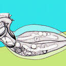
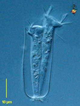
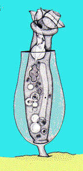
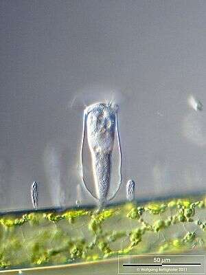
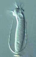
Thuricola (thurr-ick-cola) folliculata and 3 swarmers on the bottom of the lorica. The transparent lorica is equiped with a valve which closes the aperture as cell retracts. This specimen shows endosymbiotic algae. This specimen was collected in freshwater ponds near Konstanz, Germany. Differential interference contrast.
-
Originally described by Ehrenberg under the name Vaginicola decumbens.
-
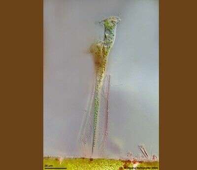
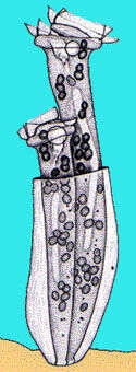
This sessile peritrich ciliat builds chitinous lorica with flap valve. The species houses symbiontic chlorellae. Multi layer image (DOF) shows ciliat and whole lorica with flap valve and epibiontic bacteria. Scale bar indicates 25 µm.See ZIP archive for more. Collected from littoral region (stand of Phragmites) of a rain storage reservoir in Kiel (Schleswig-Holstein, Germany). Images were taken using Zeiss Universal with Olympus C7070 CCD camera.
-
Pyxicola (pig-sick-cola) carteri with opened cap of the lorica. The cap is part of the cell and not of the lorica. This specimen was collected in freshwater ponds near Konstanz, Germany. Differential interference contrast.
-
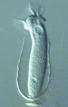
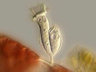
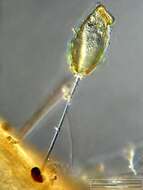
Vaginicola (vadge-in-ee-cola) is a sessile peritrich ciliate. The cells live within a lorica. often found in pairs, the cells attach to the base of the lorica by the posterior ends of the cell. they can contract into the lorica. The oral cilia form a wreath around the anterior end of the cell. No body ciliature. Differential interference contrast.
-
Pyxicola (pig-sick-cola) carteri with opened cap of the lorica. The cap is part of the cell and not of the lorica. This cell has a closed lorica. The individual can pull down and tighten the cap by contraction of the body. This specimen was collected in freshwater ponds near Konstanz, Germany. Differential interference contrast.
-
Vaginicola (vadge-in-ee-cola) is a sessile peritrich ciliate. The cells live within a lorica. often found in pairs, the cells attach to the base of the lorica by the posterior ends of the cell. they can contract into the lorica. The oral cilia form a wreath around the anterior end of the cell. No body ciliature. Phase contrast.
-
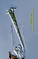
Scale bar indicates 50 µm. Collected from Bodden, the brackish waters lying between the isles of Hiddensee and Ruegen (German Baltic Sea). The image was built up using several photomicrographic frames with manual stacking technique. This image was taken using Zeiss Universal with Olympus C7070 CCD camera.
-
-
Scale bar indicates 50 µm. Collected from Bodden, the brackish waters lying between the isles of Hiddensee and Ruegen (German Baltic Sea). The image was built up using several photomicrographic frames with manual stacking technique. This image was taken using Zeiss Universal with Olympus C7070 CCD camera.
-
Differential Interference Contrast.
-
Cothurnia (co-thur-knee-a) is a sessile peritrich ciliate. The cells live within a lorica which is itself stalked. Cells attach to the base of the lorica by the posterior ends of the cell. Cells can contract into the lorica. The oral cilia form a wreath around the anterior end of the cell. One of the peritrich ciliates, distinguished by having a wreath of cilia around the anterior of the cell. Normally feeding on bacteria. No cilia on the body. Phase contrast.
-
-
-
Vaginicola (vadge-in-ick-cola) tincta, two individuals in their lorica which has a flat bottom and no stalk. This specimen was collected in a pond near Konstanz, Germany. Differential interference contrast.
-
Differential interference contrast image showing living cell and lorica with a short stalk.
-
Originally described by Ehrenberg under the name Vaginicola tinctus.
-
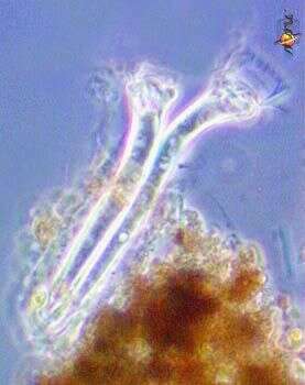
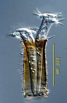
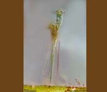
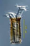
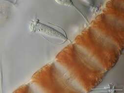
Cothurnia spec living as Aufwuchs on Ceramium diaphanum, shortly after binary fission. Now the swarmer (right cell) has to leave the lorica and build his own. Multi layer image using 8 frames generating depth of focus, stacked manually using Corel Photopaint. Collected from Bodden, the brackish waters lying between the isles of Hiddensee and Ruegen (German Baltic Sea). This image was taken using Zeiss Universal with Olympus C7070 CCD camera.
-
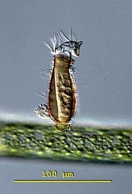
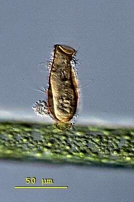
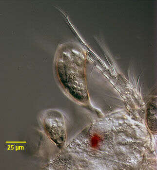
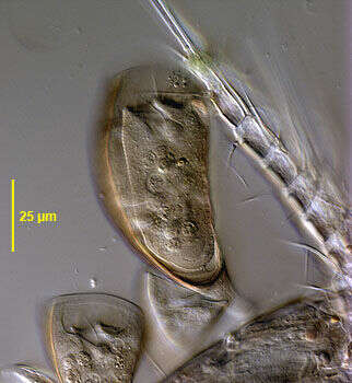
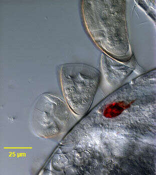
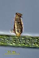
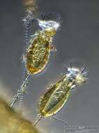
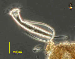
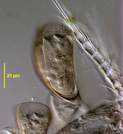
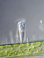
Portrait of the peritrich ciliate, Cyclodonta bipartita (Stokes, 1885) Matthes, 1958. Usually found as an epibiont of freshwater copepods. The cells are contained in a vase-shaped transparent lorica that has fine longitudinal striations. The lorica has a short, stout noncontractile stalk. The cell is attached to the posterior portion of the lorica by a series of membranes and does not protrude from the lorica. The cell body is cylindrical in cross section, rounded posteriorly and transversely truncate anteriorly. The cell surface has fine transverse striations. The macronucleus is ellipsoid. There is a single contractile vacuole. Found on the surface of a cyclopoid copepod collected from a freshwater pond near Boise, Idaho March 2005. DIC
-
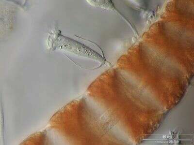
Cothurnia spec. on a young branch of the red alga Ceramium diaphanum. The optical longitudinal section of the axial cells oft the red alga thallus shows their central strand of cytoplasma. Collected from Bodden, the brackish waters lying between the isles of Hiddensee and Ruegen (German Baltic Sea). This image was taken using Zeiss Universal with Olympus C7070 CCD camera.
-
Portrait of the peritrich ciliate, Cyclodonta bipartita (Stokes, 1885) Matthes, 1958. Usually found as an epibiont of freshwater copepods. The cells are contained in a vase-shaped transparent lorica that has fine longitudinal striations. The lorica has a short, stout noncontractile stalk. The cell is attached to the posterior portion of the lorica by a series of membranes and does not protrude from the lorica. The cell body is cylindrical in cross section, rounded posteriorly and transversely truncate anteriorly. The cell surface has fine transverse striations. The macronucleus is ellipsoid. There is a single contractile vacuole. Found on the surface of a cyclopoid copepod collected from a freshwater pond near Boise, Idaho March 2005. DIC
-
Cothurnia spec. on a branch of the green alga Cladophora accompanied by colonies of Zoogloea (Eubacteria). Scale bar indicates 50 µm. Sample from Lake Constance (Bodensee, Southern Germany) near Bodman. This image was taken using Zeiss Universal with Olympus C7070 CCD camera.
-
Portrait of the peritrich ciliate, Cyclodonta bipartita (Stokes, 1885) Matthes, 1958. Usually found as an epibiont of freshwater copepods. The cells are contained in a vase-shaped transparent lorica that has fine longitudinal striations. The lorica has a short, stout, noncontractile stalk. The cell is attached to the posterior portion of the lorica by a series of membranes and does not protrude from the lorica. The cell body is cylindrical in cross section, rounded posteriorly and transversely truncate anteriorly. The cell surface has fine transverse striations. The macronucleus is ellipsoid. There is a single contractile vacuole. Found on the surface of a cyclopoid copepod collected from a freshwater pond near Boise, Idaho March 2005. DIC
-