-
-
-
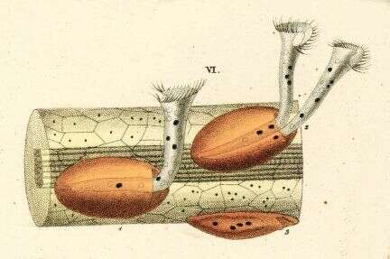
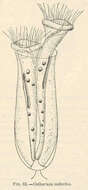
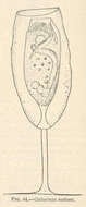
Cothurnia imberbis.
-
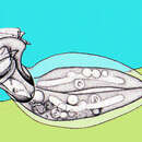
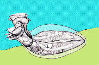
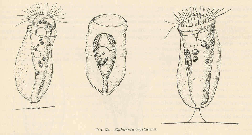
Cothurnia crystallini.
-
-
-
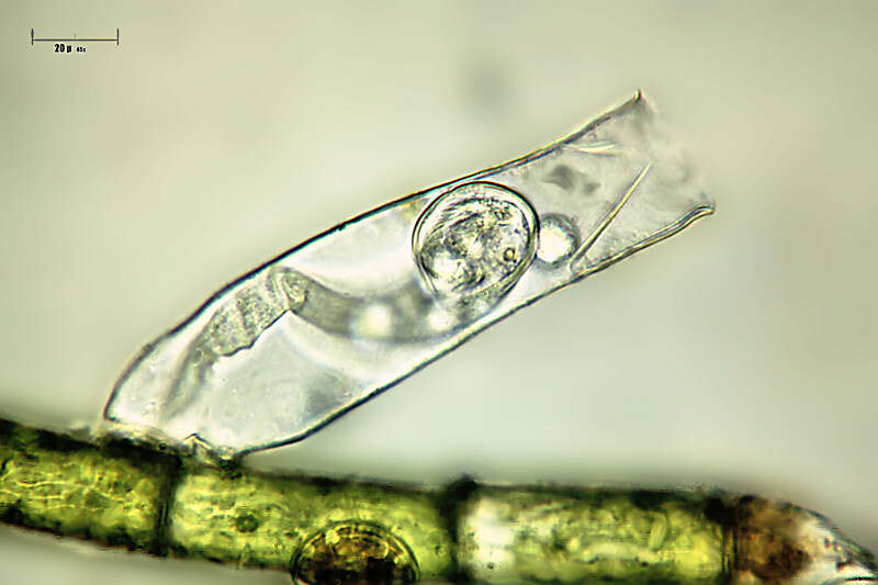
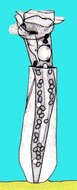
Portrait of Platycola. This peritrich ciliate resides in a simple non-valved lorica with a curved neck. The lorica adheres to the substrate along its length. Most often two individuals per lorica. From freshwater pond with abundant filamentous algae near Boise, Idaho. Oblique illumination.
-
-
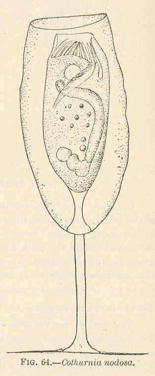
Cothurnia nodosa.
-
-
Platycola, a loricate peritrich. This one forms a flattened lorica with apical apertures through which the cells extend while feeding. The cell cannot be seen clearly. The lorica appears transparent when first formed but gets darker with age. From Lake Donghu, China. Phase contrast micrograph.
-
Thuricola (thurr-ick-owe-la) is a peritrich ciliate which lives within a lorica. Contractile and this cell has withdrawn into the lorica. A flap has closed over the contractile cell and this features distinguishes this genus. Differential interference contrast.
-
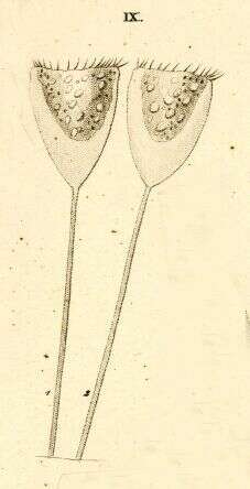
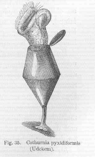
Cothurnia pyxidiformia (Udckem).
-
Platycola, a loricate peritrich. This one forms a flattened lorica with apical apertures through which the cells extend while feeding. Two contracted cells lie inside the lorica. The lorica appears transparent when first formed but gets darker with age. From Lake Donghu, China. Differential interference contrast micrograph.
-
-
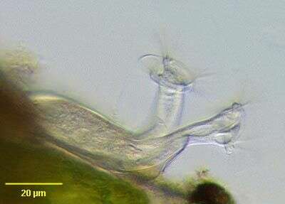
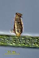
Platycola has mounted its lorica upside down on the Hyponeuston, the aquatic area closely attached to the water surface. Obviously the water´s surface tension was more attractive than the petri dish ore bunches of filamentous algae. Sample from a little creek near Kiel. This image was taken using Zeiss Universal with Olympus C7070 CCD camera.
-
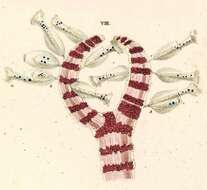
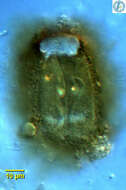
Thuricola (thurr-ick-cola) folliculata and 3 swarmers on the bottom of the lorica. The transparent lorica is equiped with a valve which closes the aperture as cell retracts. This specimen shows endosymbiotic algae. This specimen was collected in freshwater ponds near Konstanz, Germany. Differential interference contrast.
-
Originally described by Ehrenberg under the name Vaginicola decumbens.
-
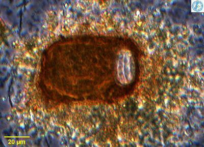
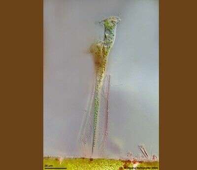
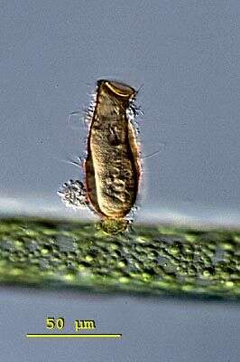
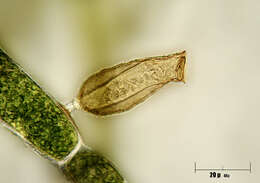
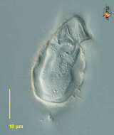
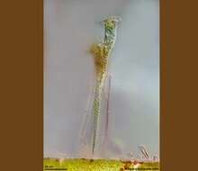
This sessile peritrich ciliat builds chitinous lorica with flap valve. The species houses symbiontic chlorellae. Multi layer image (DOF) shows ciliat and whole lorica with flap valve and epibiontic bacteria. Scale bar indicates 25 µm.See ZIP archive for more. Collected from littoral region (stand of Phragmites) of a rain storage reservoir in Kiel (Schleswig-Holstein, Germany). Images were taken using Zeiss Universal with Olympus C7070 CCD camera.
-
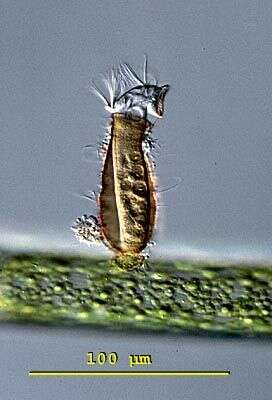
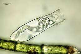
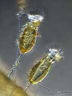
Pyxicola (pig-sick-cola) carteri with opened cap of the lorica. The cap is part of the cell and not of the lorica. This specimen was collected in freshwater ponds near Konstanz, Germany. Differential interference contrast.
-
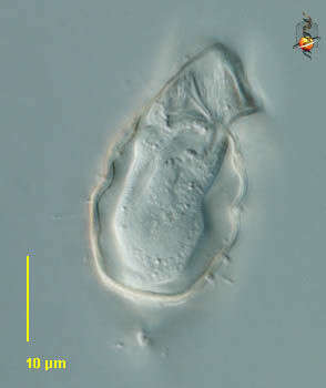
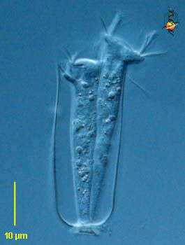
Vaginicola (vadge-in-ee-cola) is a sessile peritrich ciliate. The cells live within a lorica. often found in pairs, the cells attach to the base of the lorica by the posterior ends of the cell. they can contract into the lorica. The oral cilia form a wreath around the anterior end of the cell. No body ciliature. Differential interference contrast.
-
Pyxicola (pig-sick-cola) carteri with opened cap of the lorica. The cap is part of the cell and not of the lorica. This cell has a closed lorica. The individual can pull down and tighten the cap by contraction of the body. This specimen was collected in freshwater ponds near Konstanz, Germany. Differential interference contrast.
-
Vaginicola (vadge-in-ee-cola) is a sessile peritrich ciliate. The cells live within a lorica. often found in pairs, the cells attach to the base of the lorica by the posterior ends of the cell. they can contract into the lorica. The oral cilia form a wreath around the anterior end of the cell. No body ciliature. Phase contrast.
-
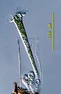
Scale bar indicates 50 µm. Collected from Bodden, the brackish waters lying between the isles of Hiddensee and Ruegen (German Baltic Sea). The image was built up using several photomicrographic frames with manual stacking technique. This image was taken using Zeiss Universal with Olympus C7070 CCD camera.