-
-
-
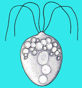
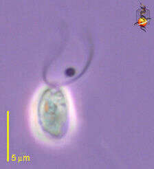
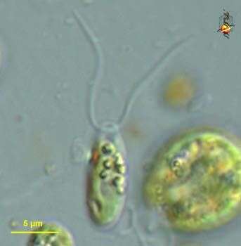
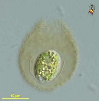
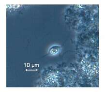
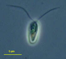
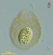
This flagellated protozoa was observed from a perchlorate treating laboratory scale anaerobic bioreactor (ORP~ -200 mV) fed with acetate as substrate. It was characterized by a pair of anterior flagella, tailing body and intracellular starch granules. Krishnakumar B and Anupama V.N. of NIIST (CSIR) are involved in the bioreactor study. The phase contrast image shows cells with intracellular starch granules and the same released to the medium.
-
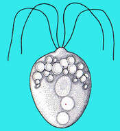
Chlamydomonas (clam-ee-doe-moan-ass), a solitary volvocid (flagellated green algal cell). Cell surrounded by a cellulosic wall, with two similar flagella emerging from near the apex. The photosynthetic pigments are located within a cup-shaped chloroplast which has a large pyrenoid with associated polysaccharide materials located posteriorly. The nucleus is located within the cup. Animations by Rosemary Arbur of flagellar beat patterns are available
here.Differential interference contrast.
-
-
Chlamydomonas (clam-ee-doe-moan-ass), a solitary volvocid (flagellated green algal cell). Cell surrounded by a cellulosic wall, this is a division form in which four daughter cells are being produced at the same time. Animations by Rosemary Arbur of flagellar beat patterns are available
here. Differential interference contrast.
-
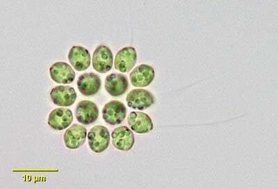
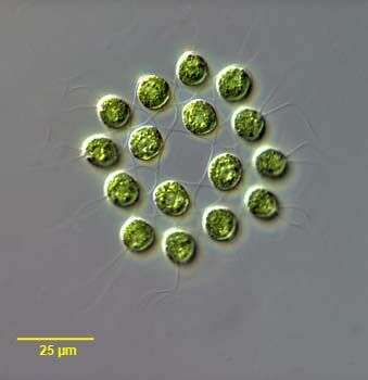
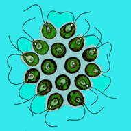
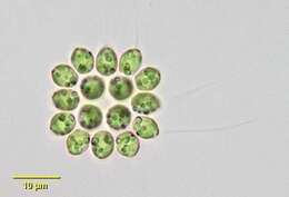
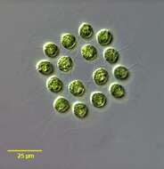
Gonium pectorale, a common colonial volvocid flagellate. Colonies usually consist of 16 cells (twelve peripheral and four central) in a plate-like arrangement of one layer. An extracellular matrix that is not easily seen connects cells. Cells have a cup-shaped chloroplast with a small anterior stigma. Lateral view of colony. From freshwater pond near Boise, Idaho. Phase contrast.
-
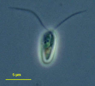
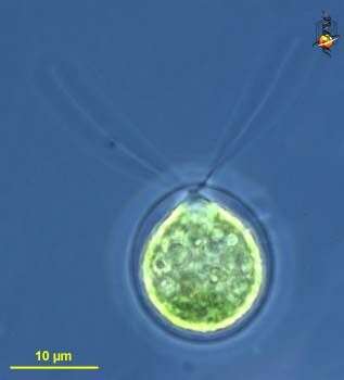
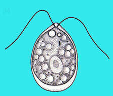
Chlamydomonas (clam-ee-doe-moan-ass), a solitary volvocid (flagellated green algal cell). Cell surrounded by a cellulosic wall, with two similar flagella emerging from near the apex. The photosynthetic pigments are located within a cup-shaped chloroplast which has a large pyrenoid with associated polysaccharide materials located posteriorly. The nucleus is located within the cup. This image shows the small red eyespot (orange colour here) and one anterior contractile vacuole. Differential interference contrast.
-
Gonium pectorale, a common colonial volvocid flagellate. Colonies usually consist of 16 cells (twelve peripheral and four central) in a plate-like arrangement of one layer. An extracellular matrix that is not easily seen connects cells. Cells have a cup-shaped chloroplast with a small anterior stigma. Anteroposterior view of colony. From freshwater pond near Boise, Idaho. Brightfield illumination.
-
Chlamydomonas (clam-ee-doe-moan-ass), a solitary volvocid (flagellated green algal cell). Cell surrounded by a cellulosic wall, with two similar flagella emerging from near the apex. The photosynthetic pigments are located within a cup-shpaed chloroplast which has a large pyrenoid with associated polysaccharide materials. Many taxa described (there are books on this genus). Eyespot located within plastid. Flagella beat with a breast-stroke pattern. Phase contrast.
-
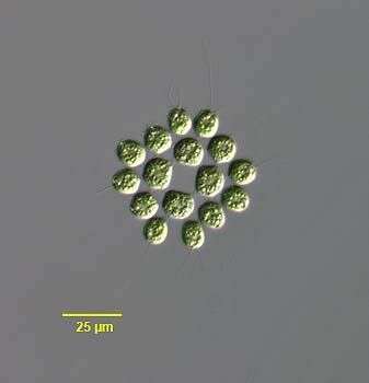
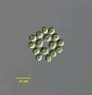
Lateral view of Gonium pectorale (Muller), a common colonial volvocid flagellate. Colonies usually consist of 16 biflagellate cells (twelve peripheral and four central) in a plate-like arrangement of one layer. An extracellular matrix that is not easily seen connects cells. Cells have a cup-shaped chloroplast with a small anterior stigma. Lateral view of colony. Collected from a freshwater pond near Boise, Idaho. Brightfield
-
Chlamydomonas (clam-ee-doe-moan-ass), a solitary volvocid (flagellated green algal cell). Cell surrounded by a cellulosic wall, with two similar flagella emerging from near the apex. The photosynthetic pigments are located within a cup-shaped chloroplast which has a large pyrenoid with associated polysaccharide materials located posteriorly. The nucleus is located within the cup. This image shows one anterior contractile vacuole. Animations by Rosemary Arbur of flagellar beat patterns are available
here.Phase contrast.
-
Anterior apical view of Gonium pectorale (Muller), a common colonial volvocid flagellate. Colonies usually consist of 16 biflagellate cells (twelve peripheral and four central) in a plate-like arrangement of one layer. An extracellular matrix (not easily seen without phase or differential intereference contrast techniques) connects cells. Cells have a cup-shaped chloroplast with a small anterior stigma. Collected from a freshwater pond near Boise, Idaho. DIC.
-
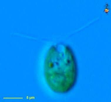
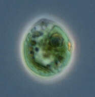
Chlamydomonas (clam-ee-doe-moan-ass), a solitary volvocid (flagellated green algal cell). Cell surrounded by a cellulosic wall. Cell damaged, no flagella. The photosynthetic pigments are located within a cup-shaped chloroplast which has a large pyrenoid with associated polysaccharide materials located posteriorly. The nucleus is located within the cup. This image shows the red eyespot to the right and two anterior contractile vacuoles. Phase contrast.
-
Anterior apical view of Gonium pectorale (Muller), a common colonial volvocid flagellate. Colonies usually consist of 16 biflagellate cells (twelve peripheral and four central) in a plate-like arrangement of one layer. An extracellular matrix that is not easily seen connects cells. Cells have a cup-shaped chloroplast with a small anterior stigma. Lateral view of colony. Collected from a freshwater pond near Boise, Idaho. DIC.
-
Chlamydomonas (clam-ee-doe-moan-ass), a solitary volvocid (flagellated green algal cell). Cell surrounded by a cellulosic wall, with two similar flagella emerging from near the apex. Elongate species. Animations by Rosemary Arbur of flagellar beat patterns are available
here. Phase contrast.
-
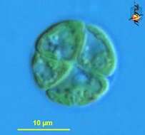
Young colonies, cell divisions are almost over, the flagellae aren't yet fully formed. Scale bar indicates 25 µm. Sample from the Lake Constance (vicinity of Bodman). The image was built up using several photomicrographic frames with manual stacking technique. Images were taken using Zeiss Universal with Olympus C7070 CCD camera.Image under Creative Commons License V 3.0 (CC BY-NC-SA).
-
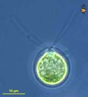
Chlamydomonas (clam-ee-doe-moan-ass), a solitary volvocid (flagellated green algal cell). Cell surrounded by a cellulosic wall. With plastid containing chlorophyll B giving the bright green colour, two similar flagella emerge from the anterior of the cell. Differential interference contrast.
-
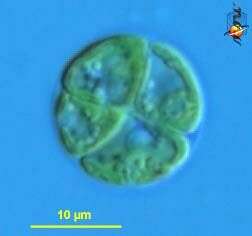
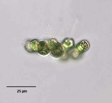
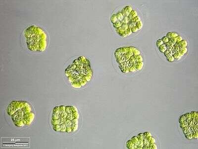
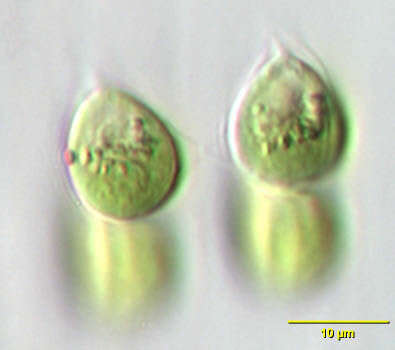
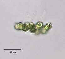
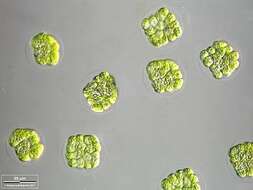
Tetrabaena socialis, a colonial volvocid flagellate. T. socialis was formerly known as Gonium sociale until the monospecific genus Tetrabaena was erected (Nozaki,H. and Itoh, M. J. Phycol. 30:353-365, 1994).The colonies are plate-shaped with four cells all oriented in the same direction. Extracellular matrix joining adjacent cells can be seen. The gelatinous envelope surrounding the whole colony is not seen in these images. The ovoid cells have two equal-length flagella, a small eyespot, large cup-shaped chloroplast and posterior round pyrenoid. From freshwater pond near Boise, Idaho. Oblique illumination.
-
Chlamydomonas (clam-ee-doe-moan-ass), a solitary volvocid (flagellated green algal cell). Cell surrounded by a cellulosic wall and enclosed in mucilagenous sheath - cells in this form were inactive as if encysted. Differential interference contrast.
-
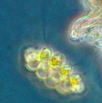
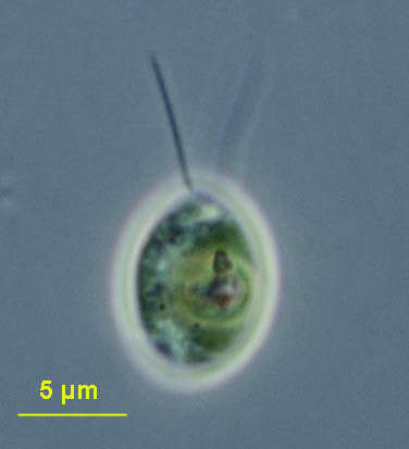
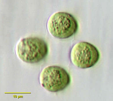
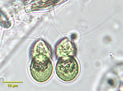
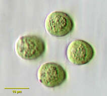
Tetrabaena, a green alga (Chlorophyta) in which four cells form a colony within a delicate mucoid matrix. Each cell with two apical flagella. This image shows an eyespot in one cell.
-
Chlamydomonas (clam-ee-doe-moan-ass), a solitary volvocid (flagellated green algal cell). Cell surrounded by a cellulosic wall. With plastid containing chlorophyll B giving the bright green colour, two similar flagella emerge from the anterior of the cell. Phase contrast.
-
Tetrabaena, a green alga (Chlorophyta) in which four cells form a colony within a delicate mucoid matrix. Each cell with two apical flagella that beat with a breast-stroke pattern.
-
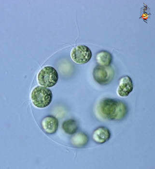
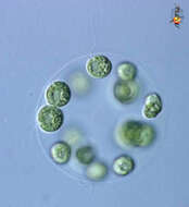
Eudorina (you-door-ine-a) is a colonial volvocid - motile green alga. In this case 16 cells are embedded in a sphere of common mucus. Each individual cell has two flagella, and a cup-shaped chloroplast - a bit like a Chlamydomonas cell. There are a number of related genera which form colonies using mucus as a common matrix, but they differ in the numbers of cells involved, how tightly packed they are, and the shape of the colony. Differential interference contrast.