-
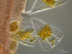
Cells at two focal levels. Material from a plankton tow off Martha's Vineyard, Massachusetts. Image by Jeff Cole.
-
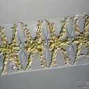
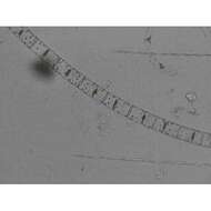
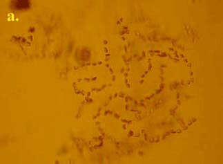
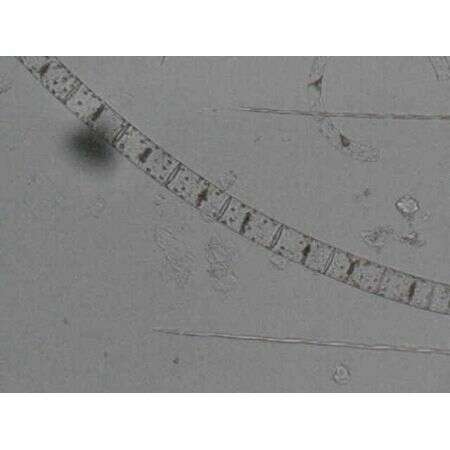
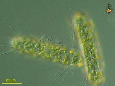
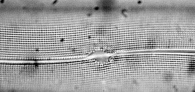
This species consists of very small cells which are united into curved chains. Cells have three short setae and one long one which causes the formation of larger secondary colonies by linking up in the centre of the colony with the long setae of other chains
-
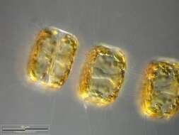
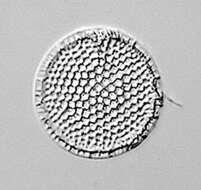
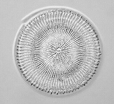
Closeup of Porosira glacialis chain. Note that the delicate spines are chitinous. Focus on frustule surface. Scale bar indicates 50 µm. The image was built up using several photomicrographic frames with manual stacking technique. Sample from North Sea near Heligoland (spring diatom bloom). Images were taken using Zeiss Universal with Olympus C7070 CCD camera.
-
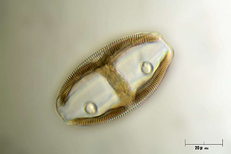
Material from the Netherlands.
-
After several minutes of observation chloroplasts are transported to the cell center, maybe to save the nucleus and most of the chloroplasts against harmful amounts of UV radiation. The image was built up using several photomicrographic frames with manual stacking technique. Scale bar indicates 50 µm. Sample from North Sea near Heligoland (spring diatom bloom). Images were taken using Zeiss Universal with Olympus C7070 CCD camera.
-
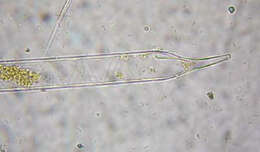
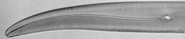
Valves are cylindrical terminating in a proboscis. Processes are absent. This is a temprate, coastal species.
-
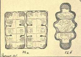
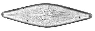
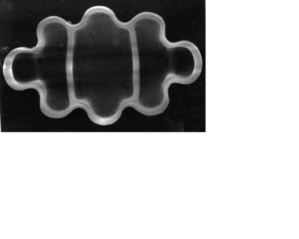
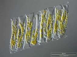
Terpsinoe americana by Lisa M. Weimer (USGS)Found in Chesapeake Bay Diatoms by Lisa M. Weimer, an online USGS publication located at http://pubs.usgs.gov/pdf/of/of99-45/diatom.pdfPlate 2Terpsinoe americana (Bail.) Rolfs, PTMC 3-P-2 422-424 cm., x 2000.
-
-
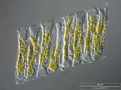
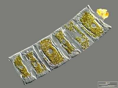
Helical arranged cell colony. Phase contrast.Scale bar indicates 50 µm. Sample from North Sea near Heligoland (spring diatom bloom). The image was built up using several photomicrographic frames with manual stacking technique. Images were taken using Zeiss Universal with Olympus C7070 CCD camera.
-
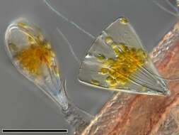
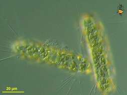
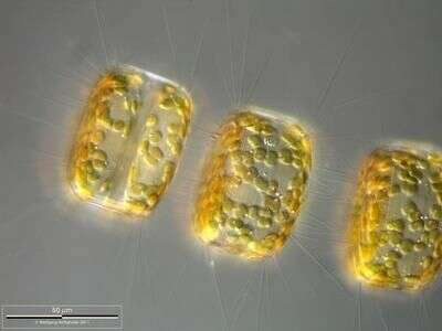
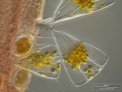
Licmophora juergensii living together with other araphid diatoms, peritrich ciliates and filamentous cyanobacteria on a red alga. They use jelly stalks for fixation on the substratum. Collected from Bodden, the brackish waters lying between the isles of Hiddensee and Ruegen (German Baltic Sea). This image was taken using Zeiss Universal with Olympus C7070 CCD camera.
-
-
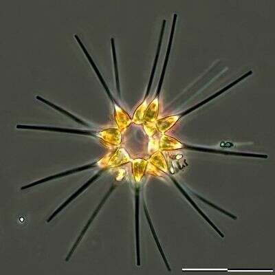
Corethron (core-eeth-ron) hystrix, centric diatom (stramenopile) with siliceous spines emerging from the border of the valves, many girdle bands (not visible) make up the body of the cylinder. With many small plastids. Phase contrast microscopy.
data on this strain.
-
Closeup of Porosira glacialis chain. Note that the delicate spines are chitinous. Focus on cell center showing cytoplasmic accumulation around the nucleus. Scale bar indicates 50 µm. The image was built up using several photomicrographic frames with manual stacking technique. Sample from North Sea near Heligoland (spring diatom bloom). Images were taken using Zeiss Universal with Olympus C7070 CCD camera.
-
Image from type material.
-
Image shows chloroplasts and nuclei. The image was built up using several photomicrographic frames with manual stacking technique. Scale bar indicates 50 µm. The image was built up using several photomicrographic frames with manual stacking technique. Sample from North Sea near Heligoland (spring diatom bloom). Images were taken using Zeiss Universal with Olympus C7070 CCD camera.
-
-
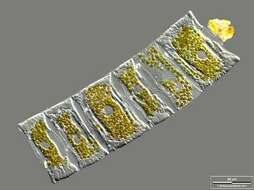
Terpsinoe americana by Josef PantocsekBeiträge zur Kenntnis der Fossilen Bacillarien Ungarns / nach dem ungarischen Manuscripte von Josef Pantocsek (1840-1916)Berlin :W. Junk,1903-1905.Band I. Plate VI
-
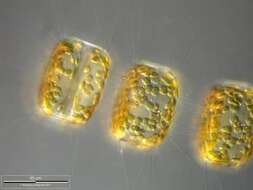
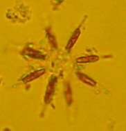
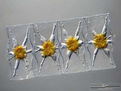
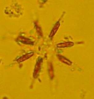
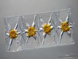
This is a northern cold water to temperate species. Like A. glacialis it can form star shaped chains.
-
On the left a Licmophora cell is visible in lateral (valvar) view, on the right we look at the girdle band (cingulim) of the cell, so this is called cingular view. Collected from Bodden, the brackish waters lying between the isles of Hiddensee and Ruegen (German Baltic Sea). This image was taken using Zeiss Universal with Olympus C7070 CCD camera.
-
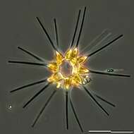
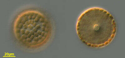
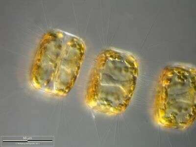
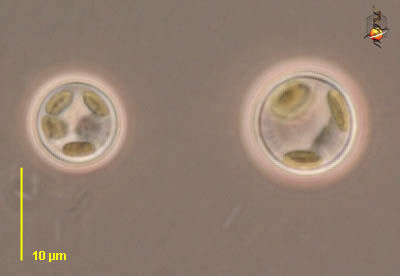
Cyclotella (sike-low-tell-a). Centric diatom, seen from valve view. Three plastid profiles are visible around the periphery of the cell. Long thin organic spines project from the cell - and are believed to have a role in flotation. The pattern of pores in the frustule is used in identification. Marine. Phase contrast.
-
Corethron (core-eeth-ron) hystrix, centric diatom (stramenopile) with siliceous spines emerging from the border of the valves, many girdle bands (not visible here) make up the body of the cylinder. This image emphasizes the plastids. Differential interference microscopy.
data on this strain.
-
-
Image from type material.
-
Image shows chloroplasts and nuclei. The image was built up using several photomicrographic frames with manual stacking technique. Scale bar indicates 50 µm. The image was built up using several photomicrographic frames with manual stacking technique. Sample from North Sea near Heligoland (spring diatom bloom). Images were taken using Zeiss Universal with Olympus C7070 CCD camera.