-
Otsego Co., Michigan
-
Otsego Co., Michigan
-
All Biocode files are based on field identifications to the best of the researcher’s ability at the time.
-
All Biocode files are based on field identifications to the best of the researcher’s ability at the time.
-
All Biocode files are based on field identifications to the best of the researcher’s ability at the time.
-
All Biocode files are based on field identifications to the best of the researcher’s ability at the time.
-
All Biocode files are based on field identifications to the best of the researcher’s ability at the time.
-
All Biocode files are based on field identifications to the best of the researcher’s ability at the time.
-
All Biocode files are based on field identifications to the best of the researcher’s ability at the time.
-
All Biocode files are based on field identifications to the best of the researcher’s ability at the time.
-
All Biocode files are based on field identifications to the best of the researcher’s ability at the time.
-
All Biocode files are based on field identifications to the best of the researcher’s ability at the time.
-
All Biocode files are based on field identifications to the best of the researcher’s ability at the time.
-
All Biocode files are based on field identifications to the best of the researcher’s ability at the time.
-
All Biocode files are based on field identifications to the best of the researcher’s ability at the time.
-
All Biocode files are based on field identifications to the best of the researcher’s ability at the time.
-
All Biocode files are based on field identifications to the best of the researcher’s ability at the time.
-
All Biocode files are based on field identifications to the best of the researcher’s ability at the time.
-
All Biocode files are based on field identifications to the best of the researcher’s ability at the time.
-
All Biocode files are based on field identifications to the best of the researcher’s ability at the time.
-
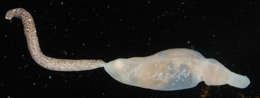
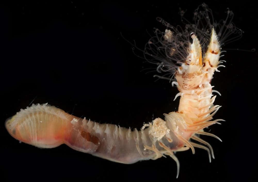
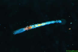
Host Ari Daniel Shapiro dives deep to discover a white worm as tall as your refrigerator that breathes through bright red feathery “lips.” This isn’t a creature from outer space. Meet Riftia, a tube worm that lives in deep-sea vents, and learn the surprising lessons this denizen of the abyss is teaching scientists about life on Earth. Photo credit: Vicki Ferrini, Marvin Lilley
Podcast transcript read moreDuration: 4:14Published: Wed, 13 Jul 2011 15:18:27 +0000
-
-
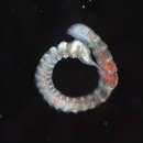
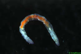
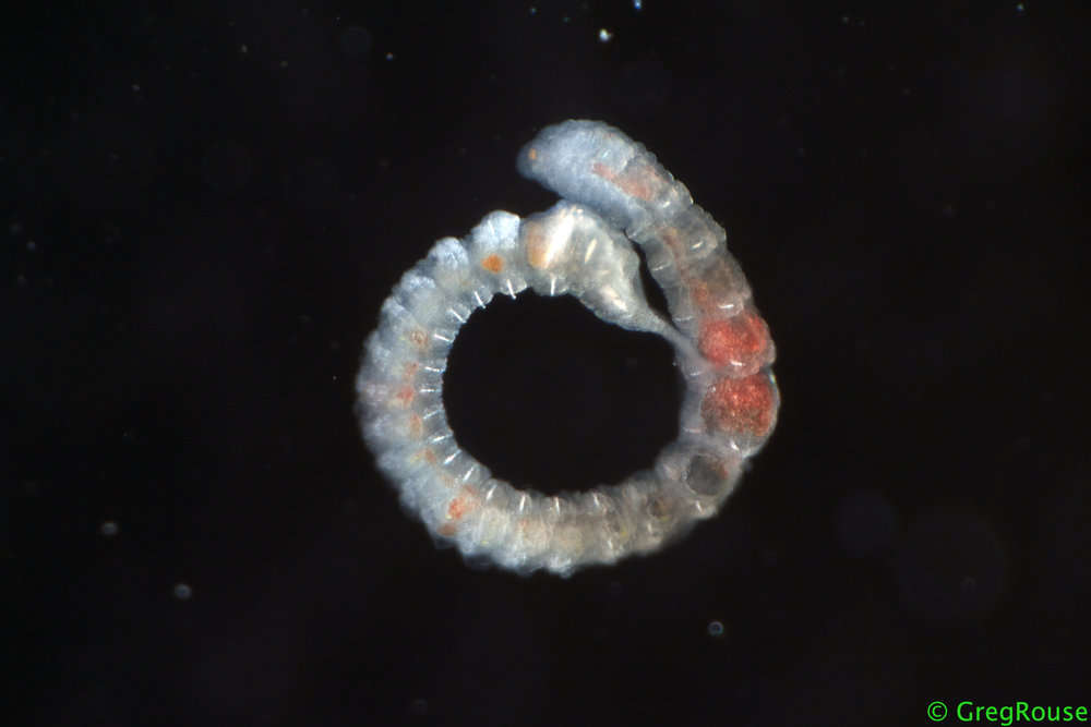
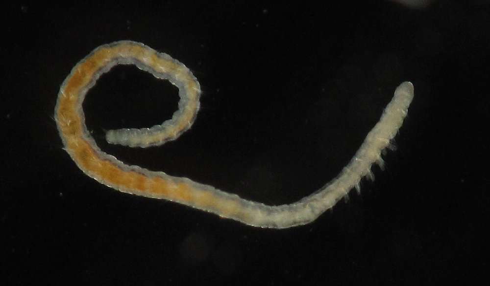
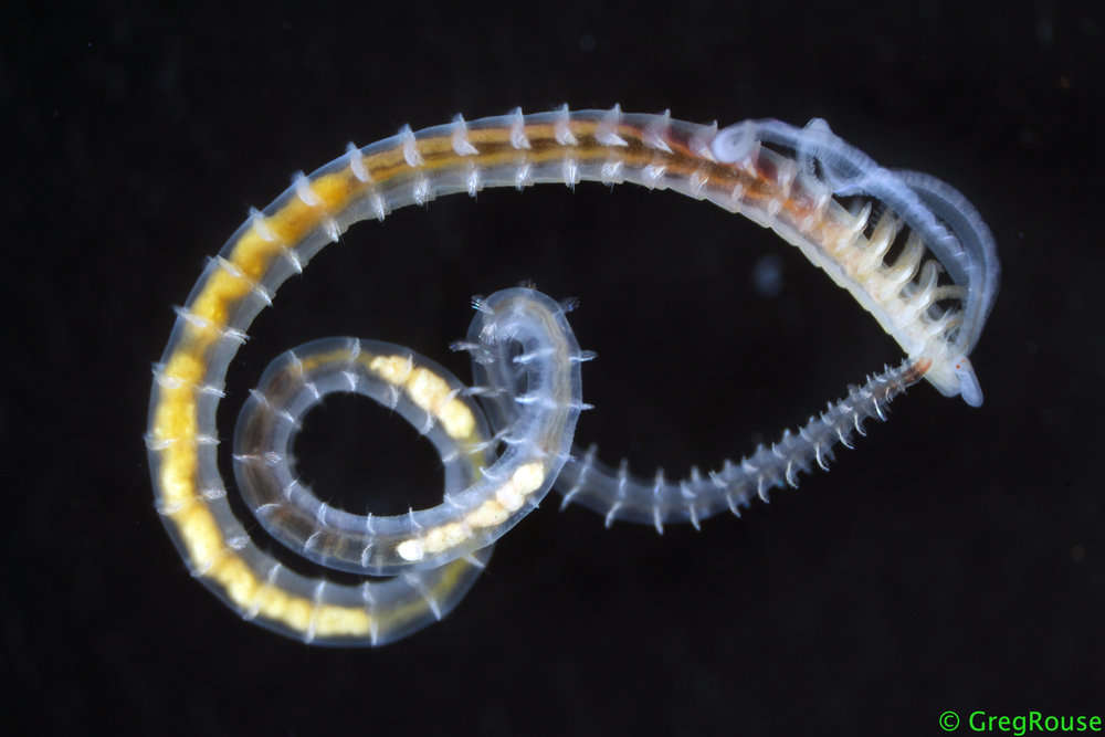
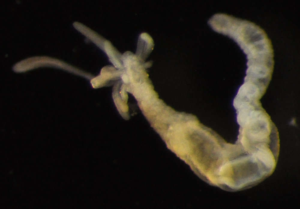
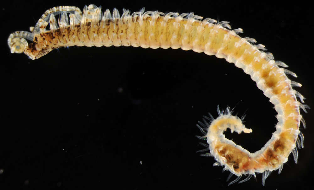
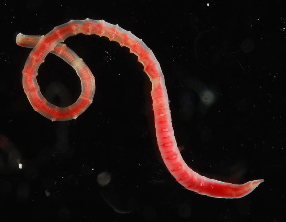
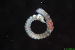
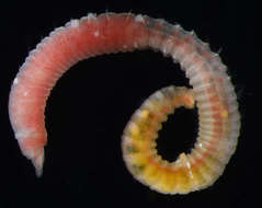
Description: Operational taxonomic unit: Capitellidae sp. A. Common and wide-spread group of polychaetes.
The dark pellets in the posterior segments are faecal pellets.
Size: approximately: 30 mm. Macrofauna (sieved on 0.5 mm). Item Type: Image Title: Capitellidae sp. Copyright: SERPENT project Species: Capitellidae sp. Behaviour: surface deposit feeder. Site: Atlantic -- Norwegian -- Dalsnuten Site Description: Seafloor Depth (m): 1452 Latitude: 66 deg 34' 33" N Longitude: 3 deg 32' 46" E Countries: Norway -- Norwegian Sector Habitat: Benthic Rig: Aker Barents Project Partners: Shell, Aker Drilling, Oceaneering ROV: Magnum 142 Deposited By: Dr K Kroeger Deposited On: 13 July 2011
-
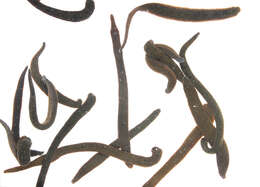
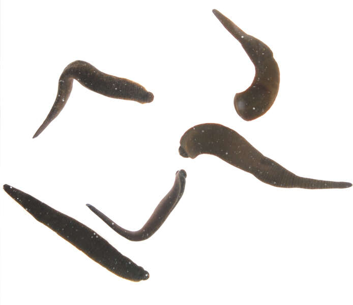
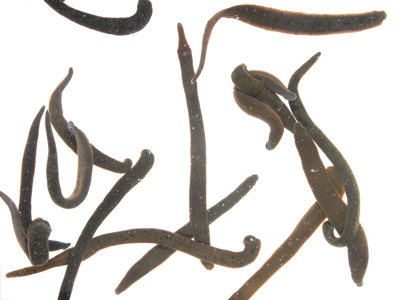
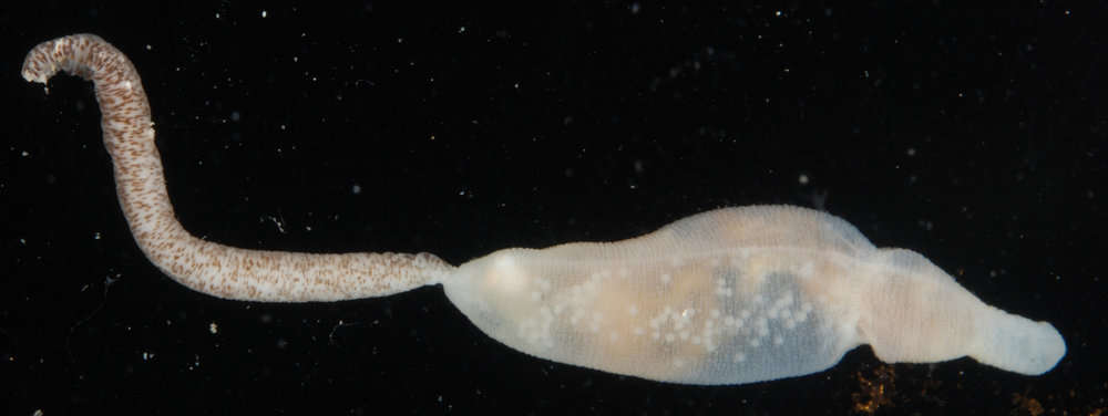
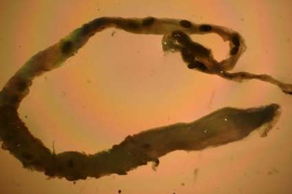
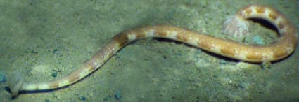
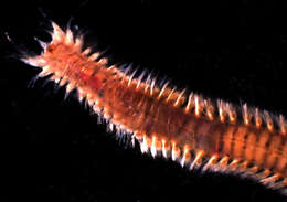
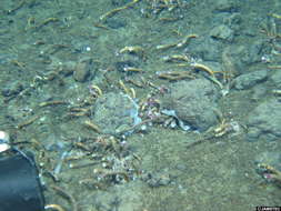
Description: Leeches rest on the seabed after feeding on fishes (Notostomum sucks the blood of rays and eelpouts).
Up to 200 mm long Item Type: Image Title: Notostomum laeve Species: Notostomum laeve Site: Atlantic -- North Sea -- West of ShetlandNorth Sea -- West of Shetland Site Description: Seafloor Depth (m): 1156 Latitude: 61 deg 16' 00" N Longitude: 2 deg 59' 00" E Countries: UK -- West of Shetland Rig: Leiv Eiriksson Project Partners: Shell ROV: Magnum 105 and Magnum 146 Deposited By: Dr Daniel Jones Deposited On: 09 May 2014