-
Xiao-Feng Xue, Hussein Sadeghi, Xiao-Yue Hong, Samira Sinaie
Zookeys
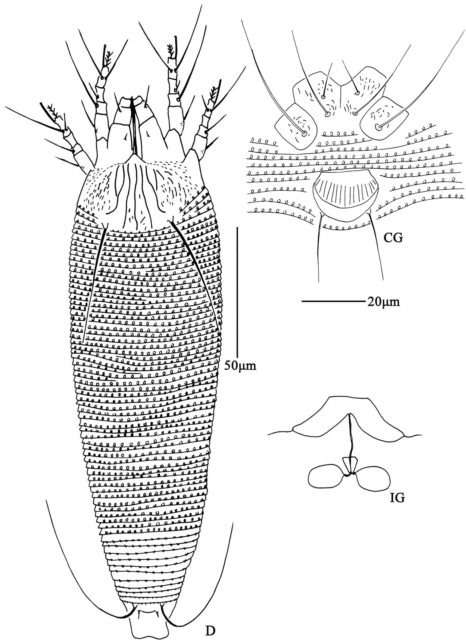
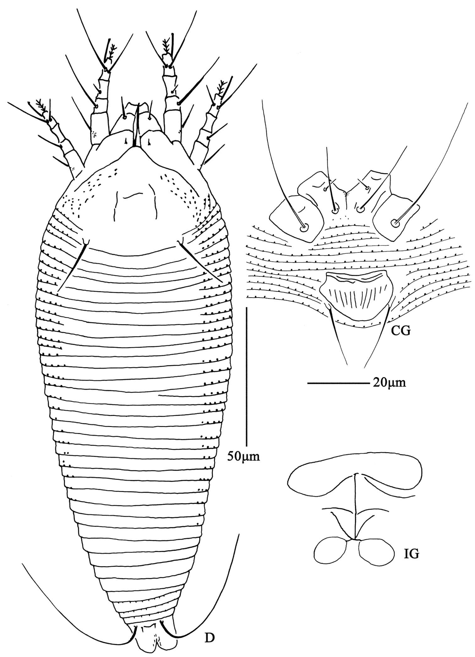
Figure 6. Aceria pulicaris sp. n. D dorsal view of female CG coxae and female genitalia IG female internal genitalia.
-
Hao-Sen Li, Xiao-Feng Xue, Xiao-Yue Hong
Zookeys
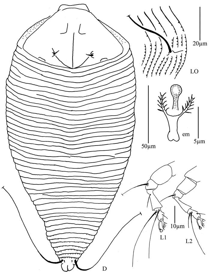
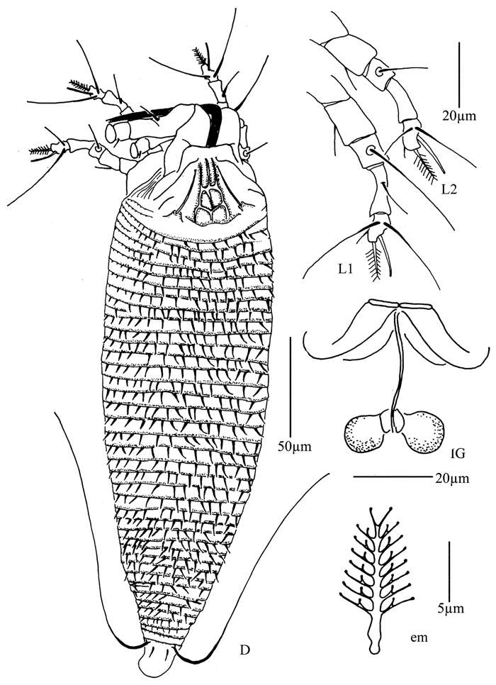
Figure 31.Diptacus mengdaensis sp. n.: D dorsal view of female LO lateral microtubercles em empodium L1 leg I L2 leg II.
-
Vladimir Pešić, Ksenia A. Semenchenko, Wonchoel Lee
Zookeys
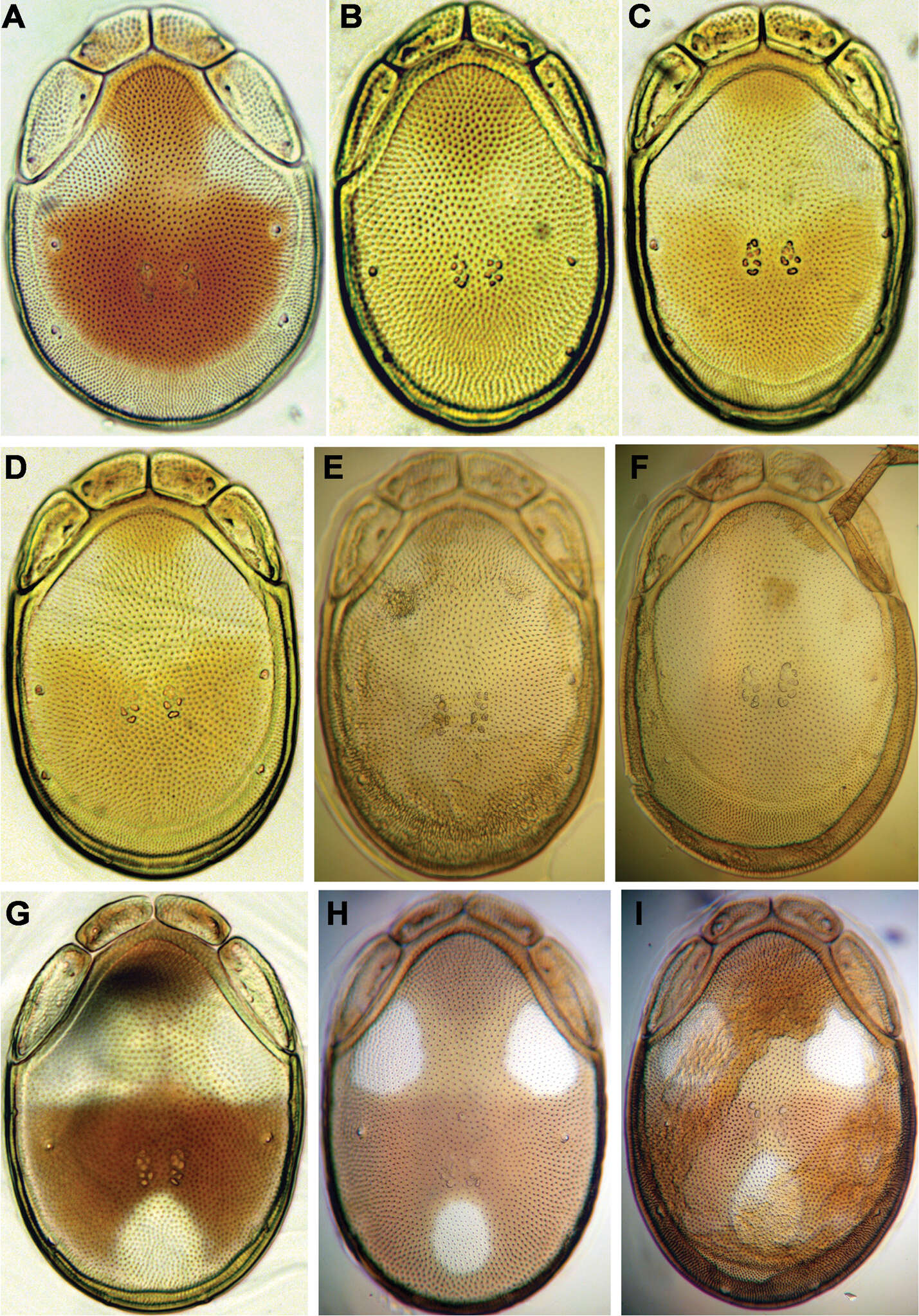
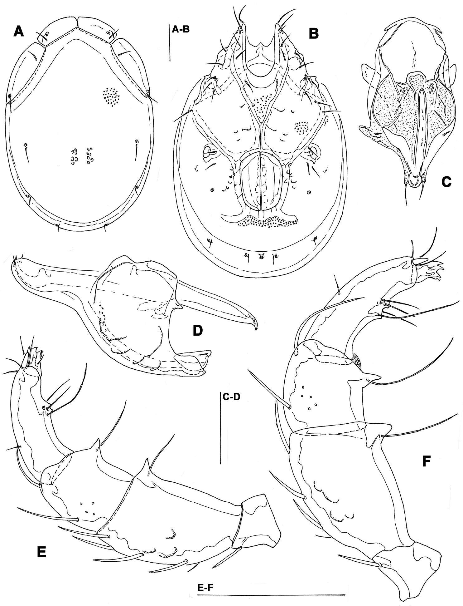
Figure 7.Photographs of dorsal shield: A Torrenticola brevirostris (Halbert, 1911), female, stream in Naebyeansan NP, Korea B Torrenticola dentifera Wiles, 1991, male, stream in Naebyeansan NP, Korea: C–F Torrenticola kimichungi sp. n. (C–D specimens from stream in Seoraksan NP, Korea, E–F specimens from Tigrovaya River, Russia): C maleholotype D–E maleparatypes F femaleparatype G–I Torrenticola nipponica (Enami, 1940) (G specimen from Dobong stream, Korea H–I specimens from Tigrovaya River, Russia): G–H male I female. Photos. V. Pešić (Figs A–D, G), K. Semenchenko (Figs E–F, H–I).
-
Yunus Esen, Vladimir Pešić, Orhan Erman, Yücel Kaya
Zookeys
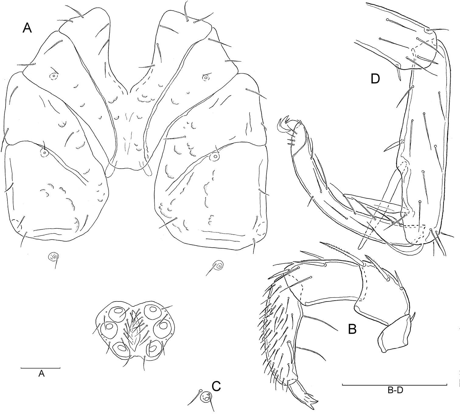
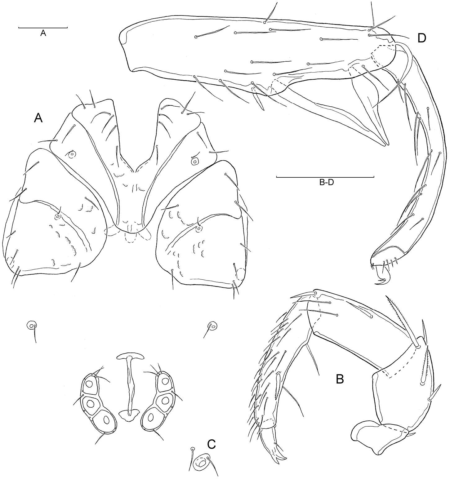
Figure 5.A–D Atractides (s. str.) nikooae Pešić, 2004, male: A Coxal and genital field B Palp, medial veiw C Vgl-1–2 D I–L-5–6 (Scale bars = 100 µm).
-
Michael J. Skvarla, J. Ray Fisher, Ashley P. G. Dowling
Zookeys
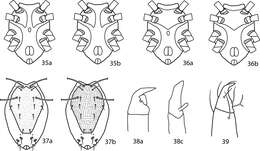
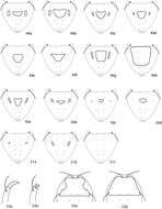
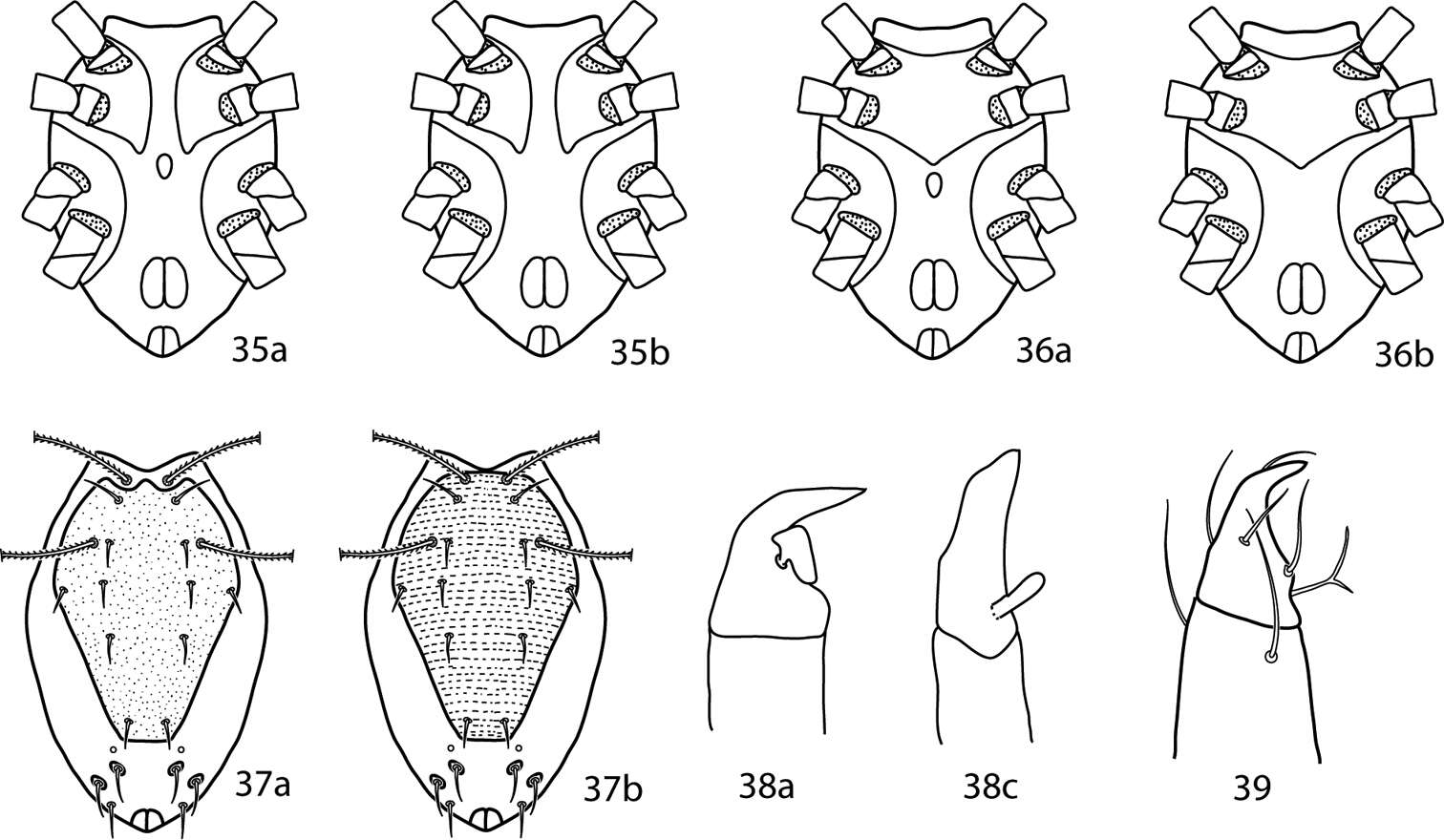
Figures 35–39.Pulaeus illustrations. 35 Genital setae in a row 36–39 Pulaeus key illustrations 36, 37 Venter, setae removed for clairity 36a Coxae I–II not coalesced medially, median platelet present 36b Coxae I–II not coalesced medially, median platelet absent 37a Coxae I–II coalesced medially, median platelet present 37b Coxae I–II coalesced medially, median platelet absent 38a Dorsal shield with punctures 38b Dorsal shield with broken striae 39a–c Pedipalp tibiotarsus 39a Tibiotarsus with elongate apophysis 39b Tibiotarsus with flat apophysis 39c Tibiotarsus with flange-like apophysis.
-
Parisa Lotfollahi, Enrico de Lillo, Karim Haddad Irani-Nejad
Zookeys
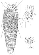
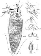
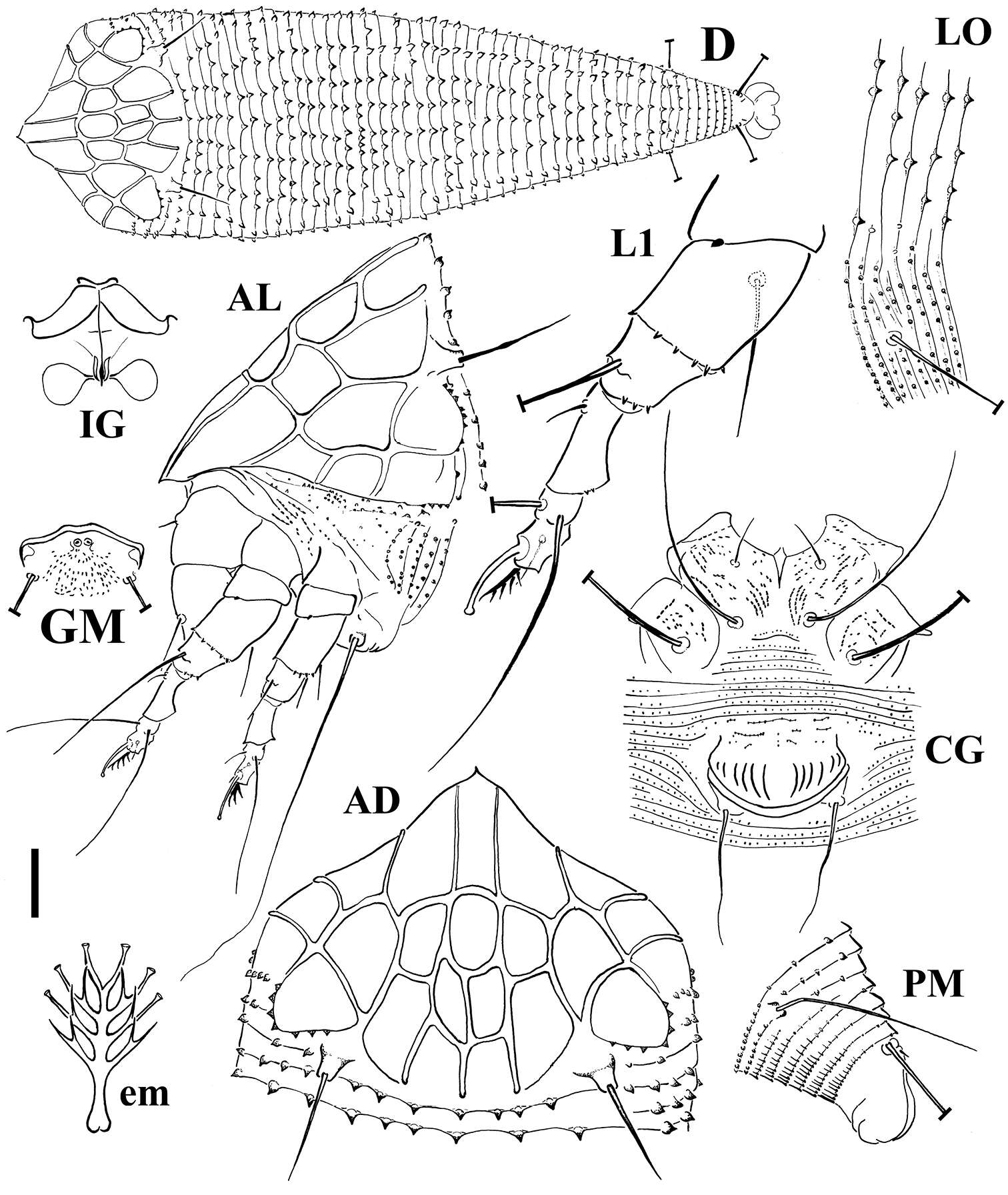
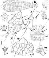
Figure 2.Schematic drawings of Echinacrus ruthenicus sp. n.: AD Dorsal view of anterior body region AL Lateral view of anterior body region CG Female coxigenital region D Dorsal view em Empodium GM Male genital region IG Internal female genitalia LO Lateral view of annuli L1 Leg I PM Lateral view of posterior opisthosoma. Scale bar: 20 μm for D; 10 μm for AD, AL, CG, IG, GM, PM; 5 μm for LO, L1; 2.5 μm for em.
-
Glenstrup Sø, Jylland, Danmark
-
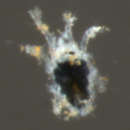
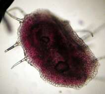
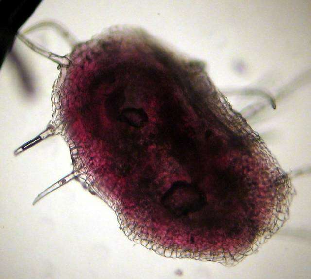
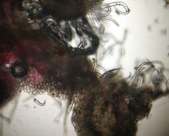
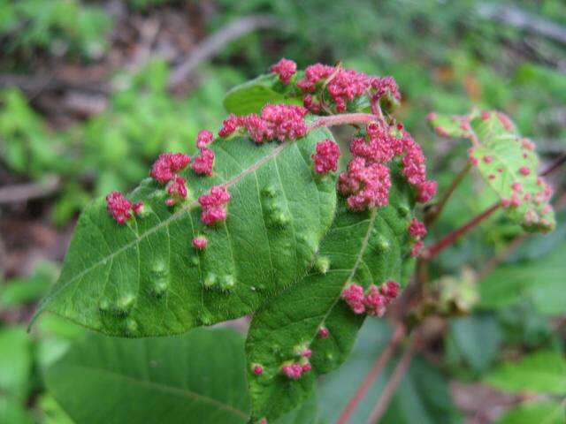
Mushroom Observer Image 103179: Aculops
-
Xiao-Feng Xue, Hussein Sadeghi, Xiao-Yue Hong, Samira Sinaie
Zookeys
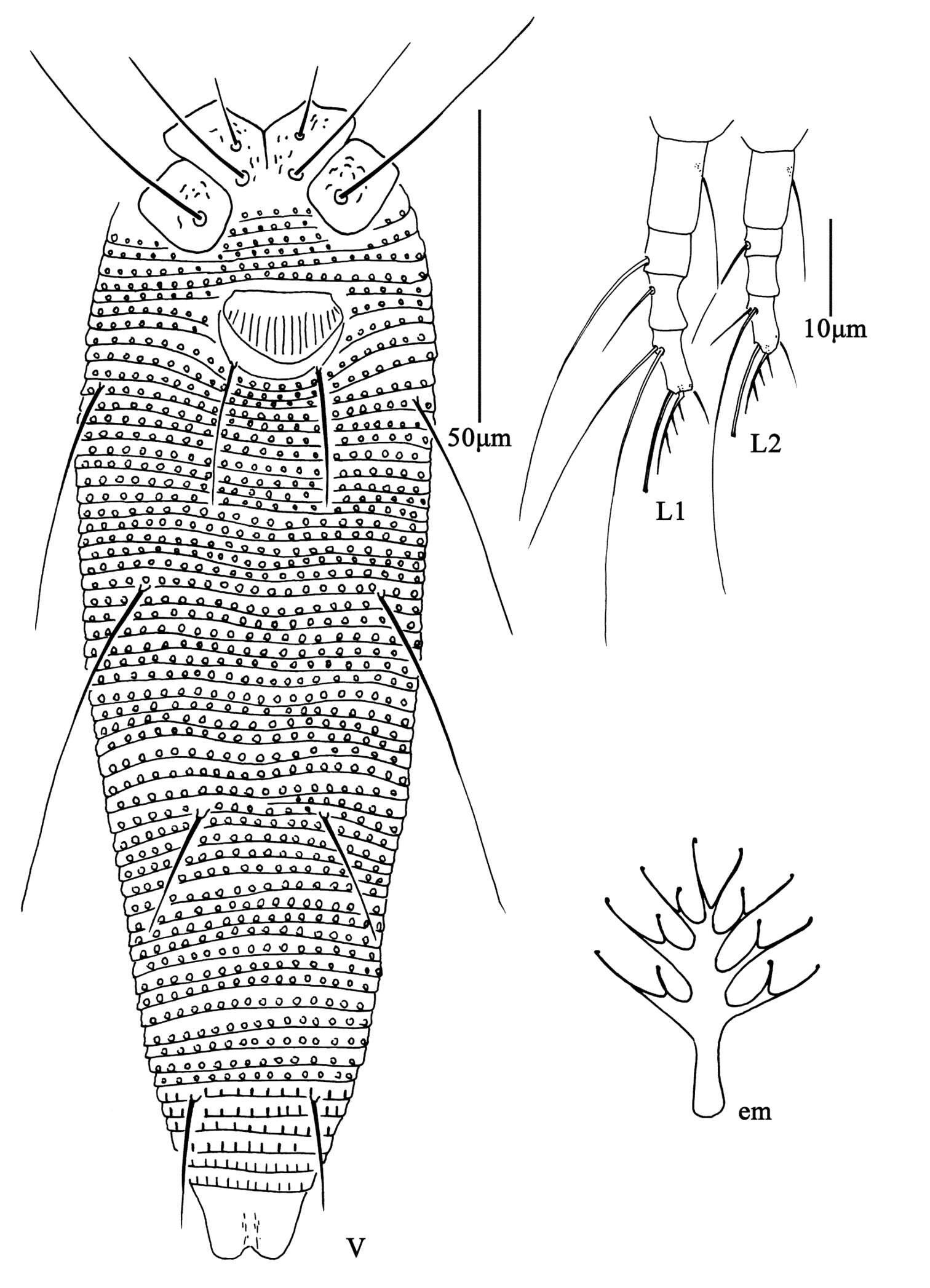
Figure 7. Aceria pulicaris sp. n. V ventral view of female em empodium L1 leg І L2 leg ІІ.
-
Hao-Sen Li, Xiao-Feng Xue, Xiao-Yue Hong
Zookeys
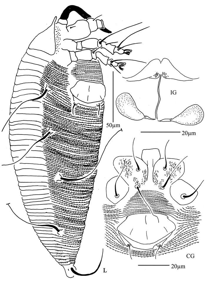
Figure 32.Diptacus mengdaensis sp. n.: L lateral view of female IG female internal genitalia CG coxae and female genitalia.
-
Vladimir Pešić, Ksenia A. Semenchenko, Wonchoel Lee
Zookeys
Figure 5.Torrenticola nipponica (Enami, 1940), male, Dobong stream, Korea: A dorsal shield B ventral shield C ejaculatory complex D gnathosoma E–F palp, medial view (E-smaller specimen, F-larger specimen). Scale bars = 100 μm.
-
Yunus Esen, Vladimir Pešić, Orhan Erman, Yücel Kaya
Zookeys
Figure 6.A–D Atractides (s. str.) nikooae Pešić, 2004, female: A Coxal and genital field B Palp, medial veiw C Vgl-1–2 D I–L-5–6 (Scale bars = 100 µm).
-
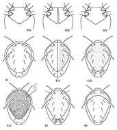
Michael J. Skvarla, J. Ray Fisher, Ashley P. G. Dowling
Zookeys
Figures 40–44.Scutopalus key illustrations. 40a Coxae I–II faintly divided 40b Coxae I–II totally divided 41 Coxae I–II fused medially 42 Dorsal shield with thick, rod-like setae present 43 Dorsal shield smooth or punctate 44a Dorsal shield rugose 44b Dorsal shield reticulate 44c Dorsal shield sparsely granulate 45a Setae mps, c1, c2, d1, e1, f1 clavate 45b Setae mps, c1, c2, d1, e1, f1 setiform 46 Setae lps, mps, c1, c2, d1, e1, f1 set on tubercles.
-
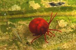
Mushroom Observer Image 103180: Aculops
-
Xiao-Feng Xue, Hussein Sadeghi, Xiao-Yue Hong, Samira Sinaie
Zookeys
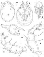
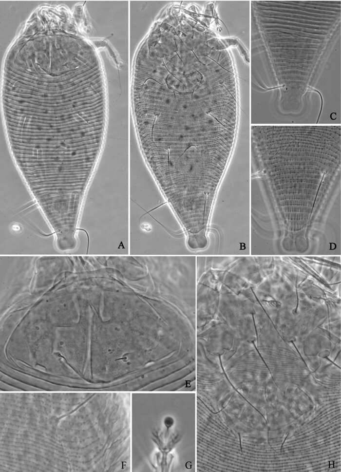
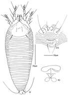
Figure 8. Aceria pulicaris sp. n. A dorsal view of female B ventral view of female C prodorsal shield D coxae and female genitalia.
-
Hao-Sen Li, Xiao-Feng Xue, Xiao-Yue Hong
Zookeys
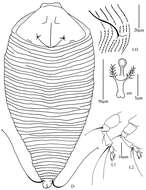
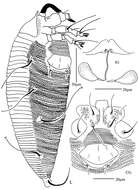
Figure 33.Diptacus mengdaensissp. n.: A dorsal view of female B ventral view of female C dorsal view of female posterior part D ventral view of female posterior part E prodorsal shield F lateral microtubercles G empodium H coxae and female genitalia.
-
Vladimir Pešić, Ksenia A. Semenchenko, Wonchoel Lee
Zookeys
Figure 6.Torrenticola nipponica (Enami, 1940), female, Tigrovaya River, Russia: A dorsal shield B ventral shield C palp, lateral view. Scale bars = 100 μm (A–B), 25 μm (C).
-
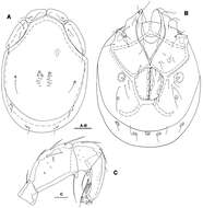
Michael J. Skvarla, J. Ray Fisher, Ashley P. G. Dowling
Zookeys
Figures 45.Scirula key illustrations. 45a Scirula impressa 45b Scirula papillata.
-
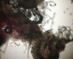
Mushroom Observer Image 103181: Aculops
-
Xiao-Feng Xue, Hussein Sadeghi, Xiao-Yue Hong, Samira Sinaie
Zookeys
Figure 12. Aculus medicager sp. n. D dorsal view of female CG coxae and female genitalia IG female internal genitalia.
-
Hao-Sen Li, Xiao-Feng Xue, Xiao-Yue Hong
Zookeys
Figure 34.Rhyncaphytoptus spinus sp. n.: D dorsal view of female L1 leg I L2 leg II IG female internal genitalia em empodium.
-
Vladimir Pešić, Ksenia A. Semenchenko, Wonchoel Lee
Zookeys
Figure 7.Photographs of dorsal shield: A Torrenticola brevirostris (Halbert, 1911), female, stream in Naebyeansan NP, Korea B Torrenticola dentifera Wiles, 1991, male, stream in Naebyeansan NP, Korea: C–F Torrenticola kimichungi sp. n. (C–D specimens from stream in Seoraksan NP, Korea, E–F specimens from Tigrovaya River, Russia): C maleholotype D–E maleparatypes F femaleparatype G–I Torrenticola nipponica (Enami, 1940) (G specimen from Dobong stream, Korea H–I specimens from Tigrovaya River, Russia): G–H male I female. Photos. V. Pešić (Figs A–D, G), K. Semenchenko (Figs E–F, H–I).
-
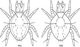
Michael J. Skvarla, J. Ray Fisher, Ashley P. G. Dowling
Zookeys
Figures 49–53.Armascirus key illustrations. 49–51 Dorsal idiosoma 49a–e Hysterosomal shield complemented with setae 50a–d Hysterosomal shield small, not complemented with setae 51a–c Hysterosomal shield absent 52a, b Pedipalp tibiotarsal claw 52a Single claw 52b Bifid claw 53a Hysterosomal plate concave on lateral edges 53b Hysterosomal plate not concave on lateral edges.
-
Mushroom Observer Image 155854: Aculops