-
Sergio Leiva, Nestor Fernandez, Pieter Theron, Christine Rollard
Zookeys
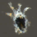
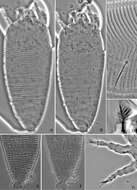
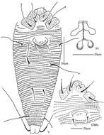
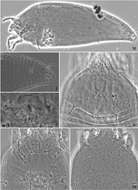
Figure 3.Agistemus aimogastaensis sp. n. Adult female, SEM. A palp, tibia and tarsus lateral view B ambulacrum leg I, lateral view. C. dorsocentral a setae D dorsolateral la setae E ps2 setae. F ps3 setae. Abreviations see material and Methods. Scale bars: B, E: 5µm; A, C, D, F: 5µm. Small stars indicate the association of eupathidia ul’, ul” and sul. Diamond indicates palp tibial claw. Double arrow, indicates claw, and special single arrow indicate capitate fan-shaped raylets.
-
Michael J. Skvarla, J. Ray Fisher, Ashley P. G. Dowling
Zookeys
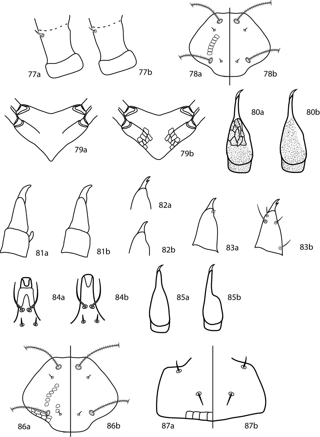
Figures 77–87.Neoscirula key illustrations 77a Pedipalp basifemoral dorsal seta spine-like 77b Pedipalp basifemoral dorsal seta simple 78a Proterosomal shield with polygonal subcuticular sculpturing present 78b Proterosomal shield with polygonal subcuticular sculpturing absent 79a Sternal shield v-shaped posteriomedially, with polygonal subcuticular sculpturing absent 79b Sternal shield rounded posteriomedially, with polygonal subcuticular sculpturing present 80a Chelicera with dorsomedial reticulations present 80b Chelicera with dorsomedial reticulations absent 81a Pedipalp genua with hook-like apophysis present 81b Pedipalp genua with hook-like apophysis absent 82a Pedipalp tibiotarsal claw with tooth present 82b Pedipalp tibiotarsal claw with tooth absent 83a Pedipalp tibiotarsus with tubercle present 83b Pedipalp tibiotarsus with tubercle absent 84a Hypognathum with ventroapical shield-like process present 84b Hypognathum with ventroapical shield-like process absent 85a Chelicera tapering gradually 85b Chelicera tapering suddenly 86a Proterosomal shield with polygonal subcuticular sculpturing present 86b Proterosomal shield with polygonal subcuticular sculpturing absent 87a Subcapitulum with row of basal subcuticular sculpturing present 87b Subcapitulum with row of basal subcuticular sculpturing absent.
-
Hao-Sen Li, Xiao-Feng Xue, Xiao-Yue Hong
Zookeys
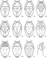
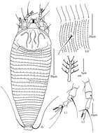
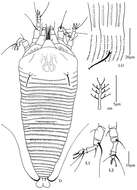
Figure 6.Proiectus xiningensis sp. n.: A dorsal view of female B ventral view of female C lateral microtubercles D empodium E dorsal view of female posterior part F ventral view of female posterior part G leg I and leg II.
-
Sergio Leiva, Nestor Fernandez, Pieter Theron, Christine Rollard
Zookeys
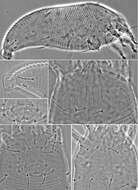
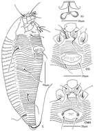
Figure 4.Agistemus aimogastaensis sp. n. Adult female, legs. All legs in dorsal view. Abbreviations: see Materials and Methods. Scale bar A–D: 50 µm.
-
Michael J. Skvarla, J. Ray Fisher, Ashley P. G. Dowling
Zookeys
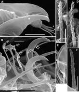
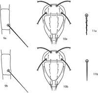
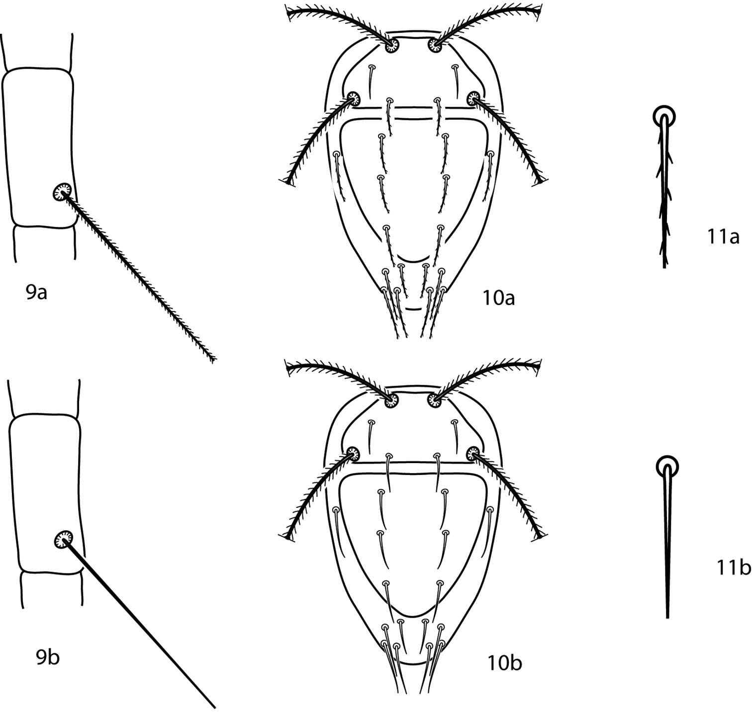
Figures 9–11.Bonzia key illustrations. 9a Setose tibial trichobothrium 9b Smooth tibial trichobothrium 10a Spiculate dorsal setae 10b Smooth dorsal setae 11a Close up of a spiculate seta 11b Close up of a smooth seta.
-
Hao-Sen Li, Xiao-Feng Xue, Xiao-Yue Hong
Zookeys
Figure 7.Proiectus xiningensis sp. n.: H lateral view of female I coxae and female genitalia J coxae and male genitalia K lateral view of female posterior part L prodorsal shield M female internal genitalia.
-
Sergio Leiva, Nestor Fernandez, Pieter Theron, Christine Rollard
Zookeys
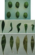
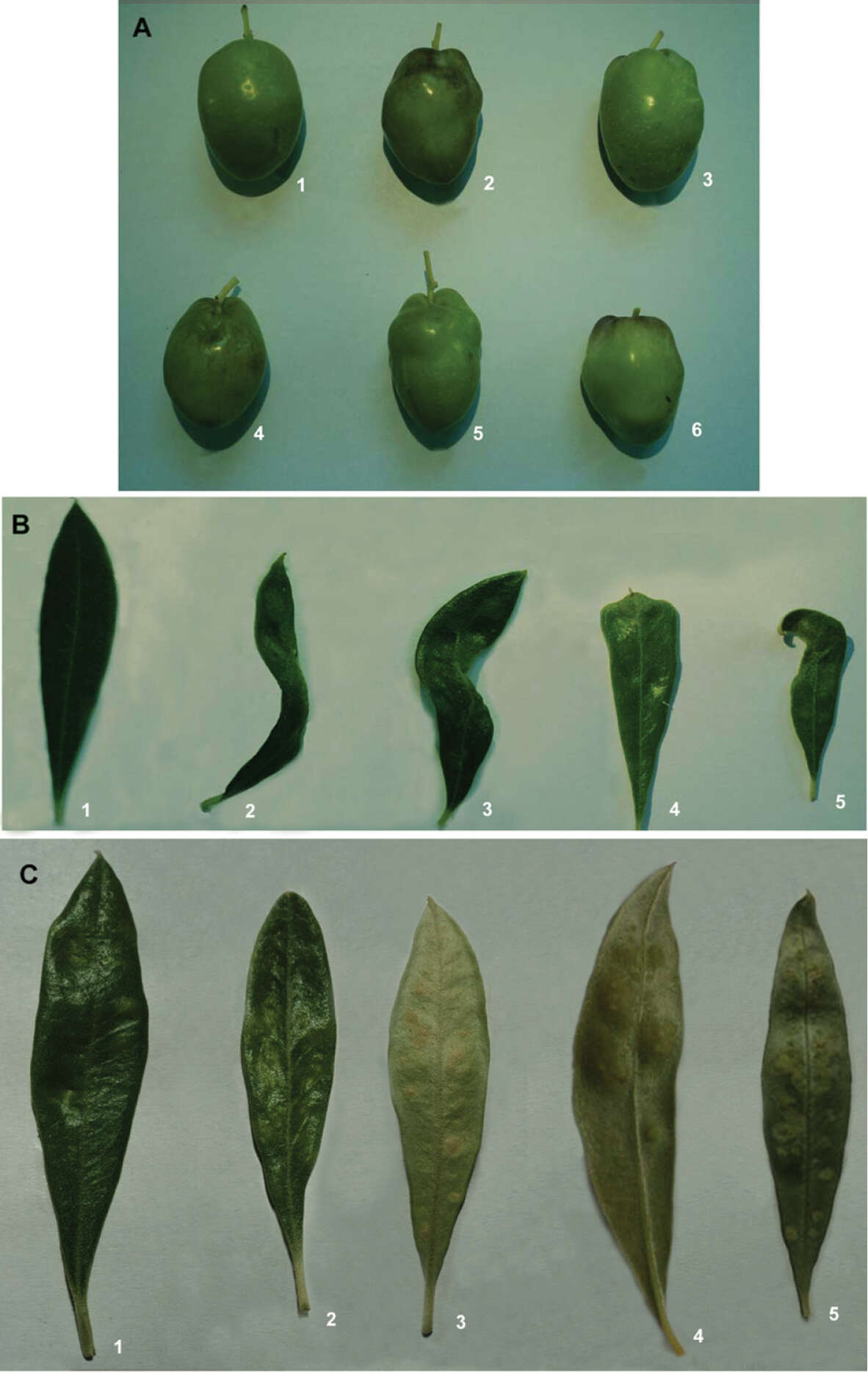
Figure 5.Malformations induced by eriophyid mites on leaves and fruit. A affected leaves B affected fruit. The upper left fruit is normal, others with malformations C young fruit attacked by Aceria oleae D detail of attack in C.
-
Michael J. Skvarla, J. Ray Fisher, Ashley P. G. Dowling
Zookeys
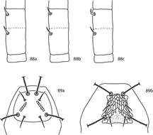
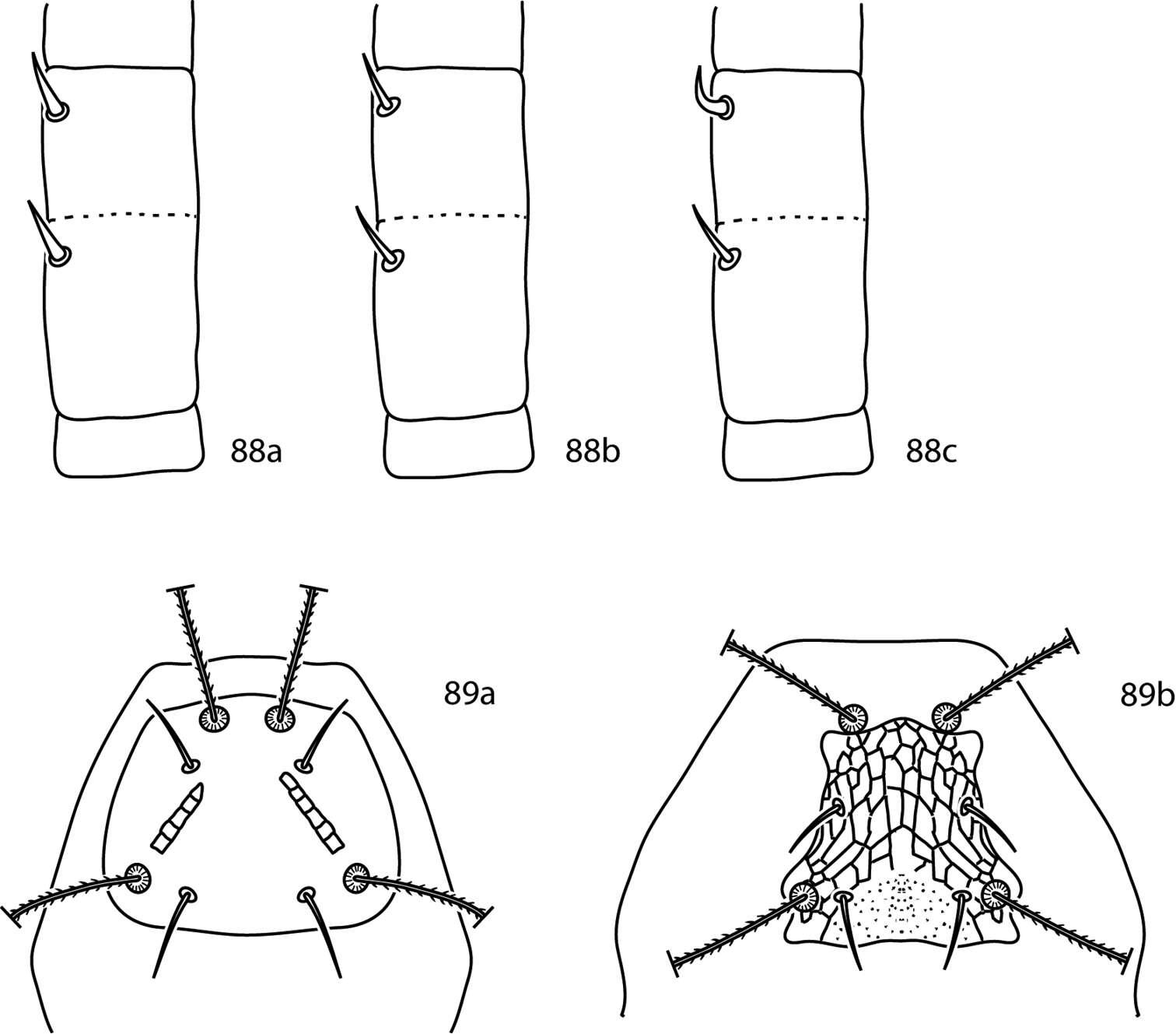
Figures 88, 89.Pseudobonzia key illustrations 88a Pedipalp basifemur and telofemur with spine-like setae on both segments 88b Pedipalp basifemur and telofemur with simple setae on both segments 88c Pedipalp with simple seta on basifemur, spine-like seta on telofemur 89a Proterosomal plate convex posteriomedially 89b Proterosomal plate not convex posteriomedially.
-
Hao-Sen Li, Xiao-Feng Xue, Xiao-Yue Hong
Zookeys
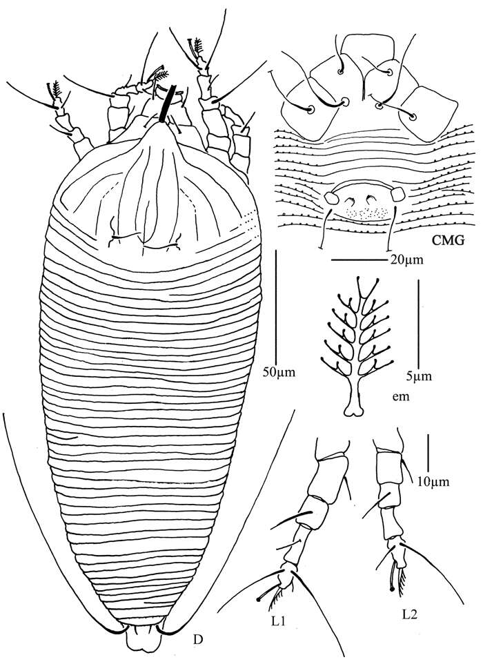
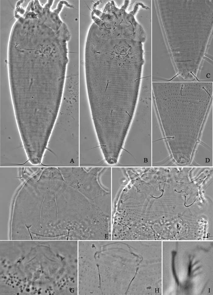
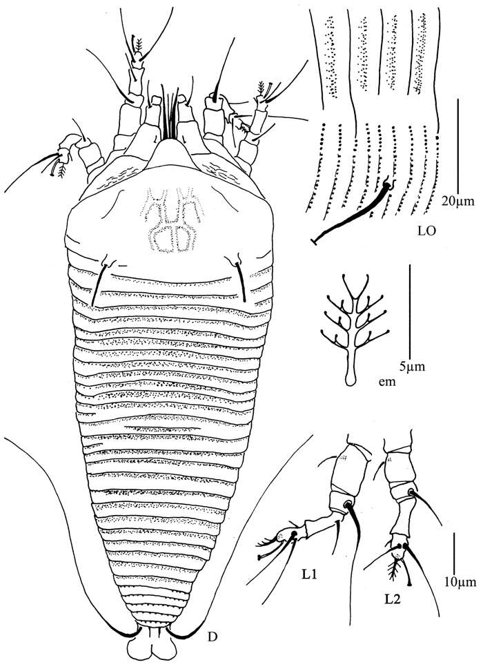
Figure 8.Phyllocoptes beishaniensis sp. n.: D dorsal view of female CMG coxae and male genitalia em empodium L1 leg I L2 leg II.
-
Michael J. Skvarla, J. Ray Fisher, Ashley P. G. Dowling
Zookeys
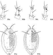
Figures 90, 91.Scutascirus key illustrations. 90a (after Corpuz-Raros and Garcia 1996). Pedipalp with tubercle not branched 90b (after Den Heyer 1980b). Pedipalp tibiotarsus with bifurcate tubercle positioned halfway along the length of the segment 90c (after Den Heyer 1980b). Pedipalp tibiotarsus with bifurcate tubercle positioned on distal third of segment 90d (after Lin et al. 2001). Pedipalp tibiotarsus with trifurcate tubercle 90a (after Den Heyer 1980b). Four pairs of dorsolateral hysterosomal plates present 91b (after Corpuz-Raros and Garcia 1996). Five pairs of dorsolateral hysterosomal plates present.
-
Hao-Sen Li, Xiao-Feng Xue, Xiao-Yue Hong
Zookeys
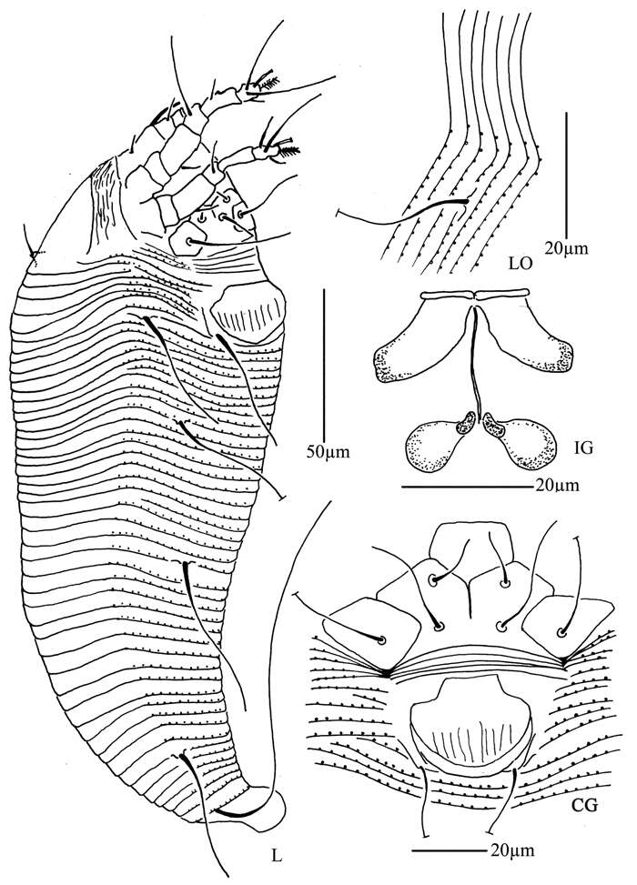
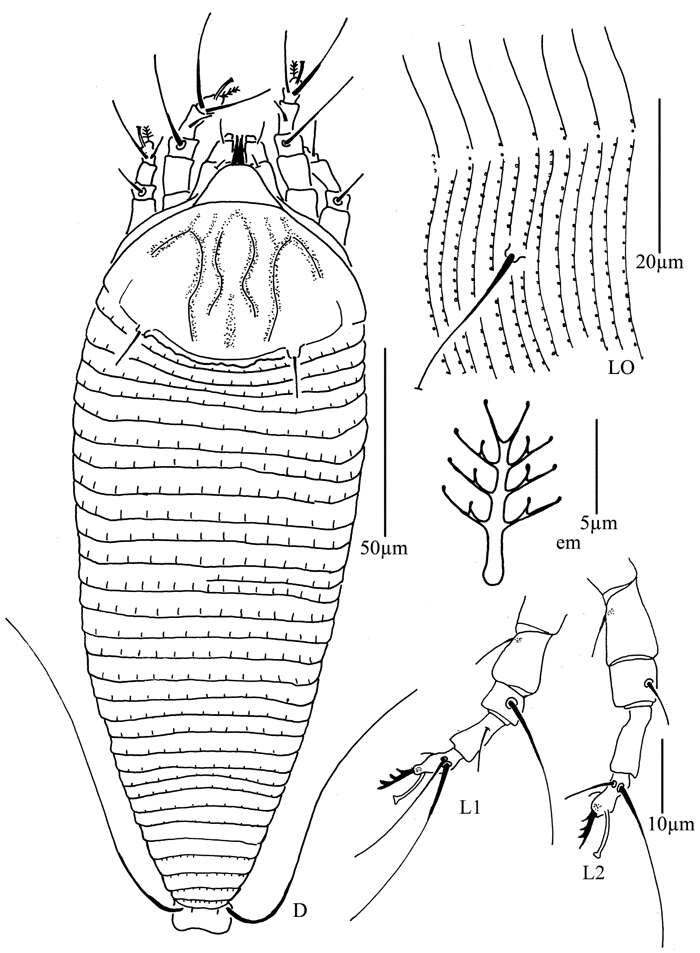
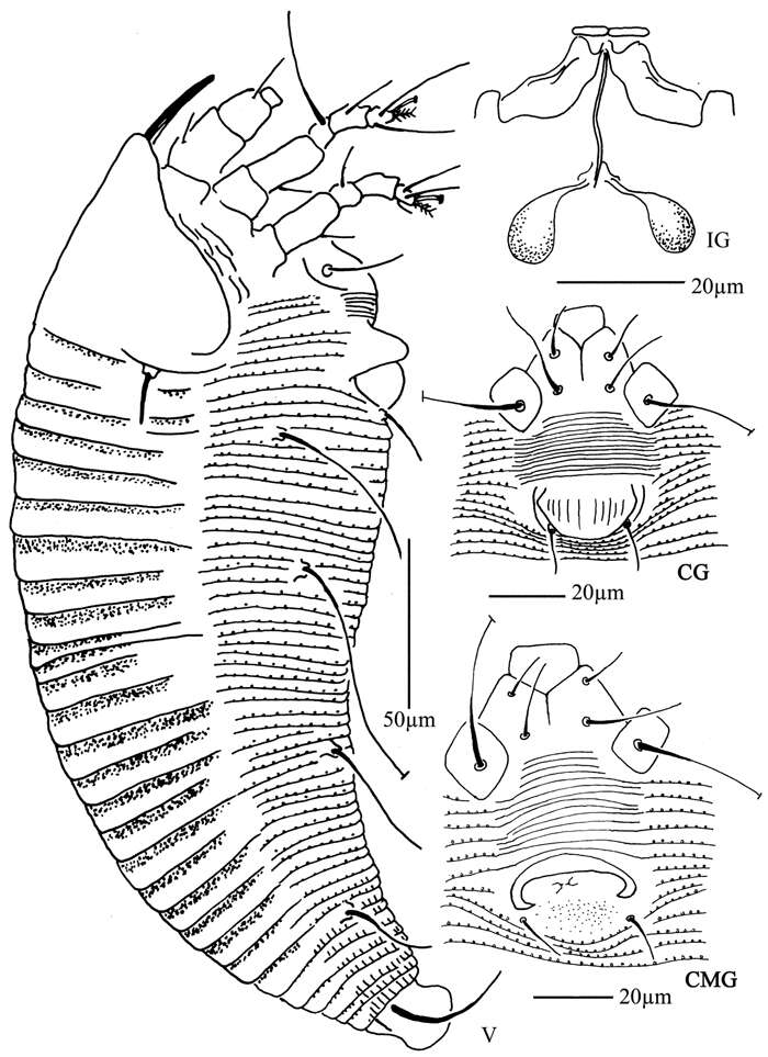
Figure 9.Phyllocoptes beishaniensis sp. n.: L lateral view of female LO lateral microtubercles IG female internal genitalia CG coxae and female genitalia
-
Michael J. Skvarla, J. Ray Fisher, Ashley P. G. Dowling
Zookeys
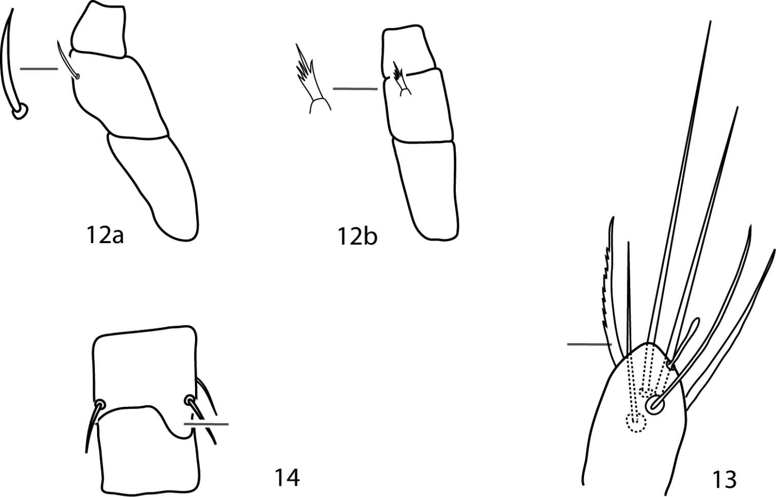
Figures 12–14.Parabonzia key illustrations. 12a Unbranched pedipalp telofemoral seta 12b Multi-branched pedipalp telofemoral seta 13 Lightly barbed pedipalp tibiotarsal sigmoid seta 14 Spur-like process on femora III.
-
Hao-Sen Li, Xiao-Feng Xue, Xiao-Yue Hong
Zookeys
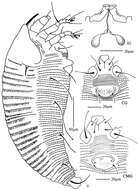
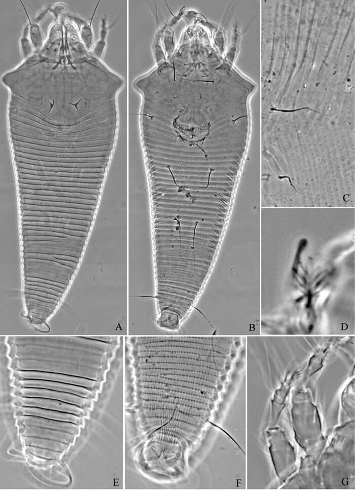
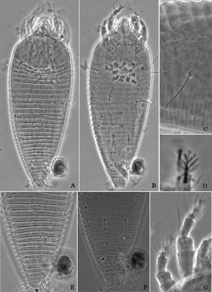
Figure 10.Phyllocoptes beishaniensis sp. n.: A dorsal view of female B ventral view of female C lateral microtubercles D empodium E dorsal view of female posterior part F ventral view of female posterior part G leg I and leg II.
-
Michael J. Skvarla, J. Ray Fisher, Ashley P. G. Dowling
Zookeys
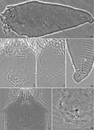
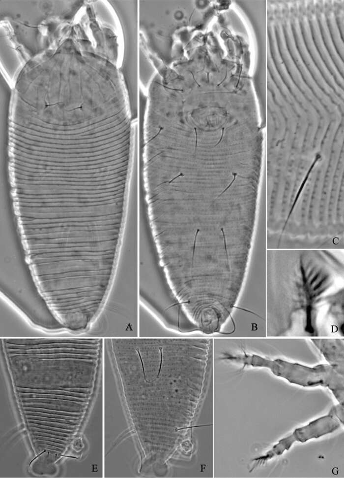
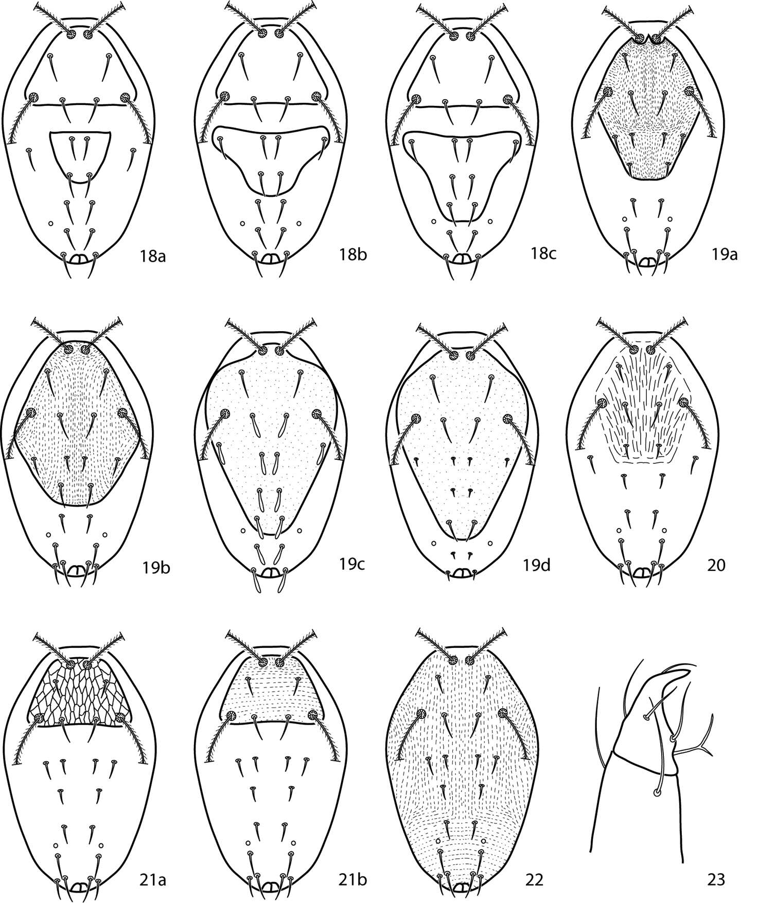
Figures 18–23.Cunaxoides key illustrations. See key for explanations.
-
Hao-Sen Li, Xiao-Feng Xue, Xiao-Yue Hong
Zookeys
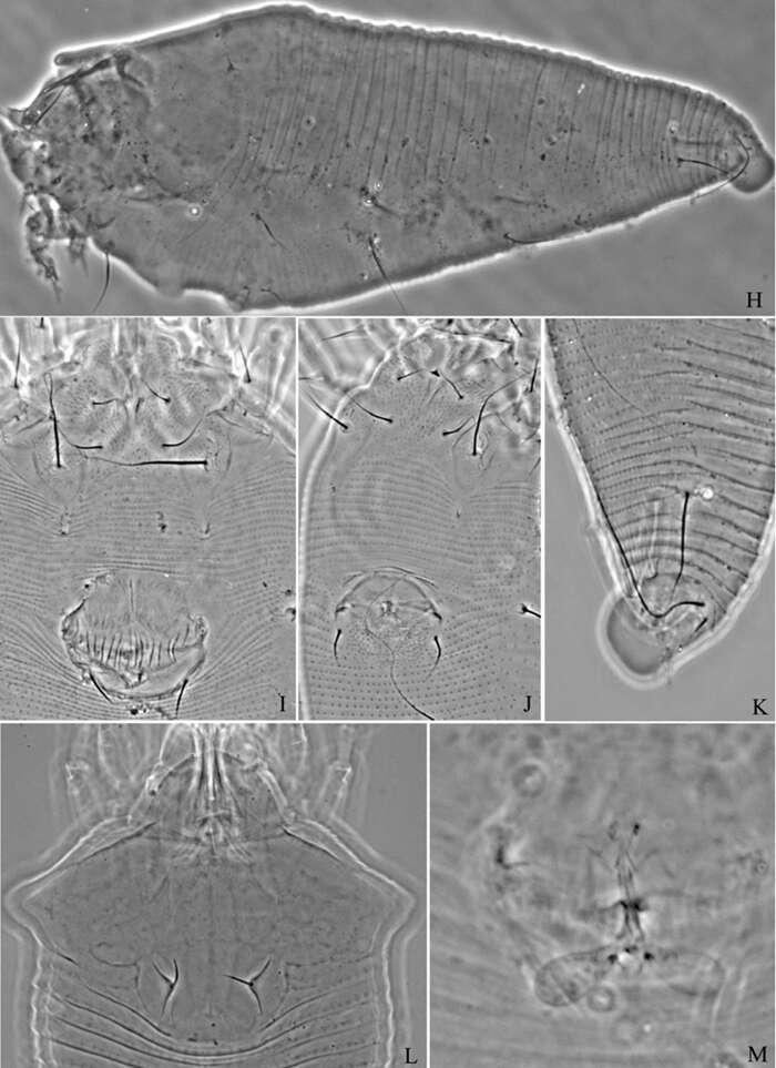
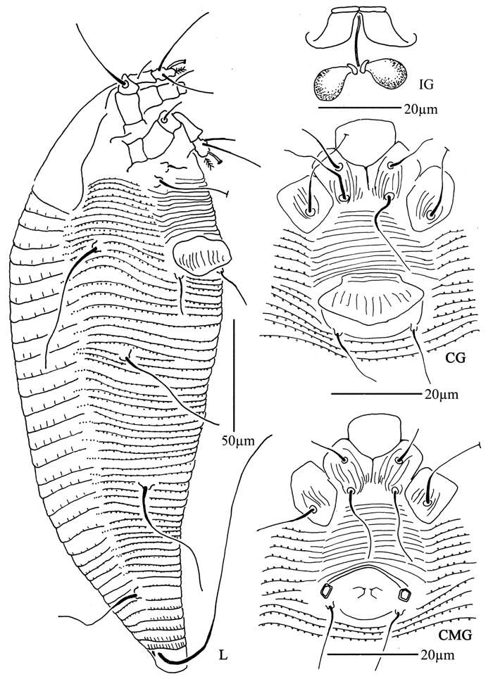
Figure 11.Phyllocoptes beishaniensis sp. n.: H lateral view of female I lateral view of female posterior part J female internal genitalia K prodorsal shield L coxae and female genitalia M coxae and male genitalia.
-
Hao-Sen Li, Xiao-Feng Xue, Xiao-Yue Hong
Zookeys
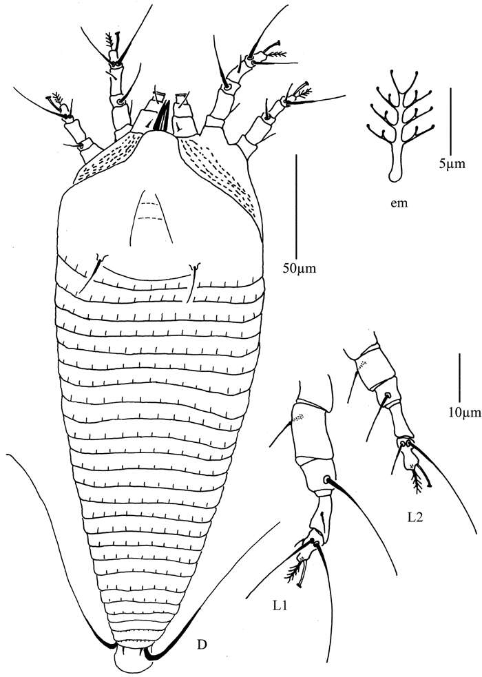
Figure 16.Tetra pruniana sp. n.: D dorsal view of female em empodium L1 leg I L2 leg II.
-
Hao-Sen Li, Xiao-Feng Xue, Xiao-Yue Hong
Zookeys
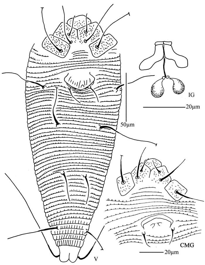
Figure 17.Tetra pruniana sp. n.: V ventral view of female IG female internal genitalia CMG coxae and male genitalia.
-
Hao-Sen Li, Xiao-Feng Xue, Xiao-Yue Hong
Zookeys
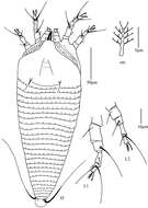
Figure 18.Tetra prunianasp. n.: A dorsal view of female B ventral view of female C dorsal view of female posterior part D ventral view of female posterior part E prodorsal shield F coxae and female genitalia G female internal genitalia H male genitalia I empodium.
-
Hao-Sen Li, Xiao-Feng Xue, Xiao-Yue Hong
Zookeys
Figure 19.Tetra pyriana sp. n.: D dorsal view of female LO lateral microtubercles em empodium L1 leg I L2 leg II.
-
Hao-Sen Li, Xiao-Feng Xue, Xiao-Yue Hong
Zookeys
Figure 20.Tetra pyriana sp. n.: L lateral view of female IG female internal genitalia CG coxae and female genitalia CMG coxae and male genitalia.
-
Hao-Sen Li, Xiao-Feng Xue, Xiao-Yue Hong
Zookeys
Figure 21.Tetra pyriana sp. n.: A dorsal view of female B ventral view of female C lateral microtubercles D empodium E dorsal view of female posterior part F ventral view of female posterior part G leg I and leg II.
-
Hao-Sen Li, Xiao-Feng Xue, Xiao-Yue Hong
Zookeys
Figure 22.Tetra pyriana sp. n.: H lateral view of female I lateral view of female posterior part J female internal genitalia K prodorsal shield L coxae and female genitalia M coxae and male genitalia.
-
Hao-Sen Li, Xiao-Feng Xue, Xiao-Yue Hong
Zookeys
Figure 23.Tetra simonia sp. n.: D dorsal view of female LO lateral microtubercles em empodium L1 leg I L2 leg II.
-
Hao-Sen Li, Xiao-Feng Xue, Xiao-Yue Hong
Zookeys
Figure 24.Tetra simonia sp. n.: L lateral view of female IG female internal genitalia CG coxae and female genitalia CMG coxae and male genitalia.