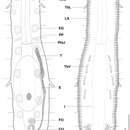
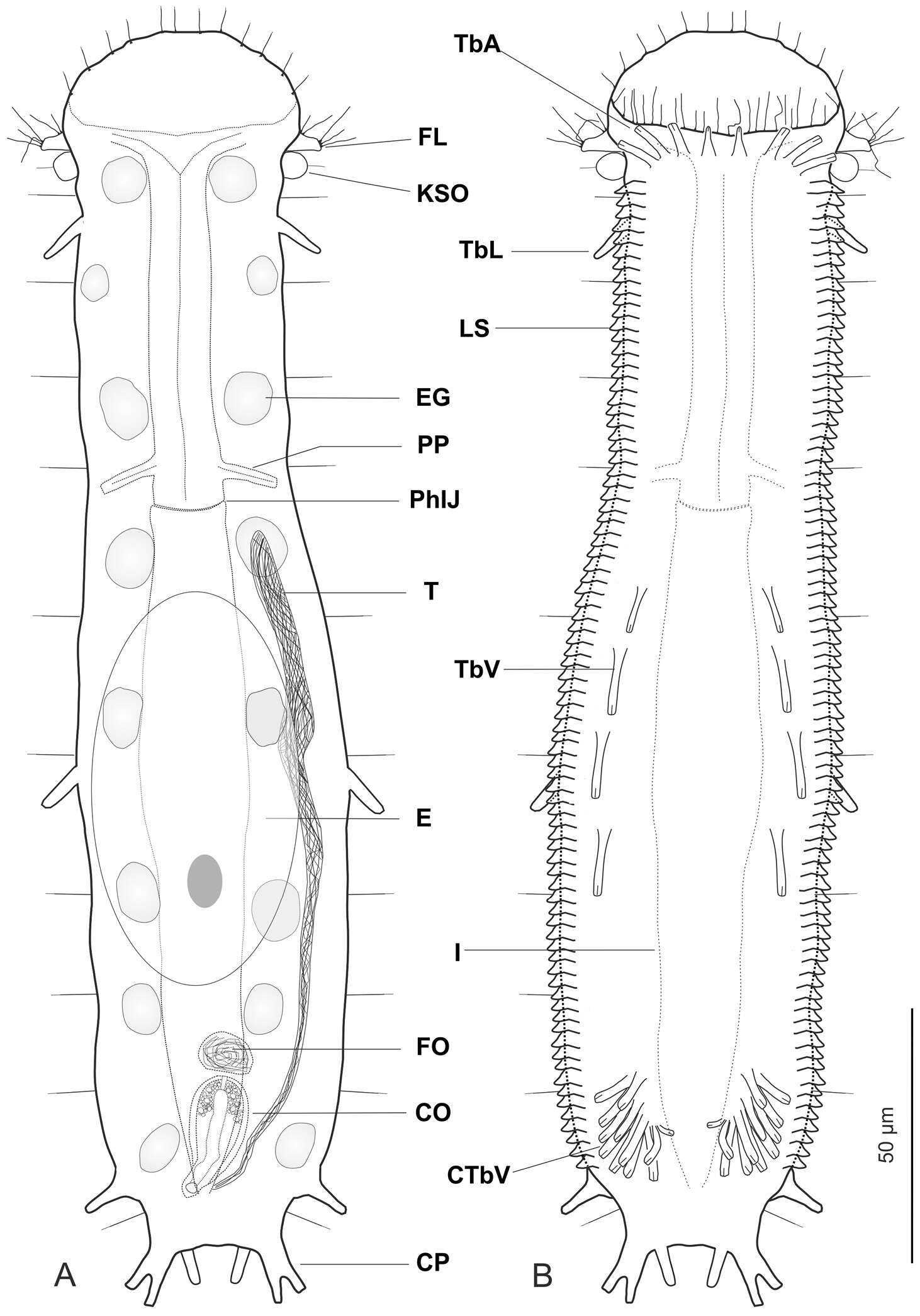
Figure 1.Ptychostomella lamelliphora sp. n. schematic drawings. A Habitus as seen from the dorsal side showing the internal anatomy B Habitus as seen from the ventral side. CO caudal organ CP caudal pedicle CTbV cluster of ventral adhesive tubes E egg EG epidermal gland FL fleshy lobe FO frontal organ I intestine KSO Knob-like sensory organ LS lamellate scales PhIJ pharyngeo-intestinal junction Pp pharyngeal pores T testicle TbA anterior adhesive tubes TbL lateral adhesive tubes TbV ventral adhesive tubes.
M. Antonio Todaro, Tobias Kånneby, Matteo Dal Zotto, Ulf Jondelius
Wikimedia Commons
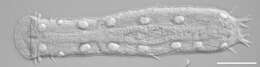
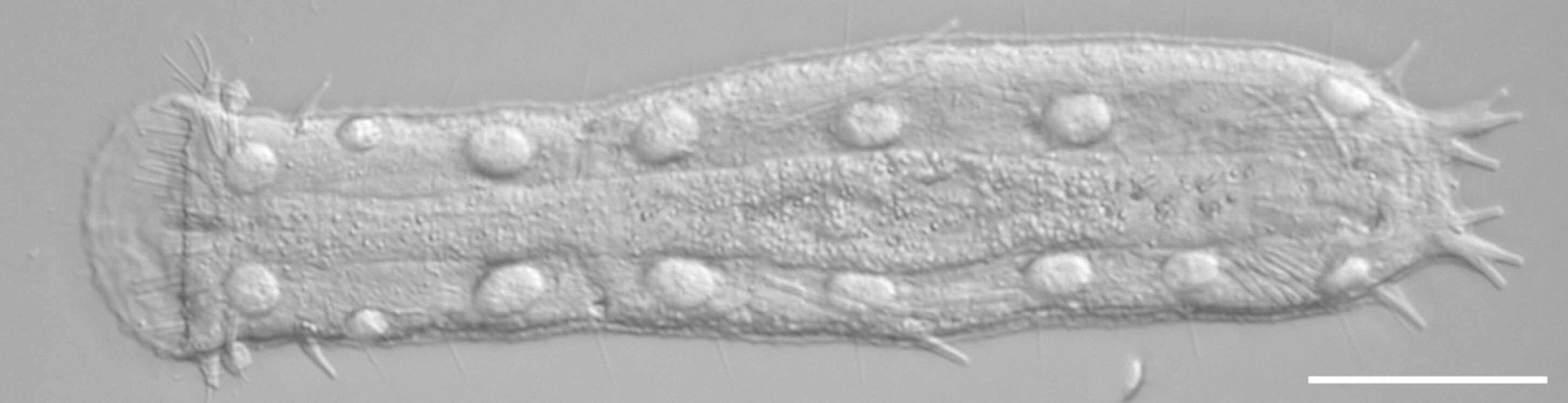
Description: English: DIC photomicrographs showing the general body shape and aspects of the cuticular covering of Ptychostomella sp. Scale bars: 50 µm. Русский: Микрофотографии брюхоресничного червя Ptychostomella sp. Дифференциальная интерференционно-контрастная микроскопия. Масштабная черта — 50 мкм. Date: March 2011. Source: Todaro, M. A., Kånneby, T., Dal Zotto, M., Jondelius, U. (2011). Phylogeny of Thaumastodermatidae (Gastrotricha: Macrodasyida) inferred from nuclear and mitochondrial sequence data. PLoS ONE 6 (3): e17892. doi:10.1371/journal.pone.0017892. Author: M. Antonio Todaro, Tobias Kånneby, Matteo Dal Zotto, Ulf Jondelius.