dcsimg

Purpureocillium lilacinum ( Catalan; Valencian )

provided by wikipedia CA

Purpureocillium lilacinum és una espècie de fongs filamentosos dins la família Ophiocordycipitaceae dins el gènere Purpureocillium, un fong comú sapròfit. S'ha aïllat de molts hàbitats incloent sòls cultivats i no cultivats, boscos, prats, deserts, sediments, estuaris i insectes. A més s'ha detectat en la rizosfera de molts cultius. El seu rang de temperatures on creix és ampli i l'òptim és entre 26 a 30 °C. també té un ampli rang de pH i pot créixer en molts substrats.[3][4] P. lilacinum mostra resultats prometedors en el control biològic de les plagues en concret dels nematodes de les arrels.

Taxonomia i filogènia

L'espècie va ser descrita pel micòleg Charles Thom el 1910, sota el nom de Penicillium lilacinum.[5] Taxonomic synonyms include Penicillium amethystinum Wehmer and Spicaria rubidopurpurea Aoki.[2] El 1974, Charles Thom va transferir-lo al gènere Paecilomyces.[3] A la dècada del 2000 es va veure que no era un gènere monofilètic,[6] i que els seus parents és propers eren Paecilomyces nostocoides, Isaria takamizusanensis iNomuraea atypicola.[7] Es va crear un nou gènere: Purpureocillium. El nom del gènere es refereix als conidis porpra.[8]

Descripció

Purpureocillium lilacinum forma un miceli dens que dóna lloc a conidiòfors. Les hifes vegetatives tenen les parets llises (hialines) i fan 2.5–4.0 µm d'amplada.

Purpureocillium lilacinum és molt adaptable depenent de la disponibilitat de nutrients en el seu ambientpot ser un fong entomopatogen,[9][10][11] micoparàsit,[12] sapròfit,[13] o nematòfag.

Patogen en humans

Purpureocillium lilacinum és una causa infreqüent de malaltia en humans.[14][15] Lamajoria dels casos es donen en pacients amb el sistema immunitari afectat o en implants de lents intraoculars.[16][17]

Agent de biocontrol

 src=
P. lilacinum usat contra Meloidogyne incognita.

Els nematodes paràsits de les plantes causen pèrdues econòmiques significatives. Purpureocillium lilacinum es va observar en ous de nematodes l'any 1966[18] i en ous de Meloidogyne incognita al Perú.[19] De vegades els fongs aïllats que funcionen in vitro o en hivernacle no ho fan en el camp.[20]

S'han estudiat molts enzims produïts per P. lilacinum per estudiar l'eficàcia en el biocontrol.

Micotoxines

La paecilotoxina és una micotoxina aïllada d'aquest fong.[21]

Referències

 src= A Wikimedia Commons hi ha contingut multimèdia relatiu a: Purpureocillium lilacinum Modifica l'enllaç a Wikidata
  1. Domsch KH, Gams W, Anderson T-H. Compendium of Soil Fungi. 2a ed.. Lubrecht & Cramer Ltd, 2007, p. 322. ISBN 978-3-9803083-8-0.
  2. 2,0 2,1 «Paecilomyces lilacinus (Thom) Samson 1974». MycoBank. International Mycological Association. [Consulta: 17 juliol 2011].
  3. 3,0 3,1 Samson RA. «Paecilomyces and some allied hyphomycetes». Studies in Mycology. Centralbureau voor Schimmelcultures [Baarn], 6, 1974, pàg. 58.
  4. Anderson T-H, Domsch KH, Gams W.. Compendium of Soil Fungi. Lubrecht & Cramer Ltd, 1995. ISBN 3-9803083-8-3.
  5. Thom C. Bulletin of the Bureau of Animal Industry US Department of Agriculture, 118, 1910, pàg. 73.
  6. Inglis PW, Tigano MS. «Identification and taxonomy of some entomopathogenic Paecilomyces spp. (Ascomycota) isolates using rDNA-ITS Sequences» (PDF). Genetics and Molecular Biology, 29, 1, 2006, pàg. 132–6.
  7. Sung G-H, Hywel-Jones NL, Sung J-M, Luangsa-ard JJ, Shrestha B, Spatafora JW. «Phylogenetic classification of Cordyceps and the clavicipitaceous fungi». Studies in Mycology, 57, 1, 2007, pàg. 5–59. DOI: 10.3114/sim.2007.57.01. PMC: 2104736. PMID: 18490993.
  8. Luangsa-Ard J, Houbraken J, van Doorn T, Hong SB, Borman AM, Hywel-Jones NL, Samson RA. «Purpureocillium, a new genus for the medically important Paecilomyces lilacinus». FEMS Microbiology Letters, 321, 2, 2011, pàg. 141–9. DOI: 10.1111/j.1574-6968.2011.02322.x. PMID: 21631575.
  9. Rombach MC, Aguda RM, Shepard BM Roberts DW. «Infection of rice brown planthopper, Nilaparvata lugens (Homoptera: Delphacidae), by field application of entomopathogenic hyphomycetes (Deuteromycotina)». Environmental Entomology, 15, 5, 1986, pàg. 1070–3. ISSN: 0046225X.
  10. Marti GA, Lastra CC, Pelizza SA, García JJ. «Isolation of Paecilomyces lilacinus (Thom) Samson (Ascomycota: Hypocreales) from the Chagas disease vector, Triatoma infestans Klug (Hemiptera: Reduviidae) in an endemic area in Argentina». Mycopathologia, 162, 5, 2006, pàg. 369–72. DOI: 10.1007/s11046-006-0072-3. PMID: 17123036.
  11. Fiedler Ż, Sosnowska D. «Nematophagous fungus Paecilomyces lilacinus (Thom) Samson is also a biological agent for control of greenhouse insects and mite pests». BioControl, 52, 4, 2007, pàg. 547–8. DOI: 10.1007/s10526-006-9052-2.
  12. Gupta SC, Leathers TD, Wicklow DT. «Hydrolytic enzymes secreted by Paecilomyces lilacinus cultured on sclerotia of Aspergillus flavus» (PDF). Applied Microbiology and Biotechnology, 39, 1, 1993, pàg. 99–103. hdl:10113/25102.
  13. Tigrano-Milani MS, Carneiro RG, de Faria MR, Frazão HS, McCoy CW. «Isozyme characterization and pathogenicity of Paecilomyces fumosoroseus and P. lilacinus to Diabrotica speciosa (Coleoptera: Chrysomelidae) and Meloidogyne javanica (Nematoda: Tylenchidae)». Biological Control, 5, 3, 1995, pàg. 378–82. DOI: 10.1006/bcon.1995.1044.
  14. Saberhagen C, Klotz SA, Bartholomew W, Drews D, Dixon A. «Infection due to Paecilomyces lilacinus: a challenging clinical identification». Clinical and Infectious Diseases, 25, 6, 1997, pàg. 1411–3. DOI: 10.1086/516136. PMID: 9431388.
  15. Westenfeld F, Alston WK, Winn WC. «Complicated soft tissue infection with prepatellar bursitis caused by Paecilomyces lilacinus in an immunocompetent host: case report and review». Journal of Clinical Microbiology, 34, 6, 1996, pàg. 1559–62. PMC: 229063. PMID: 8735119.
  16. O'Day DM «Fungal endophthalmitis caused by Paecilomyces lilacinus after intraocular lens implantation». American Journal of Ophthalmology, 83, 1, 1977, pàg. 130–1. PMID: 299984.
  17. Pettit TH, Olson RJ, Foos RY, Martin WJ «Fungal endophthalmitis following intraocular lens implantation. A surgical epidemic». Archives of Ophthalmology, 98, 6, 1980, pàg. 1025–39. PMID: 7190003.
  18. Lysek H. «Study of biology of geohelminths. II. The importance of some soil microorganisms for the viability of geohelminth eggs in the soil». Acta Universitatis Palackianae Olomucensis, 40, 1996, pàg. 83–90.
  19. Jatala P, Kaltenbach R, Bocangel M. «Biological control of Meloidogyne incognita acrita and Globodera pallida on potatoes». Journal of Nematology, 11, 1979, pàg. 303.
  20. Gomes Carniero RMD, Cayrol J. «Relationship between inoculum density of the nematophagous fungus Paecilomyces lilacinus and control of Meloidogyne arenaria on tomato». Revue Nématologique, 14, 4, 1991, pàg. 629–34.
  21. Mikami Y, Yazawa K, Fukushima K, Arai T, Udagawa S, Samson RA. «Paecilotoxin production in clinical or terrestrial isolates of Paecilomyces lilacinus strains». Mycopathologia, 108, 3, pàg. 195–9. DOI: 10.1007/BF00436225. PMID: 2615806.

Error de citació: L'etiqueta amb el nom "Bonants1995" definida a no s'utilitza en el text anterior.
Error de citació: L'etiqueta amb el nom "Jatala1986" definida a no s'utilitza en el text anterior.
Error de citació: L'etiqueta amb el nom "Khan2003" definida a no s'utilitza en el text anterior.
Error de citació: L'etiqueta amb el nom "Khan2004" definida a no s'utilitza en el text anterior.
Error de citació: L'etiqueta amb el nom "Money1998" definida a no s'utilitza en el text anterior.
Error de citació: L'etiqueta amb el nom "Park2004" definida a no s'utilitza en el text anterior.
Error de citació: L'etiqueta amb el nom "Safdar2002" definida a no s'utilitza en el text anterior.

Error de citació: L'etiqueta amb el nom "Stirling1991" definida a no s'utilitza en el text anterior.
license
cc-by-sa-3.0
copyright
Autors i editors de Wikipedia
original
visit source
partner site
wikipedia CA

Purpureocillium lilacinum: Brief Summary ( Catalan; Valencian )

provided by wikipedia CA

Purpureocillium lilacinum és una espècie de fongs filamentosos dins la família Ophiocordycipitaceae dins el gènere Purpureocillium, un fong comú sapròfit. S'ha aïllat de molts hàbitats incloent sòls cultivats i no cultivats, boscos, prats, deserts, sediments, estuaris i insectes. A més s'ha detectat en la rizosfera de molts cultius. El seu rang de temperatures on creix és ampli i l'òptim és entre 26 a 30 °C. també té un ampli rang de pH i pot créixer en molts substrats. P. lilacinum mostra resultats prometedors en el control biològic de les plagues en concret dels nematodes de les arrels.

license
cc-by-sa-3.0
copyright
Autors i editors de Wikipedia
original
visit source
partner site
wikipedia CA

Purpureocillium lilacinum

provided by wikipedia EN

Purpureocillium lilacinum is a species of filamentous fungus in the family Ophiocordycipitaceae.[3] It has been isolated from a wide range of habitats, including cultivated and uncultivated soils, forests, grassland, deserts, estuarine sediments and sewage sludge, and insects. It has also been found in nematode eggs, and occasionally from females of root-knot and cyst nematodes. In addition, it has frequently been detected in the rhizosphere of many crops. The species can grow at a wide range of temperatures – from 8 to 38 °C (46 to 100 °F) for a few isolates, with optimal growth in the range 26 to 30 °C (79 to 86 °F). It also has a wide pH tolerance and can grow on a variety of substrates.[4][5] P. lilacinum has shown promising results for use as a biocontrol agent to control the growth of destructive root-knot nematodes.

Taxonomy and phylogeny

The species was originally described by American mycologist Charles Thom in 1910, under than name Penicillium lilacinum.[6] Taxonomic synonyms include Penicillium amethystinum Wehmer and Spicaria rubidopurpurea Aoki.[2] In 1974, Robert A. Samson transferred the species to Paecilomyces.[4] Publications in the 2000s (decade) indicated that the genus Paecilomyces was not monophyletic,[7] and that close relatives were Paecilomyces nostocoides, Isaria takamizusanensis and Nomuraea atypicola.[8] The new genus Purpureocillium was created to hold the taxon. The generic name refers to the purple conidia produced by the fungus.[9]

Description

Purpureocillium lilacinum forms a dense mycelium which gives rise to conidiophores. These bear phialides from the ends of which spores are formed in long chains. Spores germinate when suitable moisture and nutrients are available. Colonies on malt agar grow rather fast, attaining a diameter of 5–7 cm within 14 days at 25 °C (77 °F), consisting of a basal felt with a floccose overgrowth of aerial mycelium; at first white, but when sporulating changing to various shades of vinaceous. The reverse side is sometimes uncolored but usually in vinaceous shades. The vegetative hyphae are smooth-walled, hyaline, and 2.5–4.0 µm wide. Conidiophores arising from submerged hyphae, 400–600 µm in length, or arising from aerial hyphae and half as long. Phialides consisting of a swollen basal part, tapering into a thin distinct neck. Conidia are in divergent chains, ellipsoid to fusiform in shape, and smooth walled to slightly roughened. Chlamydospores are absent.[4]

Life cycles

Purpureocillium lilacinum is highly adaptable in its life strategy: depending on the availability of nutrients in the surrounding microenvironments it may be entomopathogenic,[10][11][12] mycoparasitic,[13] saprophytic,[14] as well as nematophagous.

Human pathogenicity

Purpureocillium lilacinum is an infrequent cause of human disease.[15][16] Most reported cases involve patients with compromised immune systems, indwelling foreign devices, or intraocular lens implants.[17][18] Research of the last decade suggests it may be an emerging pathogen of both immunocompromised[19] as well as immunocompetent adults.[20]
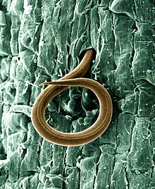
Biocontrol agent

P. lilacinum is used as a biocontrol agent for several nematode species, such as Meloidogyne incognita, shown here.

Plant-parasitic nematodes cause significant economic losses to a wide variety of crops. Chemical control is a widely used option for plant-parasitic nematode management. However, chemical nematicides are now being reappraised in respect of environmental hazard, high costs, limited availability in many developing countries or their diminished effectiveness following repeated applications.

Control of plant-parasitic nematodes

Purpureocillium lilacinum was first observed in association with nematode eggs in 1966[21] and the fungus was subsequently found parasitising the eggs of Meloidogyne incognita in Peru.[22] It has now been isolated from many cyst and root-knot nematodes and from soil in many locations.[23][24] Several successful field trials using P. lilacinum against pest nematodes were conducted in Peru.[22] The Peruvian isolate was then sent to nematologists in 46 countries for testing, as part of the International Meloidogyne project, resulting in many more field trials on a range of crops in many soil types and climates.[25] Field trials, glasshouse trials and in vitro testing of P. lilacinum continues and more isolates have been collected from soil, nematodes and occasionally from insects. Isolates vary in their pathogenicity to plant-parasitic nematodes. Some isolates are aggressive parasites while others, though morphologically indistinguishable, are less or non-pathogenic. Sometimes isolates that looked promising in vitro or in glasshouse trials have failed to provide control in the field.[26]

Enzymes

Many enzymes produced by P. lilacinum have been studied. A basic serine protease with biological activity against Meloidogyne hapla eggs has been identified.[27] One strain of P. lilacinum has been shown to produce proteases and a chitinase, enzymes that could weaken a nematode egg shell so as to enable a narrow infection peg to push through.[28]

Egg infection

Before infecting a nematode egg, P. lilacinum flattens against the egg surface and becomes closely appressed to it. P. lilacinum produces simple appressoria anywhere on the nematode egg shell either after a few hyphae grow along the egg surface, or after a network of hyphae form on the egg. The presence of appressoria appears to indicate that the egg is, or is about to be, infected. In either case, the appressorium appears the same, as a simple swelling at the end of a hypha, closely appressed to the eggshell. Adhesion between the appressorium and nematode egg surface must be strong enough to withstand the opposing force produced by the extending tip of a penetration hypha.[29] When the hypha has penetrated the egg, it rapidly destroys the juvenile within, before growing out of the now empty egg shell to produce conidiophores and to grow towards adjacent eggs.

Mycotoxins

Paecilotoxin is a mycotoxin isolated from the fungus.[30] Its significance is unknown. Khan et al. (2003) tested one strain of P. lilacinum for the production of paecilotoxin and were unable to show toxin production in that strain, suggesting that toxin synthesis may vary among isolates.[31][32]

References

  1. ^ Domsch KH, Gams W, Anderson TH, eds. (2007). Compendium of Soil Fungi (2nd ed.). Lubrecht & Cramer Ltd. p. 322. ISBN 978-3-9803083-8-0.
  2. ^ a b "Paecilomyces lilacinus (Thom) Samson 1974". MycoBank. International Mycological Association. Retrieved 2011-07-17.
  3. ^ Spatafora; et al. (2015). "New 1F1N Species Combinations in Ophiocordycipitaceae (Hypocreales)". IMA Fungus. 6 (2): 357–362. doi:10.5598/imafungus.2015.06.02.07. PMC 4681259. PMID 26734546.
  4. ^ a b c Samson RA. (1974). "Paecilomyces and some allied hyphomycetes". Studies in Mycology. 6: 58.
  5. ^ Anderson TH, Domsch KH, Gams W (1995). Compendium of Soil Fungi. Lubrecht & Cramer Ltd. ISBN 978-3-9803083-8-0.
  6. ^ Thom C. (1910). Bulletin of the Bureau of Animal Industry US Department of Agriculture. 118: 73. {{cite journal}}: Missing or empty |title= (help)
  7. ^ Inglis PW, Tigano MS (2006). "Identification and taxonomy of some entomopathogenic Paecilomyces spp. (Ascomycota) isolates using rDNA-ITS Sequences" (PDF). Genetics and Molecular Biology. 29 (1): 132–6. doi:10.1590/s1415-47572006000100025.
  8. ^ Sung GH, Hywel-Jones NL, Sung JM, Luangsa-ard JJ, Shrestha B, Spatafora JW (2007). "Phylogenetic classification of Cordyceps and the clavicipitaceous fungi". Studies in Mycology. 57 (1): 5–59. doi:10.3114/sim.2007.57.01. PMC 2104736. PMID 18490993.
  9. ^ Luangsa-Ard J, Houbraken J, van Doorn T, Hong SB, Borman AM, Hywel-Jones NL, Samson RA (2011). "Purpureocillium, a new genus for the medically important Paecilomyces lilacinus". FEMS Microbiology Letters. 321 (2): 141–9. doi:10.1111/j.1574-6968.2011.02322.x. PMID 21631575.
  10. ^ Rombach MC, Aguda RM, Shepard BM, Roberts DW (1986). "Infection of rice brown planthopper, Nilaparvata lugens (Homoptera: Delphacidae), by field application of entomopathogenic hyphomycetes (Deuteromycotina)". Environmental Entomology. 15 (5): 1070–3. doi:10.1093/ee/15.5.1070. ISSN 0046-225X.
  11. ^ Marti GA, Lastra CC, Pelizza SA, García JJ (2006). "Isolation of Paecilomyces lilacinus (Thom) Samson (Ascomycota: Hypocreales) from the Chagas disease vector, Triatoma infestans Klug (Hemiptera: Reduviidae) in an endemic area in Argentina". Mycopathologia. 162 (5): 369–72. doi:10.1007/s11046-006-0072-3. hdl:11336/81093. PMID 17123036. S2CID 25132425.
  12. ^ Fiedler Ż, Sosnowska D (2007). "Nematophagous fungus Paecilomyces lilacinus (Thom) Samson is also a biological agent for control of greenhouse insects and mite pests". BioControl. 52 (4): 547–8. doi:10.1007/s10526-006-9052-2. S2CID 39116161.
  13. ^ Gupta SC, Leathers TD, Wicklow DT (1993). "Hydrolytic enzymes secreted by Paecilomyces lilacinus cultured on sclerotia of Aspergillus flavus" (PDF). Applied Microbiology and Biotechnology. 39 (1): 99–103. doi:10.1007/bf00166856. hdl:10113/25102. S2CID 20548355. Archived from the original (PDF) on 2012-04-25. Retrieved 2018-07-23.
  14. ^ Tigrano-Milani MS, Carneiro RG, de Faria MR, Frazão HS, McCoy CW (1995). "Isozyme characterization and pathogenicity of Paecilomyces fumosoroseus and P. lilacinus to Diabrotica speciosa (Coleoptera: Chrysomelidae) and Meloidogyne javanica (Nematoda: Tylenchidae)". Biological Control. 5 (3): 378–82. doi:10.1006/bcon.1995.1044.
  15. ^ Saberhagen C, Klotz SA, Bartholomew W, Drews D, Dixon A (1997). "Infection due to Paecilomyces lilacinus: a challenging clinical identification". Clinical Infectious Diseases. 25 (6): 1411–3. doi:10.1086/516136. PMID 9431388.
  16. ^ Westenfeld F, Alston WK, Winn WC (1996). "Complicated soft tissue infection with prepatellar bursitis caused by Paecilomyces lilacinus in an immunocompetent host: case report and review". Journal of Clinical Microbiology. 34 (6): 1559–62. doi:10.1128/JCM.34.6.1559-1562.1996. PMC 229063. PMID 8735119.
  17. ^ O'Day DM (1977). "Fungal endophthalmitis caused by Paecilomyces lilacinus after intraocular lens implantation". American Journal of Ophthalmology. 83 (1): 130–1. doi:10.1016/0002-9394(77)90206-9. PMID 299984.
  18. ^ Pettit TH, Olson RJ, Foos RY, Martin WJ (1980). "Fungal endophthalmitis following intraocular lens implantation. A surgical epidemic". Archives of Ophthalmology. 98 (6): 1025–39. doi:10.1001/archopht.1980.01020031015002. PMID 7190003.
  19. ^ Safdar A. (2002). "Progressive cutaneous hyalohyphomycosis due to Paecilomyces lilacinus: rapid response to treatment with caspofungin and itraconazole". Clinical Infectious Diseases. 34 (10): 1415–7. doi:10.1086/340260. PMID 11981740.
  20. ^ Carey J, D’Amico R, Sutton DA, Rinaldi MG. Paecilomyces lilacinus vaginitis in an immunocompetent patient. Emerg Infect Dis [serial online] 2003 Sep
  21. ^ Lysek H. (1996). "Study of biology of geohelminths. II. The importance of some soil microorganisms for the viability of geohelminth eggs in the soil". Acta Universitatis Palackianae Olomucensis. 40: 83–90.
  22. ^ a b Jatala P, Kaltenbach R, Bocangel M (1979). "Biological control of Meloidogyne incognita acrita and Globodera pallida on potatoes". Journal of Nematology. 11: 303.
  23. ^ Stirling, GR (1991). Biological Control of Plant Parasitic Nematodes. UK: CABI Publishing. p. 282.
  24. ^ Stirling GR, West LM (1991). "Fungal parasites of root-knot nematode eggs from tropical and sub-tropical regions of Australia". Australasian Plant Pathology. 20 (4): 149–54. doi:10.1071/APP9910149. S2CID 34778287.
  25. ^ Jatala P. (1986). "Biological control of plant-parasitic nematodes". Annual Review of Phytopathology. 24: 453–89. doi:10.1146/annurev.py.24.090186.002321.
  26. ^ Gomes Carniero RMD, Cayrol J (1991). "Relationship between inoculum density of the nematophagous fungus Paecilomyces lilacinus and control of Meloidogyne arenaria on tomato". Revue de Nématologie. 14 (4): 629–34.
  27. ^ Bonants PJM; Fitters PFL; Thijs H; den Belder E; Waalwijk C; Henfling JWDM. (1995). "A basic serine protease from Paecilomyces lilacinus with biological activity against Meloidogyne hapla eggs". Microbiology. 141 (Pt 4): 775–84. doi:10.1099/13500872-141-4-775. PMID 7773385.
  28. ^ Khan A, Williams KL, Nevalainen HK (2004). "Effects of Paecilomyces lilacinus protease and chitinase on the eggshell structures and hatching of Meloidogyne javanica juveniles". Biological Control. 31 (3): 346–52. doi:10.1016/j.biocontrol.2004.07.011.
  29. ^ Money NP. (1998). "Mechanics of invasive fungal growth and the significance of turgor in plant infection". Molecular genetics of host-specific toxins in plant disease. Netherlands: Kluwer Academic Publishers. pp. 261–71.
  30. ^ Mikami Y, Yazawa K, Fukushima K, Arai T, Udagawa S, Samson RA (December 1989). "Paecilotoxin production in clinical or terrestrial isolates of Paecilomyces lilacinus strains". Mycopathologia. 108 (3): 195–9. doi:10.1007/BF00436225. PMID 2615806. S2CID 681219.
  31. ^ Khan A, Williams K, Nevalainen H (2003). "Testing the nematophagous bioloigcal control strain Paecilomyces lilacinus 251 for paecilotoxin production". FEMS Microbiology Letters. 227 (1): 107–11. doi:10.1016/S0378-1097(03)00654-2. PMID 14568155.
  32. ^ Park JO, Hargreaves JR, McConville EJ, Stirling GR, Ghisalberti EL (2004). "Production of leucinostatins and nematicidal activity of Australian isolates of Paecilomyces lilacinus (Thom) Samson". Letters in Applied Microbiology. 38 (4): 271–6. doi:10.1111/j.1472-765X.2004.01488.x. PMID 15214724.

license
cc-by-sa-3.0
copyright
Wikipedia authors and editors
original
visit source
partner site
wikipedia EN

Purpureocillium lilacinum: Brief Summary

provided by wikipedia EN

Purpureocillium lilacinum is a species of filamentous fungus in the family Ophiocordycipitaceae. It has been isolated from a wide range of habitats, including cultivated and uncultivated soils, forests, grassland, deserts, estuarine sediments and sewage sludge, and insects. It has also been found in nematode eggs, and occasionally from females of root-knot and cyst nematodes. In addition, it has frequently been detected in the rhizosphere of many crops. The species can grow at a wide range of temperatures – from 8 to 38 °C (46 to 100 °F) for a few isolates, with optimal growth in the range 26 to 30 °C (79 to 86 °F). It also has a wide pH tolerance and can grow on a variety of substrates. P. lilacinum has shown promising results for use as a biocontrol agent to control the growth of destructive root-knot nematodes.

license
cc-by-sa-3.0
copyright
Wikipedia authors and editors
original
visit source
partner site
wikipedia EN

Paecilomyces lilacinus ( Portuguese )

provided by wikipedia PT

Paecilomyces lilacinus é um fungo filamentoso saprófita comum. Tem sido isolado numa grande variedade de habitats incluindo solos cultivados e não cultivados, florestas, pradarias, desertos, sedimentos estuarinos e em lamas de esgoto. Foi também encontrado em ovos de nemátodes, e ocasionalmente em fêmeas de nemátodes das galhas e quistos das raízes. Adicionalmente, é frequentemente detectado na rizosfera de muitas culturas. A espécie pode desenvolver-se num amplo intervalo de temperatura – dos 8°C aos 38°C para alguns isolados, tendo crescimento óptimo no intervalo entre os 26°C e os 30°C. Tem também uma grande tolerância ao pH e pode crescer sobre uma grande variedade de substratos.[1][2] P. lilacinus mostrou resultados promissores para o seu uso como agente de controlo biológico contra os nemátodes das galhas das raízes.

Descrição

P. lilacinus forma um micélio denso que produz conidióforos. Estes sustentam as fiálides em cujas extremidades se formam longas cadeias de esporos. Os esporos germinam quando as condições de humidade e nutrição são adequadas. Colónias em agar de malte crescem bastante depressa, atingindo um diâmetro de 5 a 7 cm em 14 dias a 25°C, consistindo duma camada basal sobre a qual se forma um micélio aéreo flocoso; inicialmente apresenta cor branca, mas quando em esporulação passa a exibir um tom vináceo. O reverso é por vezes incolor mas geralmente apresenta-se em tons vináceos. As hifas vegetativas têm paredes lisas, hialinas e 2.5–4.0 µm de largura. Os conidióforos com origem em hifas submersas têm 400–600 µm de comprimento e os que têm origem em hifas aéreas metade desse comprimento. As fiálides consistem de uma parte basal inchada, que afunila num distinto pescoço delgado. Os conídios encontram-se dispostos em cadeias divergentes, de forma elipsóide a fusiforme, e com paredes lisas a ligeiramente rugosas. Clamidiósporos estão ausentes.[1]

Ciclos de vida

P. lilacinus é altamente adaptável quanto à sua estratégia de vida: dependendo da disponibilidade de nutrientes no microambiente que o rodeia, pode ser entomopatogénico,[3][4][5] micoparasita,[6] saprófita,[7] bem como nematófago.

Patogenicidade humana

P. lilacinus é uma causa pouco frequente de doença humana.[8][9] A maioria dos casos relatados envolve pacientes com sistema imunitário comprometido, introdução de corpos estranhos, ou implantes de lentes intra-oculares.[10][11] As pesquisas da última década sugerem que pode ser um patógeno emergente tanto em adultos imunocomprometidos[12] como em adultos imunocompetentes..[13]

Agente de controlo biológico

Os nemátodes parasitas de plantas causam perdas económicas significativas a uma grande variedade de culturas. O controlo químico é uma opção amplamente utilizada no controlo de nemátodes parasitas de plantas. Porém, os nematicidas químicos encontram-se em reavaliação no que toca ao seu risco ambiental, elevados custos, disponibilidade limitada em países em desenvolvimento e à sua eficácia diminuída após várias aplicações.

Controlo de nemátodes parasitas de plantas

P. lilacinus foi observado pela primeira vez em ovos de nemátodes em 1966[14] e mais tarde foi encontrado parasitando ovos de Meloidogyne incognita no Peru.[15] Foi entretanto isolado a partir de muitos nemátodes de quisto e galha e em solos de muitos locais.[16][17] Vários ensaios de campo bem-sucedidos de uso de P. lilacinus contra nemátodes foram feitos no Peru.[15] O isolado peruano foi então enviado para nematólogos de 46 países para análise, como parte dum projecto internacional, resultando daí muitos outros ensaios de campo em várias culturas em muitos tipos de solo e clima diferentes.[18] Ensaios de campo e de estufa e análise in vitro de P. lilacinus continuam a ser feitos e mais isolados foram obtidos de solo, nemátodes e ocasionalmente insectos. Os isolados variam na sua patogenicidade relativamente aos nemátodes parasitas de plantas. Alguns isolados são parasitas agressivos, enquanto outros, apesar de morfologicamente indistinguíveis, são menos ou mesmo não-patogénicos. Por vezes, isolados que parecem promissores em ensaios in vitro ou de estufa, falharam quando utilizados em ensaios de campo.[19]

Enzimas

Foram estudadas muitas enzimas produzidas por P. lilacinus. Foi identificada uma serino-protease com actividade biológica contra ovos de Meloidogyne hapla.[20] Foi demonstrado que uma estirpe de P. lilacinus produz proteases e uma quitinase, enzimas que poderiam enfraquecer o exterior dos ovos de nemátodes permitindo a penetração de uma cunha de infecção.[21]

Infecção de ovos

Antes de infectar um ovo de nemátode, P. lilacinus achata-se contra o ovo fixando-se neste. P. lilacinus produz apressórios simples em qualquer ponto do revestimento do ovo de nemátode quer após o crescimento de algumas hifas ao longo da superfície do ovo, ou após a formação de uma malha de hifas no ovo. A presença de apressórios parece indicar que o ovo está, ou prestes a estar, infectado. Em qualquer dos casos, o apressório tem o mesmo aspecto, o de um simples inchaço na extremidade de uma hifa, unido ao revestimento exterior do ovo. A adesão entre o apressório e a superfície do ovo de nemátode tem de ser suficientemente forte para resistir à força oposta produzida pela extremidade extensível de uma hifa de penetração.[22] Quando a hifa penetra o ovo, destrói rapidamente o juvenil no seu interior, antes de crescer para o exterior do ovo para produzir conidióforos e para continuar o seu crescimento em direcção aos ovos adjacentes.

Referências

  1. a b Samson, RA (1974). «Paecilomyces and some allied hyphomycetes». Baarn: Centralbureau voor Schimmelcultures. Studies in Mycology. 6. 119 páginas
  2. Anderson, Traute-Heidi; Domsch, K. H.; Gams, W. (1995). Compendium of Soil Fungi. [S.l.]: Lubrecht & Cramer Ltd. ISBN 3-9803083-8-3 !CS1 manut: Nomes múltiplos: lista de autores (link)
  3. Rombach MC, Aguda RM, Shepard BM Roberts DW. (1986). Infection of rice brown planthopper, Nilaparvata lugens (Homoptera: Delphacidae), by field application of entomopathogenic hyphomycetes (Deuteromycotina). Environ Entomol 15:1070–3.
  4. Marti GA, Lastra CC, Pelizza SA, García JJ (2006). «Isolation of Paecilomyces lilacinus (Thom) Samson (Ascomycota: Hypocreales) from the Chagas disease vector, Triatoma infestans Klug (Hemiptera: Reduviidae) in an endemic area in Argentina». Mycopathologia. 162 (5): 369–72. PMID 17123036. doi:10.1007/s11046-006-0072-3 !CS1 manut: Nomes múltiplos: lista de autores (link)
  5. Fiedler, Z. and Sosnowska, D. (2007) Nematophagous fungus Paecilomyces lilacinus (Thom) Samson is also a biological agent for control of greenhouse insects and mite pests. BioControl 52, 547–558
  6. Gupta, S.C., Leathers, T.D. and Wicklow, D.T. (1993) Hydrolytic enzymes secreted by Paecilomyces lilacinuscultured on sclerotia of Aspergillus flavus. Appl Microbiol Biotechnol 39, 99–103.
  7. Tigano-Milani, M.S., Carneiro, R.G., de Faria, M.R., Frazão, H.S. and McCoy, C.W. (1995) Isozyme characterization and pathogenicity of Paecilomyces fumosoroseus and P. lilacinus to Diabrotica speciosa (Coleoptera: Chrysomelidae) and Meloidogyne javanica (Nematoda: Tylenchidae). Biol Control 5, 378–382
  8. Saberhagen C, Klotz SA, Bartholomew W, Drews D, Dixon A (1997). «Infection due to Paecilomyces lilacinus: a challenging clinical identification». Clin. Infect. Dis. 25 (6): 1411–3. PMID 9431388 !CS1 manut: Nomes múltiplos: lista de autores (link)
  9. Westenfeld F, Alston WK, Winn WC (1996). «Complicated soft tissue infection with prepatellar bursitis caused by Paecilomyces lilacinus in an immunocompetent host: case report and review». J. Clin. Microbiol. 34 (6): 1559–62. PMC . PMID 8735119 !CS1 manut: Nomes múltiplos: lista de autores (link)
  10. O'Day DM (1977). «Fungal endophthalmitis caused by Paecilomyces lilacinus after intraocular lens implantation». Am. J. Ophthalmol. 83 (1): 130–1. PMID 299984
  11. Pettit TH, Olson RJ, Foos RY, Martin WJ (1980). «Fungal endophthalmitis following intraocular lens implantation. A surgical epidemic». Arch. Ophthalmol. 98 (6): 1025–39. PMID 7190003 !CS1 manut: Nomes múltiplos: lista de autores (link)
  12. Safdar A (2002). «Progressive cutaneous hyalohyphomycosis due to Paecilomyces lilacinus: rapid response to treatment with caspofungin and itraconazole». Clin. Infect. Dis. 34 (10): 1415–7. PMID 11981740. doi:10.1086/340260
  13. Carey J, D’Amico R, Sutton DA, Rinaldi MG. Paecilomyces lilacinus vaginitis in an immunocompetent patient. Emerg Infect Dis [serial online] 2003 Sep
  14. Lysek, H. «Study of biology of geohelminths. II. The importance of some soil microorganisms for the viability of geohelminth eggs in the soil». Acta Universitatis Palackianae Olomucensis. 40: 83–90
  15. a b Jatala, P; Kaltenbach R and Bocangel M. «Biological control of Meloidogyne incognita acrita and Globodera pallida on potatoes». Journal of Nematology. 11. 303 páginas A referência emprega parâmetros obsoletos |coautor= (ajuda)
  16. Stirling, GR (1991). Biological Control of Plant Parasitic Nematodes. UK: CABI Publishing. 282 páginas
  17. Stirling, GR; West LM. «Fungal parasites of root-knot nematode eggs from tropical and sub-tropical regions of Australia». Australasian Plant Pathology. 20: 149–154. doi:10.1071/APP9910149 A referência emprega parâmetros obsoletos |coautor= (ajuda)
  18. Jatala, P (1986). «Biological control of plant-parasitic nematodes». Annual Review of Phytopathology. 24: 453–489. doi:10.1146/annurev.py.24.090186.002321
  19. Gomes Carniero, RMD; Cayrol J. «Relationship between inoculum density of the nematophagous fungus Paecilomyces lilacinus and control of Meloidogyne arenaria on tomato». Revue Nématologique. 14 (4): 629–634 A referência emprega parâmetros obsoletos |coautor= (ajuda)
  20. Bonants, PJM; Fitters PFL, Thijs H, den Belder E, Waalwijk C and Henfling J W DM (1995). «A basic serine protease from Paecilomyces lilacinus with biological activity against Meloidogyne haplaeggs». Microbiology. 141: 775–784. PMID 7773385 A referência emprega parâmetros obsoletos |coautor= (ajuda)
  21. Khan, A; Williams KL, and Nevalainen HKM (2004). «Effects of Paecilomyces lilacinus protease and chitinase on the eggshell structures and hatching of Meloidogyne javanica juveniles». Biological Control. 31: 346–352. doi:10.1016/j.biocontrol.2004.07.011 A referência emprega parâmetros obsoletos |coautor= (ajuda)
  22. Money, NP (1998). Mechanics of invasive fungal growth and the significance of turgor in plant infection. In: Molecular genetics of host-specific toxins in plant disease. Netherlands: Kluwer Academic Publishers. pp. 261–271
  • Este artigo foi inicialmente traduzido, total ou parcialmente, do artigo da Wikipédia em inglês, cujo título é «Paecilomyces lilacinus», especificamente .
 title=
license
cc-by-sa-3.0
copyright
Autores e editores de Wikipedia
original
visit source
partner site
wikipedia PT

Paecilomyces lilacinus: Brief Summary ( Portuguese )

provided by wikipedia PT

Paecilomyces lilacinus é um fungo filamentoso saprófita comum. Tem sido isolado numa grande variedade de habitats incluindo solos cultivados e não cultivados, florestas, pradarias, desertos, sedimentos estuarinos e em lamas de esgoto. Foi também encontrado em ovos de nemátodes, e ocasionalmente em fêmeas de nemátodes das galhas e quistos das raízes. Adicionalmente, é frequentemente detectado na rizosfera de muitas culturas. A espécie pode desenvolver-se num amplo intervalo de temperatura – dos 8°C aos 38°C para alguns isolados, tendo crescimento óptimo no intervalo entre os 26°C e os 30°C. Tem também uma grande tolerância ao pH e pode crescer sobre uma grande variedade de substratos. P. lilacinus mostrou resultados promissores para o seu uso como agente de controlo biológico contra os nemátodes das galhas das raízes.

license
cc-by-sa-3.0
copyright
Autores e editores de Wikipedia
original
visit source
partner site
wikipedia PT

Paecilomyces lilacinus ( Szl )

provided by wikipedia SZL

Paecilomyces lilacinus je grzib[2], co go nojprzōd ôpisoł Thom, a terŏźnõ nazwã doł mu Samson 1974. Paecilomyces lilacinus nŏleży do zorty Paecilomyces i familije Trichocomaceae.[3][4] Żŏdne podgatōnki niy sōm wymianowane we Catalogue of Life.[3]

Przipisy

  1. Thom (1910), In: Bulletin of the U.S. Department of Agriculture, Bureau Animal Industry 118:73
  2. Samson (1974), In: Stud. Mycol. 6:58
  3. 3,0 3,1 Bisby F.A., Roskov Y.R., Orrell T.M., Nicolson D., Paglinawan L.E., Bailly N., Kirk P.M., Bourgoin T., Baillargeon G., Ouvrard D. (red.): Species 2000 & ITIS Catalogue of Life: 2019 Annual Checklist.. Species 2000: Naturalis, Leiden, the Netherlands., 2019. [dostymp 24 września 2012].
  4. Species Fungorum. Kirk P.M., 2010-11-23
license
cc-by-sa-3.0
copyright
Wikipedia authors and editors
original
visit source
partner site
wikipedia SZL

Paecilomyces lilacinus: Brief Summary ( Szl )

provided by wikipedia SZL

Paecilomyces lilacinus je grzib, co go nojprzōd ôpisoł Thom, a terŏźnõ nazwã doł mu Samson 1974. Paecilomyces lilacinus nŏleży do zorty Paecilomyces i familije Trichocomaceae. Żŏdne podgatōnki niy sōm wymianowane we Catalogue of Life.

license
cc-by-sa-3.0
copyright
Wikipedia authors and editors
original
visit source
partner site
wikipedia SZL

Paecilomyces nostocoides ( Szl )

provided by wikipedia SZL

Paecilomyces nostocoides je grzib[1], co go ôpisoł M.T. Dunn 1983. Paecilomyces nostocoides nŏleży do zorty Paecilomyces i familije Trichocomaceae.[2][3] Żŏdne podgatōnki niy sōm wymianowane we Catalogue of Life.[2]

Przipisy

  1. M.T. Dunn (1983), In: Mycologia 75(1):179
  2. 2,0 2,1 Bisby F.A., Roskov Y.R., Orrell T.M., Nicolson D., Paglinawan L.E., Bailly N., Kirk P.M., Bourgoin T., Baillargeon G., Ouvrard D. (red.): Species 2000 & ITIS Catalogue of Life: 2019 Annual Checklist.. Species 2000: Naturalis, Leiden, the Netherlands., 2019. [dostymp 24 września 2012].
  3. Species Fungorum. Kirk P.M., 2010-11-23
license
cc-by-sa-3.0
copyright
Wikipedia authors and editors
original
visit source
partner site
wikipedia SZL

Paecilomyces nostocoides: Brief Summary ( Szl )

provided by wikipedia SZL

Paecilomyces nostocoides je grzib, co go ôpisoł M.T. Dunn 1983. Paecilomyces nostocoides nŏleży do zorty Paecilomyces i familije Trichocomaceae. Żŏdne podgatōnki niy sōm wymianowane we Catalogue of Life.

license
cc-by-sa-3.0
copyright
Wikipedia authors and editors
original
visit source
partner site
wikipedia SZL

Purpureocillium lilacinum ( Russian )

provided by wikipedia русскую Википедию
Царство: Грибы
Подцарство: Высшие грибы
Отдел: Аскомицеты
Подотдел: Pezizomycotina
Подкласс: Hypocreomycetidae
Порядок: Гипокрейные
Семейство: Ophiocordycipitaceae
Род: Purpureocillium
Вид: Purpureocillium lilacinum
Международное научное название

Purpureocillium lilacinum (Thom) Luangsa-ard et al., 2011

Commons-logo.svg
Изображения
на Викискладе
NCBI 33203EOL 1004980MB 519530

Purpureocíllium lilácinum (лат.)вид несовершенных грибов (половая стадия не известна), относящийся к роду Purpureocillium семейства Ophiocordycipitaceae. Типовой вид рода. Ранее включался в состав рода Пециломицес (Paecilomyces) как Paecilómyces lilácinus.

Описание

Колонии на агаре с солодовым экстрактом (MEA) быстрорастущие, на 7-е сутки 2,5—3,5 см в диаметре, плотные, нередко с шерстистым воздушным мицелием, белые, затем винно-розовые. Реверс сиреневый, иногда неокрашенный.

Конидиеносцы отходят от субстратного мицелия, иногда образуют синнемы до 2 мм длиной, несут мутовки веточек с пучками из 2—4 фиалид. Фиалиды в основании вздутые, суженные в короткую шейку, 6—9 × 2,5—3 мкм. Конидии в длинных цепочках, иногда собранных в рыхлые колонки, эллипсоидальные до веретеновидных, иногда едва шероховатые, 2—4 × 2—3 мкм, сиреневые в массе. У агара образуются одиночные или в мутовках фиалиды, несущие слизистые головки конидий; эти конидии цилиндрические, иногда несколько изогнутые, 2—14 × 1,5—2,5 мкм.

Экология и значение

Преимущественно почвенный гриб. Изредка у пациентов с ослабленной имунной системой вызывает микозы, наиболее часто — синуситы.

Таксономия

Purpureocillium lilacinum (Thom) Luangsa-ard, Houbraken, Hywel-Jones & Samson, FEMS Microbiol. Lett. 321: 144 (2011). — Penicillium lilacinum Thom, Bull. Bur. Anim. Ind. U.S. Dep. Agric. 118: 73 (1910).

Синонимы

  • Paecilomyces lilacinus (Thom) Samson, 1974
  • Paecilomyces nostocoides M.T.Dunn, 1983
  • Penicillium lilacinum Thom, 1910

Примечания

license
cc-by-sa-3.0
copyright
Авторы и редакторы Википедии

Purpureocillium lilacinum: Brief Summary ( Russian )

provided by wikipedia русскую Википедию

Purpureocíllium lilácinum (лат.) — вид несовершенных грибов (половая стадия не известна), относящийся к роду Purpureocillium семейства Ophiocordycipitaceae. Типовой вид рода. Ранее включался в состав рода Пециломицес (Paecilomyces) как Paecilómyces lilácinus.

license
cc-by-sa-3.0
copyright
Авторы и редакторы Википедии