-
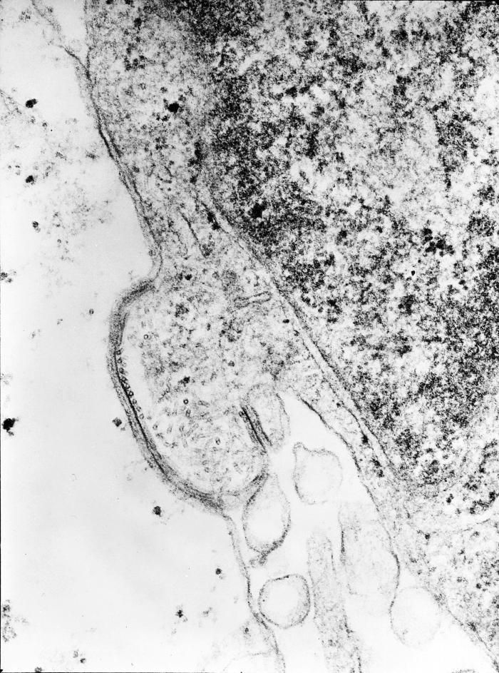
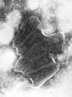
This transmission electron micrograph (TEM) revealed the presence of Lagos bat virus (LBV) virions, and an intracytoplasmic inclusion body in this tissue sample. LBV is a Rhabdoviridae family member, and a member of the genus, Lyssavirus.Created: 1975
-
This negatively-stained transmission electron micrograph (TEM) revealed the presence of numerous negative-sense, single-stranded RNA ((-) ssRNA) Flanders virus virions. Note the bullet-like shape of these virions, which are very similar to other Rhabdoviruses, i.e., see PHIL 1876 depicting a TEM revealing the bullet-shaped rabies virus virions.Created: 1975
-
This negatively-stained transmission electron micrograph (TEM) revealed the presence of numerous bovine ephemeral fever virus virions, which are members of the Rhabdoviridae family of viruses, and the genus Ephemerovirus, infecting animals as well as plants.Created: 1975
-
This negatively-stained transmission electron micrograph (TEM) revealed the presence of two Piry virus virions. Note the bullet-like shape of the small 155nm x 162nm virions. Normally, under electron microscopic examination, the virions are observed as being discoidal or spheroidal in shape, and only rarely as bullet-shaped, as was the case here.Created: 1975
-
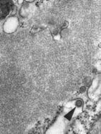
This negatively-stained transmission electron micrograph (TEM) revealed the presence of numerous Piry virus virions, many of which could be seen as they were budding from the host cell, thereby, becoming free to migrate throughout the hosts system. Note the bullet-like shape of the small 155nm x 162nm virions, as theyre freed from the host cell. Normally, under electron microscopic examination, the virions are observed as being discoidal or spheroidal in shape, and only rarely as bullet-shaped, as was the case here.Created: 1975
-
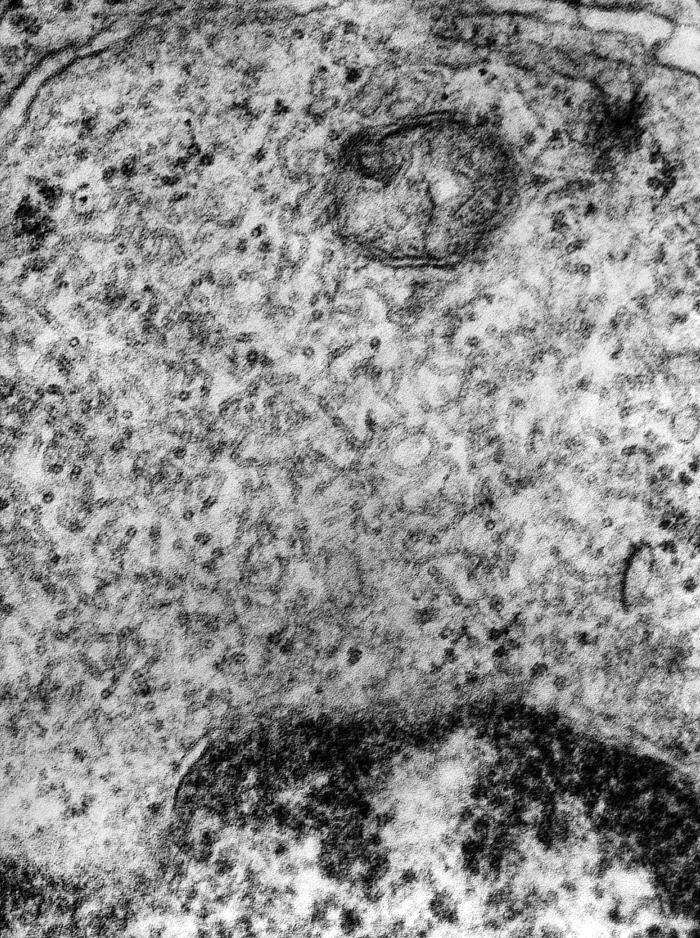
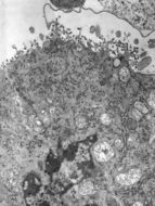
This transmission electron micrograph (TEM) revealed some of the internal cross-sectional structural morphology of a rabies virion (arrow) in this central nervous system tissue specimen. The virion is adjacent to a Negri body, which is pathognomonic in the positive diagnosis for Rabies. The virus infects the central nervous system, causing encephalopathy and ultimately death. Rabies virus belongs to the order Mononegavirales, viruses with a nonsegmented, negative-sense single-stranded RNA ((-) ssRNA) genomes. Within this group, viruses with a distinct "bullet" shape are classified in the Rhabdoviridae family, which includes at least three genera of animal viruses, Lyssavirus, Ephemerovirus, and Vesiculovirus. The genus Lyssavirus includes rabies virus, Lagos bat, Mokola virus, Duvenhage virus, European bat virus 1 & 2 and Australian bat virus.Created: 1975
-
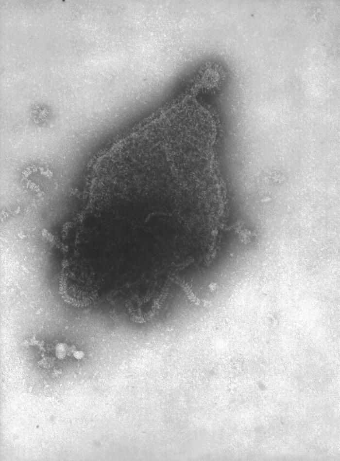
This transmission electron micrograph (TEM) revealed some of the nucleocapsid morphologic features displayed by the human parainfluenza virus Type-4a (HPIV-4), a member of the Paramyxoviridae family. These viruses possess a genome consisting of negative-sense single-stranded RNA ((-) ssRNA).Each of the four HPIVs has different clinical and epidemiologic features. The most distinctive clinical feature of HPIV-1 and HPIV-2 is croup (i.e., laryngotracheobronchitis); HPIV-1 is the leading cause of croup in children, whereas HPIV-2 is less frequently detected. Both HPIV-1 and -2 can cause other upper and lower respiratory tract illnesses. HPIV-3 is more often associated with bronchiolitis and pneumonia. HPIV-4 is infrequently detected, possibly because it is less likely to cause severe disease. The incubation period for HPIVs is generally from 1 to 7 days.Created: 1975
-
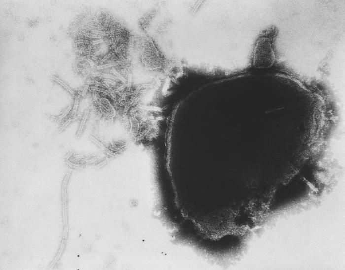
This transmission electron micrograph (TEM) revealed the presence of the human parainfluenza type 4A virus (HPIV-4A), which like the mumps virus, is also a Paramyxoviridae family member, and a member of the genus, Rubulavirus.Created: 1975
-
This transmission electron micrograph (TEM) revealed the presence of the human parainfluenza type 4A virus (HPIV-4A), which like the mumps virus, is also a Paramyxoviridae family member, and a member of the genus, Rubulavirus.Created: 1975
-
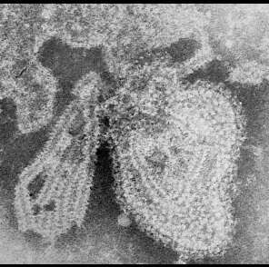
This transmission electron micrograph (TEM) revealed the presence of numerous paramyxovirus virions, which in this instance, were responsible for a case of the mumps. Paramyxoviruses are members of the family, Paramyxoviridae, and those that cause mumps in humans belong to the genus, Rubulavirus. The virus itself can present itself in a number of morphologic shapes, including spherical, and stand-like, or filamentous, ranging from 150nm to 200nm in diameter, and 1000nm to 10000nm in length. At its core lies a non-segmented, negative-sense RNA genome.Created: 1975
-
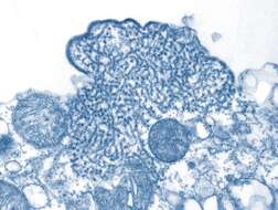
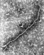
Under a highly magnified view of 168,000x this transmission electron micrographic (TEM) image revealed ultrastructural details of a Nipah virus nucleocapsid, a virus which was named for the location in Malaysia where it was first isolated.Created: 1999
-

Description: English: This electron micrograph depicts the Respiratory Syncytial Virus (RSV) pathogen. RSV is a negative-sense, enveloped RNA virus. The virion is variable in shape and size, with a diameter ranging between 120 and 300 nm, and is unstable in the environment surviving only a few hours on environmental surfaces. Polski: RSV w elektronowym mikroskopie transmisyjnym. Norsk bokmål: RS-virus. Date: 1981. Source: : This media comes from the
Centers for Disease Control and Prevention's
Public Health Image Library (PHIL), with identification number
#276. Note: Not all PHIL images are public domain; be sure to check copyright status and credit authors and content providers.
العربية |
Deutsch |
English |
македонски |
slovenščina |
+/−. Originally from
en.wikipedia; description page is/was
here. Author: Photo Credit: Content Providers(s): CDC/ Dr. Erskine Palmer. Permission(
Reusing this file): PD-USGov-HHS-CDC English: None - This image is in the public domain and thus free of any copyright restrictions. As a matter of courtesy we request that the content provider be credited and notified in any public or private usage of this image.
-
Description: This transmission electron micrograph (TEM) depicted a number of Nipah virus virions that had been isolated from a patient's cerebrospinal fluid (CSF) specimen. Nipah virus is a member of the family Paramyxoviridae, and is related, but not identical, to Hendra virus. Nipah virus was initially isolated in 1999 upon examining samples from an outbreak of encephalitis and respiratory illness among adult men in Malaysia and Singapore. Hendra virus, formerly called equine morbillivirus, is also a member of the family Paramyxoviridae. The virus was first isolated in 1994 from specimens obtained during an outbreak of respiratory and neurologic disease in horses and humans in Hendra, a suburb of Brisbane, Australia. Date: 22 May 2008. Source:
[1] (CDC). Author: CDC/ C. S. Goldsmith, P. E. Rollin. Permission(
Reusing this file): public domain.
-
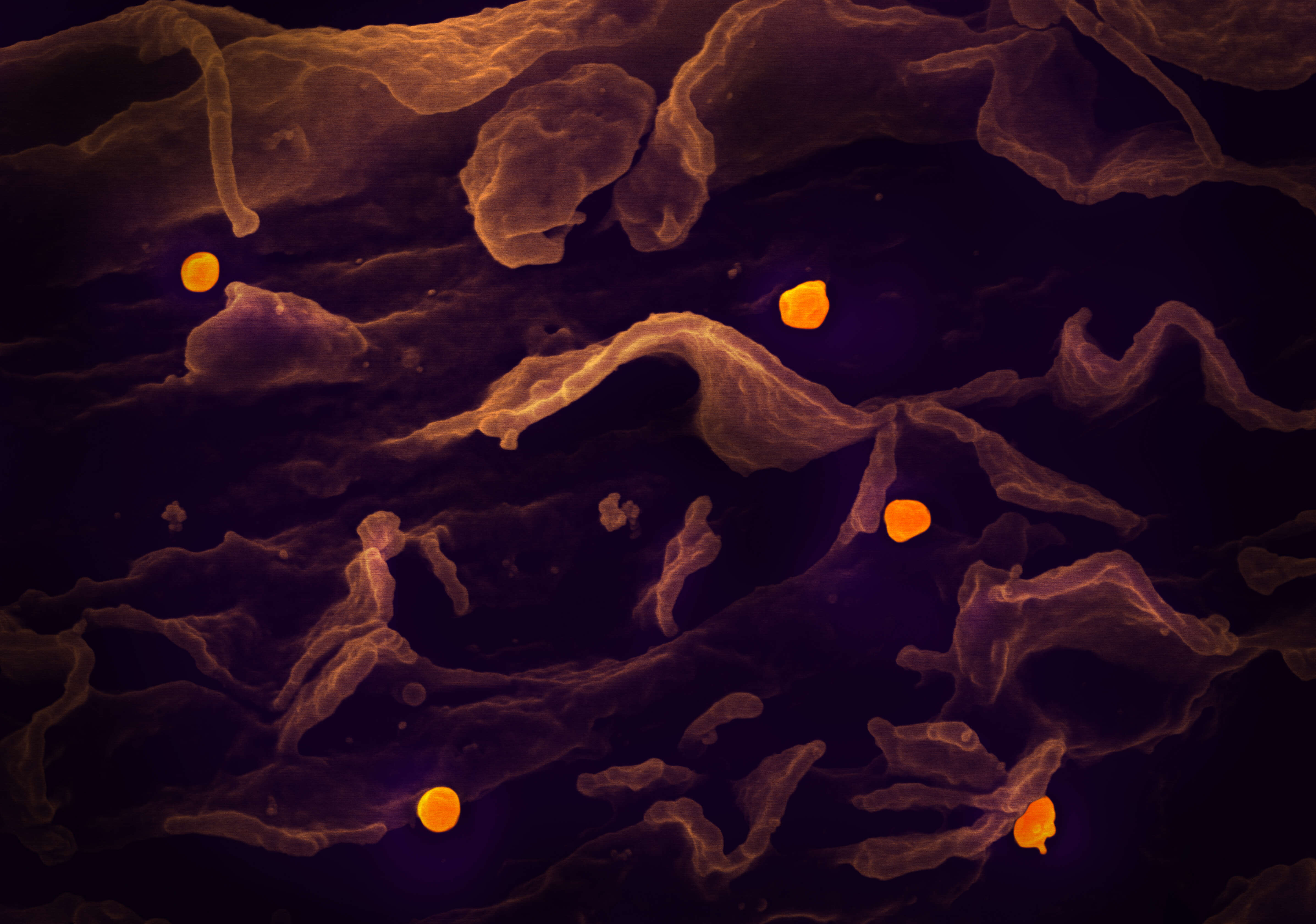
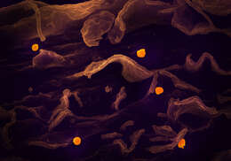
Description: In this image: A scanning electron micrograph shows the Nipah virus (yellow) budding from the surface of a cell. The experimental antiviral drug remdesivir completely protected four African green monkeys from a lethal dose of Nipah virus, according to a new study in Science Translational Medicine from National Institutes of Health scientists and colleagues. First identified in 1999 in Malaysia, Nipah virus is an emerging pathogen found primarily in Bangladesh and India. The virus is spread to humans by fruit bats; person-to-person transmission also occurs. Nipah virus can cause neurological and respiratory disease; the mortality rate is about 70%. Delayed relapse, manifesting as brain inflammation or encephalitis, can occur. An outbreak in May 2018 in India resulted in 23 cases and 21 deaths. Read more:
www.nih.gov/news-events/news-releases/experimental-drug-c.. Credit: National Institute of Allergy and Infectious Diseases, NIH. Date: 8 May 2019, 11:52. Source:
Nipah Virus. Author:
NIH Image Gallery from Bethesda, Maryland, USA.
-
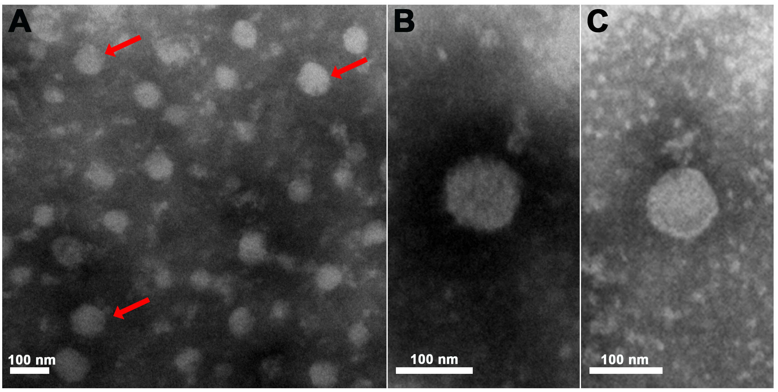
Description: English: Electron micrographs of purified Pteromalus puparum negative-strand RNA virus 1 (PpNSRV-1) particles from follicular cells. (A) Electron micrographs of purified PpNSRV-1 virions. (B) and (C) are the magnification pictures. Red arrows indicated viral particles. Date: 9 March 2017. Source: Wang F, Fang Q, Wang B, Yan Z, Hong J, Bao Y, et al. (2017) A novel negative-stranded RNA virus mediates sex ratio in its parasitoid host. PLoS Pathog 13(3): e1006201.
https://doi.org/10.1371/journal.ppat.1006201. Author: Fei Wang, Qi Fang, Beibei Wang, Zhichao Yan, Jian Hong, Yiming Bao, Jens H. Kuhn, John H. Werren, Qisheng Song, Gongyin Ye.
-
Description: English: This 1976 negative stained transmission electron micrograph (TEM) depicted the ultrastructural features displayed by the mumps virus. Deutsch: Aus dem Virion freigesetzte Anteile des helikalen Kapsids. Polski: Mikrofotografia wirusa świnki. Español: Imagen de microscopio electrónico de transmisión (MET) del virus de las paperas. Türkçe: Kabakulak virüsünün TEM mikrografı. Date: 1974. Source: : This media comes from the
Centers for Disease Control and Prevention's
Public Health Image Library (PHIL), with identification number
#1874. Note: Not all PHIL images are public domain; be sure to check copyright status and credit authors and content providers.
العربية |
Deutsch |
English |
македонски |
slovenščina |
+/−. Author: Photo Credit: Content Providers: CDC/ Dr. F. A. Murphy. Permission(
Reusing this file): PD-USGov-HHS-CDC English: None - This image is in the public domain and thus free of any copyright restrictions. As a matter of courtesy we request that the content provider be credited and notified in any public or private usage of this image.
-
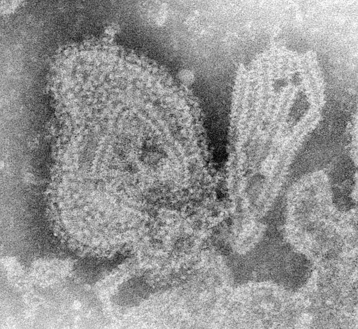
Summary.mw-parser-output table.commons-file-information-table,.mw-parser-output.fileinfotpl-type-information{border:1px solid #a2a9b1;background-color:#f8f9fa;padding:5px;font-size:95%;border-spacing:2px;box-sizing:border-box;margin:0;width:100%}.mw-parser-output table.commons-file-information-table>tbody>tr,.mw-parser-output.fileinfotpl-type-information>tbody>tr{vertical-align:top}.mw-parser-output table.commons-file-information-table>tbody>tr>td,.mw-parser-output table.commons-file-information-table>tbody>tr>th,.mw-parser-output.fileinfotpl-type-information>tbody>tr>td,.mw-parser-output.fileinfotpl-type-information>tbody>tr>th{padding:4px}.mw-parser-output.fileinfo-paramfield{background:#ccf;text-align:right;padding-right:0.4em;width:15%;font-weight:bold}.mw-parser-output.commons-file-information-table+table.commons-file-information-table,.mw-parser-output.commons-file-information-table+div.commons-file-information-table>table{border-top:0;padding-top:0;margin-top:-8px}@media only screen and (max-width:719px){.mw-parser-output table.commons-file-information-table,.mw-parser-output.commons-file-information-table.fileinfotpl-type-information{border-spacing:0;padding:0;word-break:break-word;width:100%!important}.mw-parser-output.commons-file-information-table>tbody,.mw-parser-output.fileinfotpl-type-information>tbody{display:block}.mw-parser-output.commons-file-information-table>tbody>tr>td,.mw-parser-output.commons-file-information-table>tbody>tr>th,.mw-parser-output.fileinfotpl-type-information>tbody>tr>td,.mw-parser-output.fileinfotpl-type-information>tbody>tr>th{padding:0.2em 0.4em;text-align:left;text-align:start}.mw-parser-output.commons-file-information-table>tbody>tr,.mw-parser-output.fileinfotpl-type-information>tbody>tr{display:flex;flex-direction:column}.mw-parser-output.commons-file-information-table+table.commons-file-information-table,.mw-parser-output.commons-file-information-table+div.commons-file-information-table>table{margin-top:-1px}.mw-parser-output.fileinfo-paramfield{box-sizing:border-box;flex:1 0 100%;width:100%}} Description: Italiano: Ingrandimento del virus della parotite endemica. Date: 1976. Source:
Public Health Image Library (PHIL) (
home page). Author: CDC/ Dr. F. A. Murphy.
-

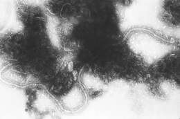
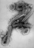
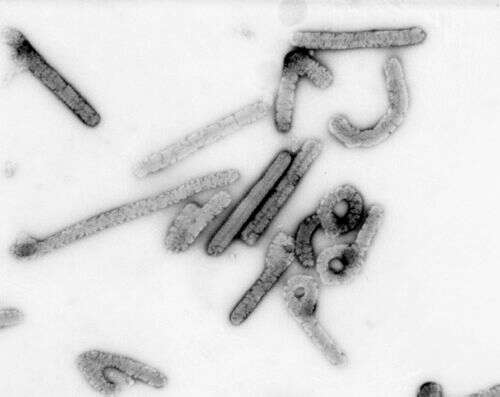
Description: English: This negative stained transmission electron micrograph (TEM) depicts a number of filamentous Marburg virions, which had been cultured on Vero cell cultures, and purified on sucrose, rate-zonal gradients. Note the virus’s morphologic appearance with its characteristic “Shepherd’s Crook” shape; Magnified approximately 100,000x. Marburg hemorrhagic fever is a rare, severe type of hemorrhagic fever which affects both humans and non-human primates. Caused by a genetically unique zoonotic (that is, animal-borne) RNA virus of the filovirus family, its recognition led to the creation of this virus family. The four species of Ebola virus are the only other known members of the filovirus family. Marburg virus was first recognized in 1967, when outbreaks of hemorrhagic fever occurred simultaneously in laboratories in Marburg and Frankfurt, Germany and in Belgrade, Yugoslavia (now Serbia). Deutsch: Marburg-Virus. Français : Vue de particules virales de Marburg au microscope électronique ; on voit la structure typique des filovirus, ainsi que les filaments caractéristiques en forme de crochets. Agrandissement 100 000x. Polski: Fotografia z elektronowego mikroskopu transmisyjnego ukazująca wiriony wirusa Marburg. Wirus Marburg. Svenska: Marburgvirus. Date: 1981. Source: : This media comes from the
Centers for Disease Control and Prevention's
Public Health Image Library (PHIL), with identification number
#275. Note: Not all PHIL images are public domain; be sure to check copyright status and credit authors and content providers.
العربية |
Deutsch |
English |
македонски |
slovenščina |
+/−. Author: Photo Credit: Content Providers(s): CDC/ Dr. Erskine Palmer, Russell Regnery, Ph.D. Permission(
Reusing this file): PD-USGov-HHS-CDC English: None - This image is in the public domain and thus free of any copyright restrictions. As a matter of courtesy we request that the content provider be credited and notified in any public or private usage of this image. Other versions:
.
-
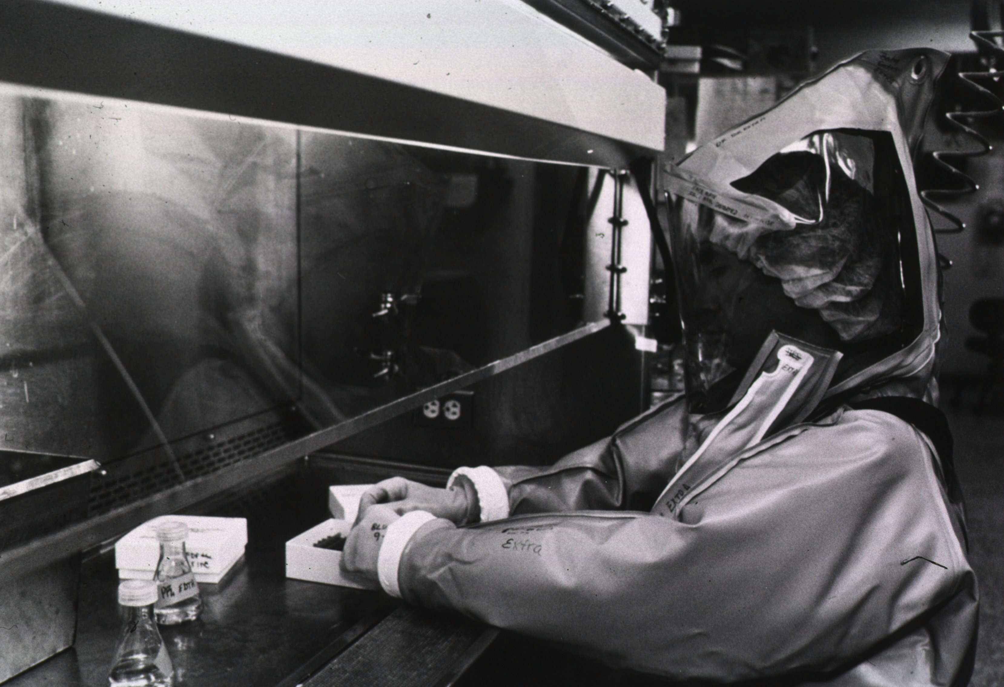
Description: English: "Researchers, such as this man in the Centers for Disease Control and Prevention's new maximum containment virology laboratory, use the most advanced technology available to study dangerous organisms like the Lassa, Machupo, Ebola, and AIDS viruses that cause deadly diseases for which no cure or vaccine exists". Date: 1987. Source:
National Library of Medicine. Author: Unknown photographer, Centers for Disease Control.
-
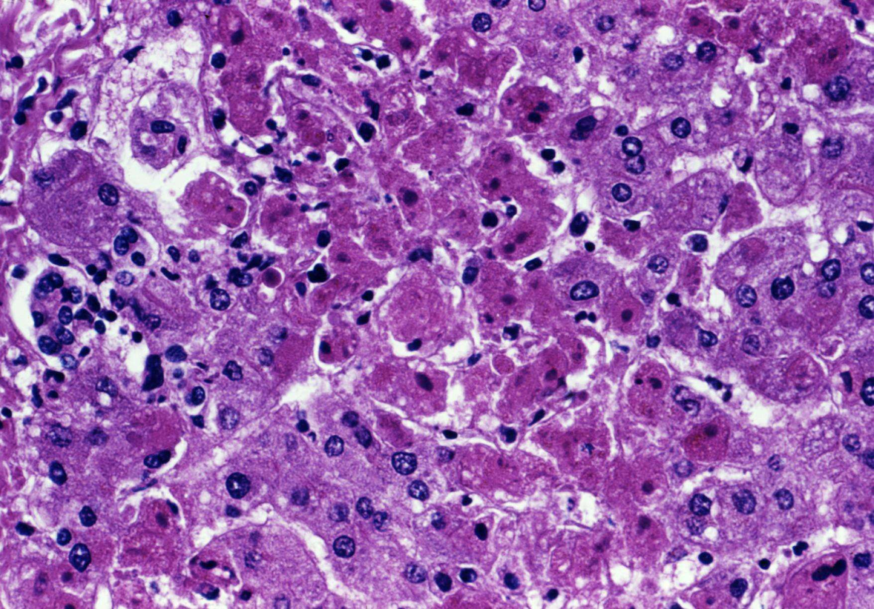
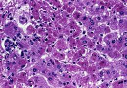
Description: English: This photomicrograph revealed the cytoarchitectural histopathologic changes detected in a liver sample from a Marburg patient (case #1) who was treated in Johannesburg, South Africa in 1975. As the disease progresses, symptoms of Marburg disease can become increasingly severe and may include liver failure, jaundice, inflammation of the pancreas, severe weight loss, delirium, shock, massive hemorrhaging, and multi-organ dysfunction. Date: 1975. Source: : This media comes from the
Centers for Disease Control and Prevention's
Public Health Image Library (PHIL), with identification number
#6559. Note: Not all PHIL images are public domain; be sure to check copyright status and credit authors and content providers.
العربية |
Deutsch |
English |
македонски |
slovenščina |
+/−. Originally uploaded to
en.wikipedia; description page is/was
here. Author: Photo Credit: Content Providers(s): CDC/ Dr. J. Lyle Conrad. Permission(
Reusing this file): PD-USGov-HHS-CDC English: None - This image is in the public domain and thus free of any copyright restrictions. As a matter of courtesy we request that the content provider be credited and notified in any public or private usage of this image.
-
-
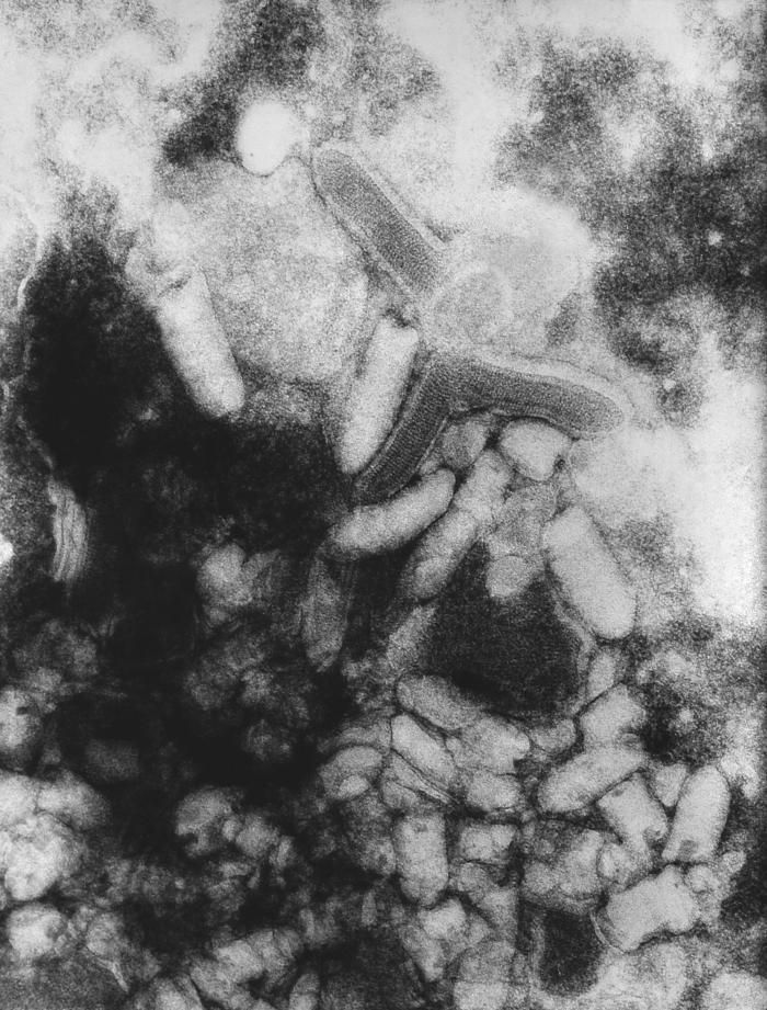
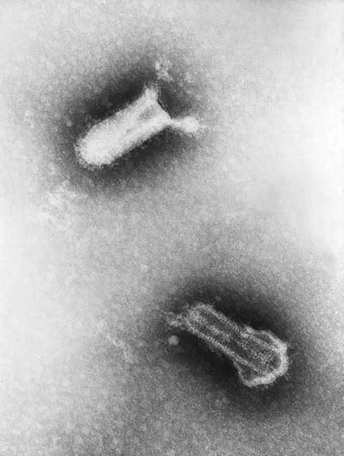
Description: English: Electron micrograph of the Marburg virus. Marburg virus, first recognized in 1967, causes a severe type of hemorrhagic fever, which affects humans, as well as non-human primates. Date: 1975. Source: : This media comes from the
Centers for Disease Control and Prevention's
Public Health Image Library (PHIL), with identification number
#1873. Note: Not all PHIL images are public domain; be sure to check copyright status and credit authors and content providers.
العربية |
Deutsch |
English |
македонски |
slovenščina |
+/−. Author: Photo Credit: Content Providers(s): CDC/Dr. Fred Murphy; J. Nakano. Permission(
Reusing this file): PD-USGov-HHS-CDC English: None - This image is in the public domain and thus free of any copyright restrictions. As a matter of courtesy we request that the content provider be credited and notified in any public or private usage of this image. Other versions:
.
-


![[1]](http://phil.cdc.gov/PHIL_Images/8948/8948_lores.jpg)