NMNH Prorocentrum arenarium type specimen
الوصف:
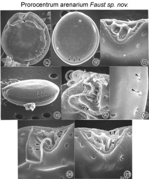
Figs. 14-19. Prorocentrum arenarium sp. nov. FIG.14. Cells in right valve view are round to oval. Cell distorted due to preparation. The periflagellar area is a broad, V-shaped depression. The longitudinal short flagellum is visible (arrowhead). Marginal poroids (arrows). FIG.15. The valve surface in left valve view is smooth and scattered with valve and marginal poroids (arrows). FIG. 6. The oblique ventral view is ellipsoid. The intercalary band is smooth. The marginal poroids are evenly spaced (arrows). FIG.17. The periflagellar area is triangular and unornamented. It has a large flagellar pore (F) and one smaller auxiliary pore (A). The apical platelets appear vertical when viewed from the anterior end of the cell. FIG. 18. Both flagella emerge from the flagellar pore (F): longitudinal flagellum (arrowheads), transverse flagellum (arrows). FIG. 9. At higher magnification, valve and marginal poroids are kidney-shaped to oblong (arrowheads) and are similar in size.FIGS. 20-21. Prorocentrum arenarium sp. nov. with peduncle-like structures. FIG. 20. The periflagellar area exhibits a short flagellum 11µm long (arrows), and a short, curved tubular structure 2-3 µm long (arrowheads), both emerging from the flagellar pore (F). The width of the curved tubular structure and flagellum are the same. FIG. 21. Both tubular structures emerge from the flagellar pore (F); one is straight, 2 µm long (arrow), and the other curved, 3µm long (arrowheads). The auxiliary pore (A) is to the left of the flagellar pore. EMu: HOLOTYPE SEM NEGATIVE #133024; SEM stub # 133; Field # 556-92; Accession # 407166; Catalog # 97; Figure # 14.
مشمول على الصفحات التالية:
هذه الصورة ليست واردة في أي مجموعات.
معلومات المصدر
- ترخيص
- cc-by-nc-sa-3.0
- حقوق النشر
- National Museum of Natural History, Smithsonian Institution
- النص الأصلي
- ملف الوسائط الأصلي
- زيارة المصدر
- موقع الشريك
- NMNH Marine Dinoflagellates
- ID