Plate 19
الوصف:
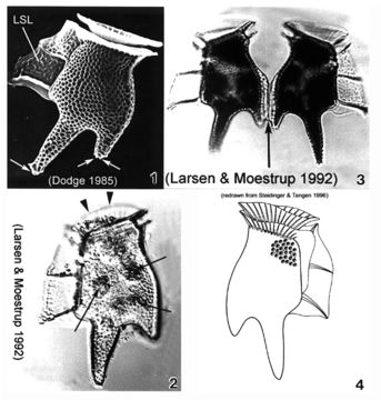
Plate 19. Dinophysis tripos. Fig. 1. SEM: lateral view. Cell large, oblong and heavily areolated. Hypothecal projections with toothed posterior ends (arrows). Left sulcal list (LSL) large, wide and reticulated. Figs. 2,3. LM: lateral view. Fig. 2. Anterior cingular list (ACL) projected anteriorly obscuring low epitheca (arrowheads). Narrow cingulum. Chloroplasts visible (arrows). Fig. 3. Paired cells. Hypothecal projection on dorsal margin sometimes seen with a narrow list (arrow) connecting two daughter cells during cell division. Fig. 4. Line drawing.
مشمول على الصفحات التالية:
- Life
- Cellular
- Eukaryota (حقيقيات النوى)
- SAR (Stramenopiles, Alveolates, Rhizaria)
- Alveolata (طلائعيات سناخية)
- Dinophyceae (سوطيات دوارة)
- Dinophysiales
- Dinophysiaceae
- Dinophysis
- Dinophysis tripos
- Dinoflagellata
هذه الصورة ليست واردة في أي مجموعات.
معلومات المصدر
- ترخيص
- cc-publicdomain
- الاقتباس الببليوغرافي
- Faust, Maria A. and Rose A. Gulledge. Identifying Harmful Marine Dinoflagellates. Smithsonian Contributions from the United States National Herbarium, volume 42: 1-144 (including 48 plates, 1 figure and 1 table).
- النص الأصلي
- ملف الوسائط الأصلي
- زيارة المصدر
- موقع الشريك
- NMNH Marine Dinoflagellates
- ID