-
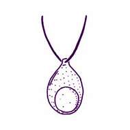
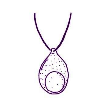
Telonema subtile Griessmann, 1913. Cell outline is oval-ovoid. Cells are about 8 microns long, anteriorly narrow and posteriorly broad with a short anterior neck. Two flagella insert below the neck, are acronematic and are slightly longer than the cell. The nucleus is centrally located. The cells swim backward with the flagella, which point behind the swimming cells. Food materials are shown in the posterior end of the cell.
-
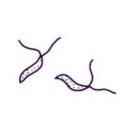
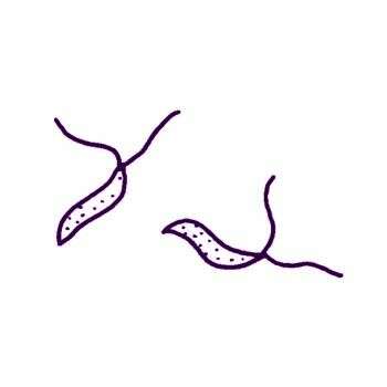
Amphimonas ankyromonadides Namyslowski, 1913. Cells are 5 microns long, 2 microns wide and slightly S-shaped, not metabolic. The cytoplasm is homogeneous, without vacuoles and granulation, the two flagella are of equal length and are about the cell length. This taxon is of dubious status because the description is inadequate.
-
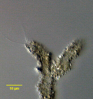
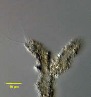
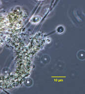
In vivo portrait of Cladomonas fruticulosa (Stein, 1878), a colonial flagellate of uncertain affinity. Colonies are formed by arborizing mucus tubes with adherent debris. The lumens of the tubes intercommunicate thus more than one cell can exit the colony through the same tube. The cells rest at the ends of the tubes with their two long (2 ½ cell length), equal flagella protruding. The tube branch on the viewer's right is vacant. Cells have a single posterior contractile vacuole. The rapidly beating flagella are held relatively straight. The cells are rounded and very deformable showing considerable metaboly. When disturbed, cells rapidly exit the tubes by turning 180 degrees to exit cell first. After leaving their tubes cells can swim fairly rapidly in the direction of their flagellae. Colonies often adhere to the surface film. Viewed by light microscopy, the cells of C. fruticulosa are indistinguishable from those of Spongomonas and Rhipidodendron. They clearly differ from the cells of another similar sized tube-dwelling flagellate, Siphomonas, the cells of which have one long and one short flagellum. Cyst formation has not been described for C. fruticulosa. Collected from a freshwater irrigation canal near Boise, Idaho November 2004. DIC.
-
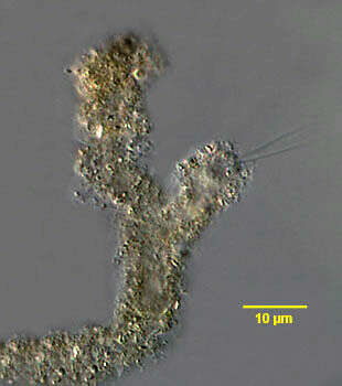
In vivo portrait of Cladomonas fruticulosa (Stein, 1878), a colonial flagellate of uncertain affinity. Colonies are formed by arborizing mucus tubes with adherent debris. The lumens of the tubes intercommunicate thus more than one cell can exit the colony through the same tube. The cells rest at the ends of the tubes with their two long (2 ½ cell length), equal flagella protruding. Cells have a single posterior contractile vacuole. The rapidly beating flagella are held relatively straight. The cells are rounded and very deformable showing considerable metaboly. When disturbed, cells rapidly exit the tubes by turning 180 degrees to exit cell first. After leaving their tubes cells can swim fairly rapidly in the direction of their flagellae. Colonies often adhere to the surface film. Viewed by light microscopy, the cells of C. fruticulosa are indistinguishable from those of Spongomonas and Rhipidodendron. They clearly differ from the cells of another similar sized tube-dwelling flagellate, Siphomonas, the cells of which have one long and one short flagellum. Cyst formation has not been described for C. fruticulosa. Collected from a freshwater irrigation canal near Boise, Idaho November 2004. DIC.
-
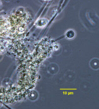
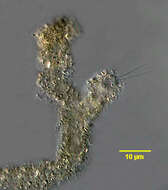
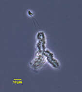
In vivo portrait of Cladomonas fruticulosa (Stein, 1878), a colonial flagellate of uncertain affinity in the process of exiting its mucus tube. Colonies are formed by arborizing mucus tubes with adherent debris. The lumens of the tubes intercommunicate thus more than one cell can exit the colony through the same tube. The cells rest at the ends of the tubes with their two long (2 ½ cell length), equal flagella protruding. Cells have a single posterior contractile vacuole. The rapidly beating flagella are held relatively straight. The cells are rounded and very deformable showing considerable metaboly. When disturbed, cells rapidly exit the tubes by turning 180 degrees to exit cell first. After leaving their tubes cells can swim fairly rapidly in the direction of their flagellae. Colonies often adhere to the surface film. Viewed by light microscopy, the cells of C. fruticulosa are indistinguishable from those of Spongomonas and Rhipidodendron. They clearly differ from the cells of another similar sized tube-dwelling flagellate, Siphomonas, the cells of which have one long and one short flagellum. Cyst formation has not been described for C. fruticulosa. Collected from a freshwater irrigation canal near Boise, Idaho November 2004. Phase contrast.
-
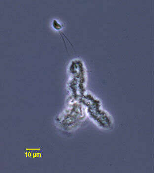
Portrait of Cladomonas fruticulosa (Stein, 1878), a colonial flagellate of uncertain affinity. This cell is free-swimming after fleeing its branched mucus tube (center). Here havin just backed out of its tube the cell will turn to swim with flagella forward. Colonies are formed by arborizing mucus tubes with adherent debris. The lumens of the tubes intercommunicate thus more than one cell can exit the colony through the same tube. The cells rest at the ends of the tubes with their two long (2 ½ cell length), equal flagella protruding. Cells have a single posterior contractile vacuole. The rapidly beating flagella are held relatively straight. The cells are rounded and very deformable showing considerable metaboly. When disturbed, cells rapidly exit the tubes by turning 180 degrees to exit cell first. After leaving their tubes cells can swim fairly rapidly in the direction of their flagellae. Colonies often adhere to the surface film. Viewed by light microscopy, the cells of C. fruticulosa are indistinguishable from those of Spongomonas and Rhipidodendron. They clearly differ from the cells of another similar sized tube-dwelling flagellate, Siphomonas, the cells of which have one long and one short flagellum. Cyst formation has not been described for C. fruticulosa. Collected from a freshwater irrigation canal near Boise, Idaho November 2004. Phase contrast.