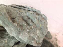
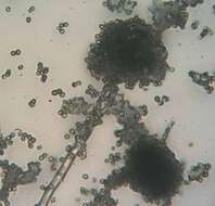
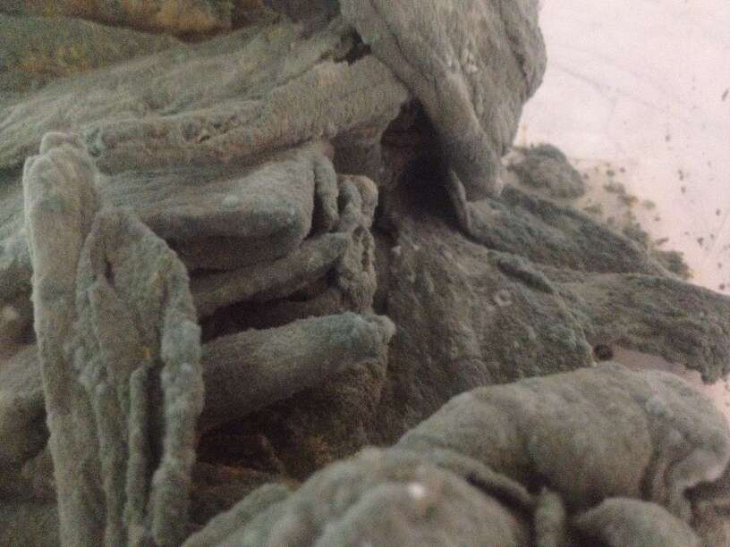
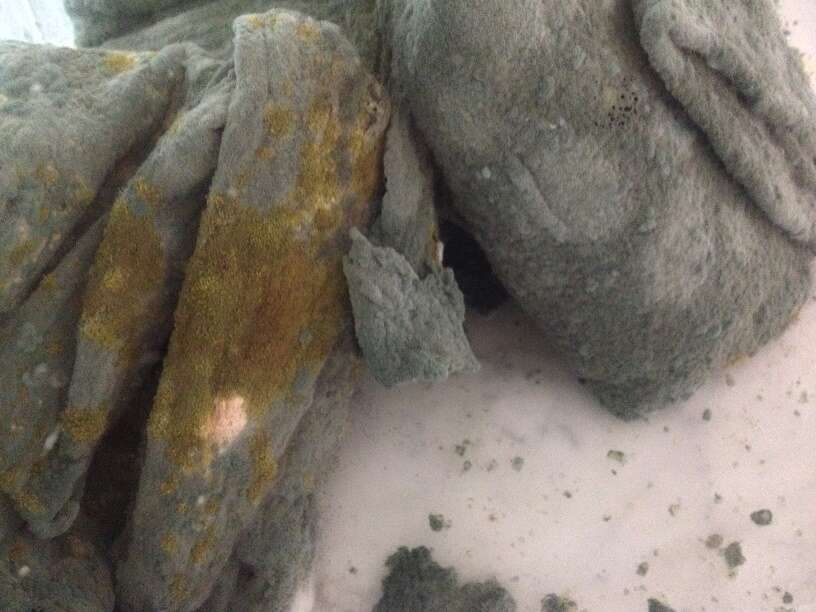
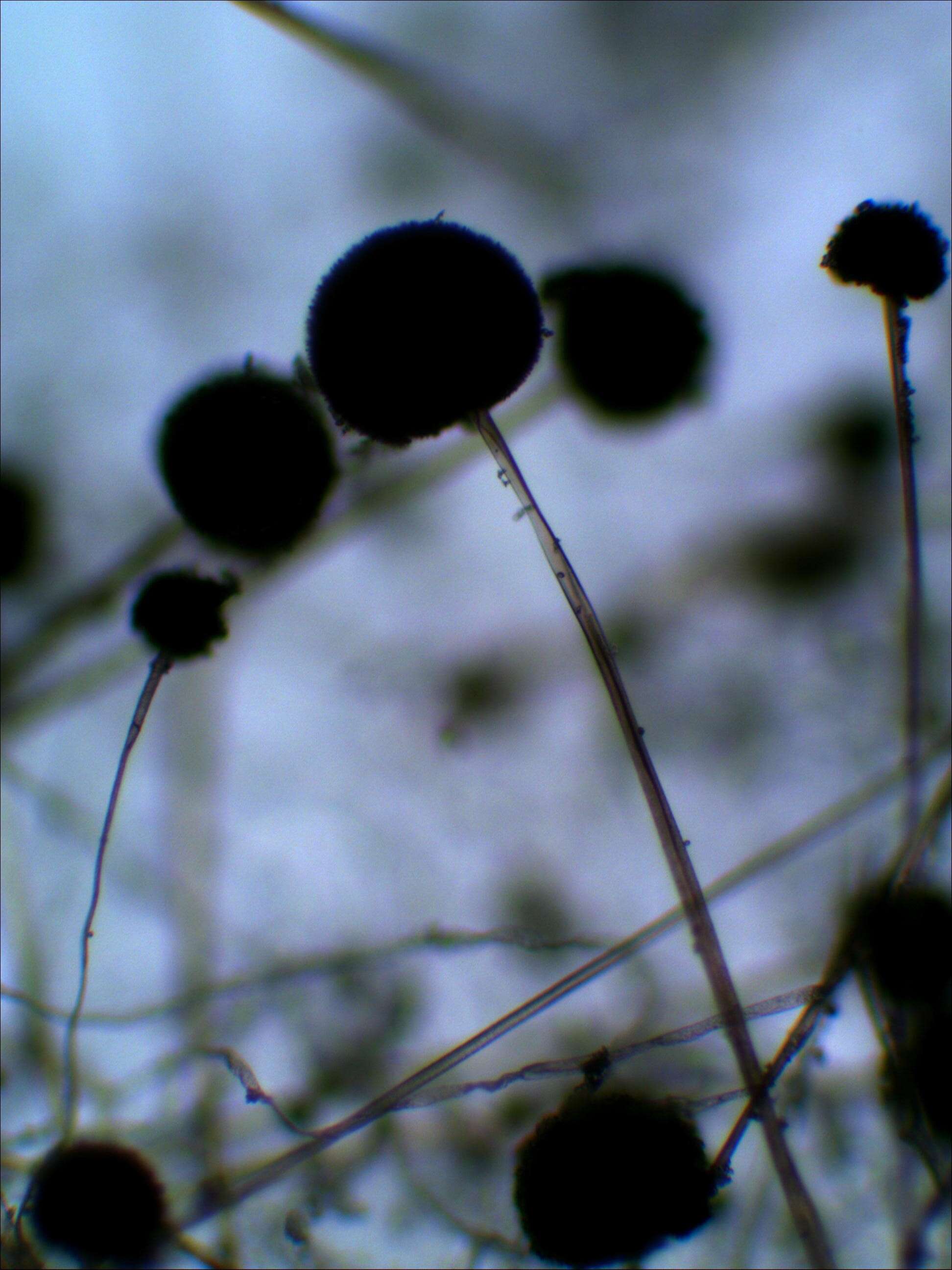
Description: فارسی: کپک نان که مدتی نزدیک به یک ماه در فضای باز رها شدهاست. Date: 5 April 2015, 15:59:15. Source: Own work. Author: Leyth. Camera location 35° 20′ 41.52″ N, 59° 13′ 44.94″ E: View all coordinates using: OpenStreetMap - Google Earth: 35.344867; 59.229150.
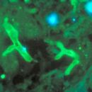
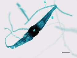
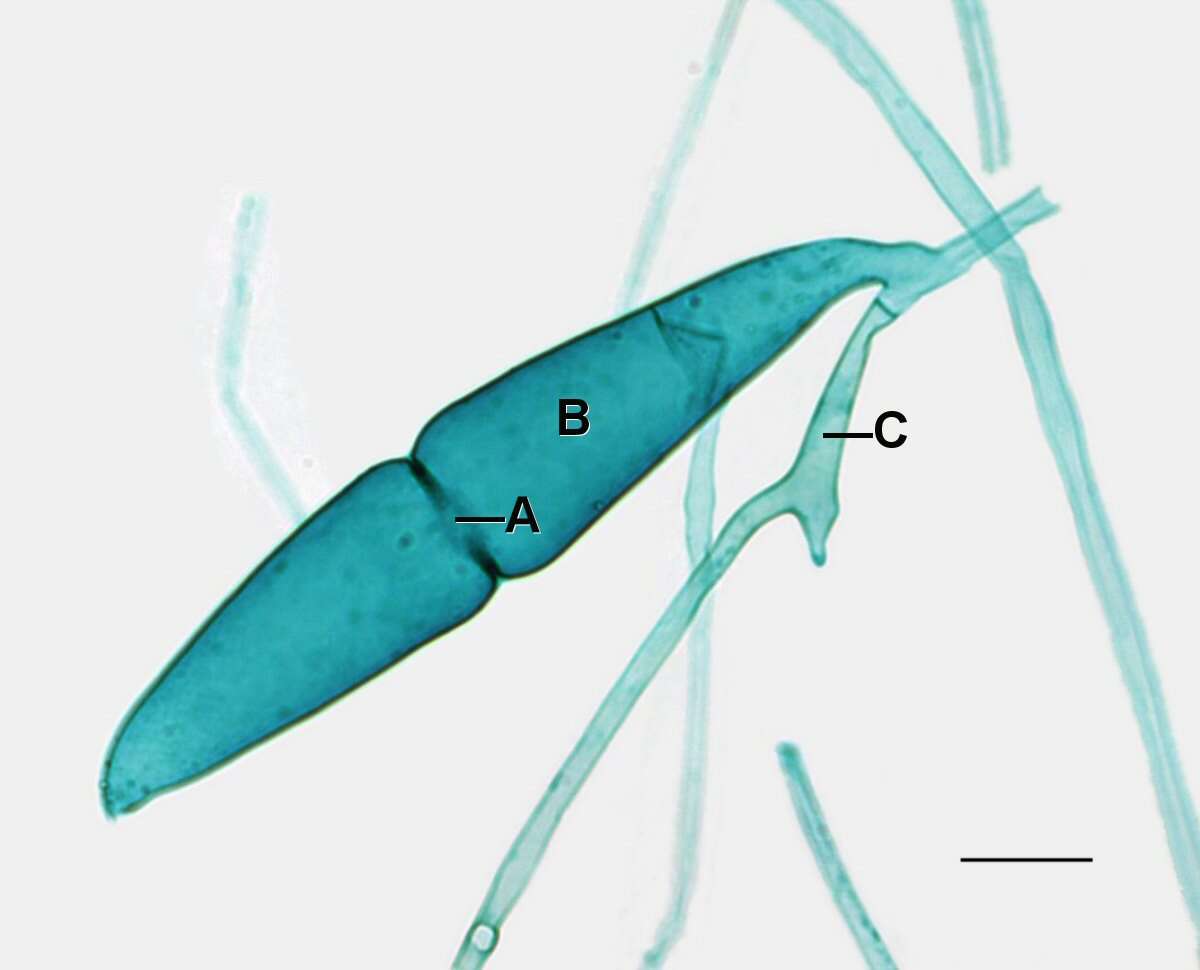
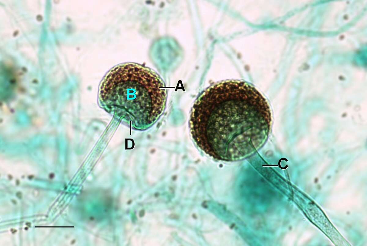
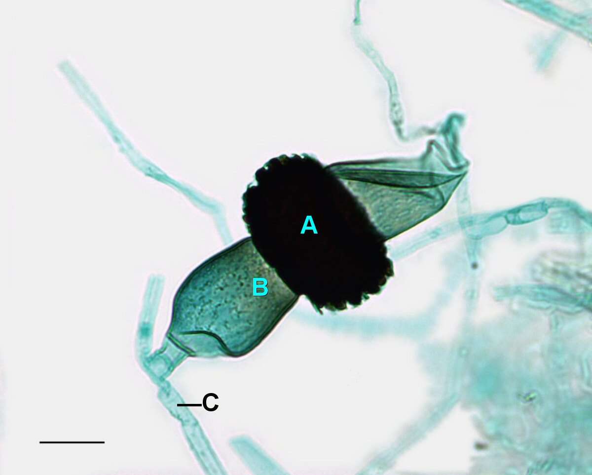
Description: English: Light microscopy of Rhizopus showing the connection of two young gametangia that had recently touched and began plasogamy. The young gametangia on the left has lost its hypha in the preparation process of the slide. A=Fusion septum, B=Suspensor cell, C=Hypha.Scale bar = 0.1mm. Date: 26 May 2014, 11:15:15. Source: Jon Houseman and Matthew Ford. Author: Jon Houseman. Other versions: Original (unlabeled). : This is a retouched picture, which means that it has been digitally altered from its original version. Modifications: Balance (Color, brightness, and contrast) and adjust background color.
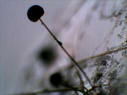
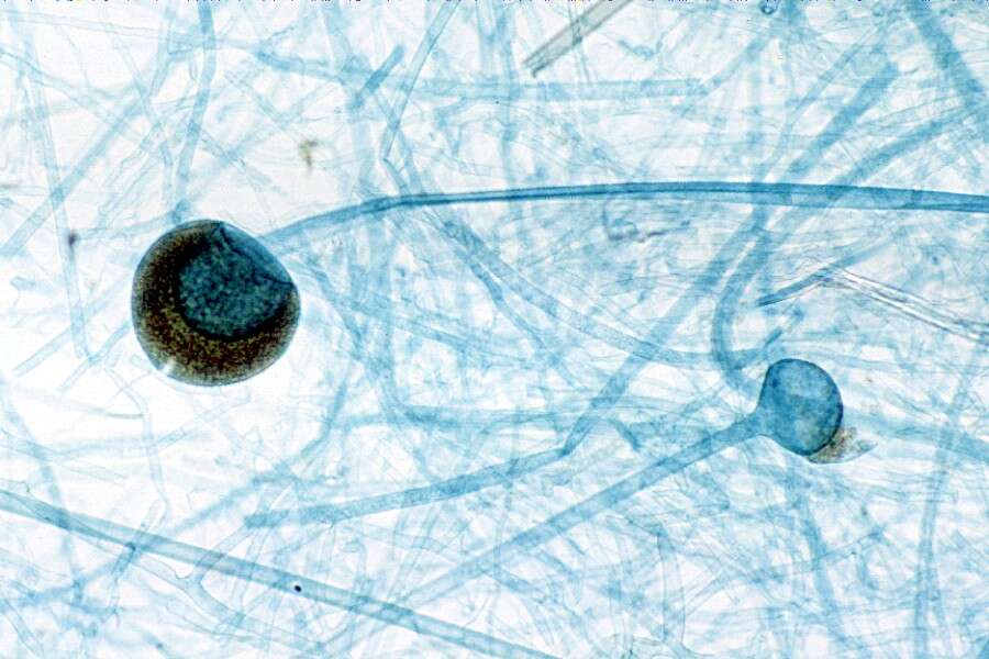
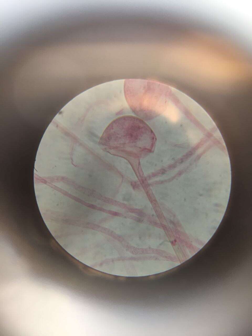
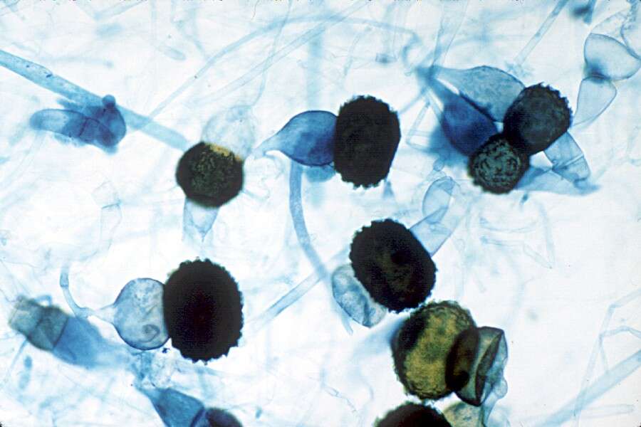
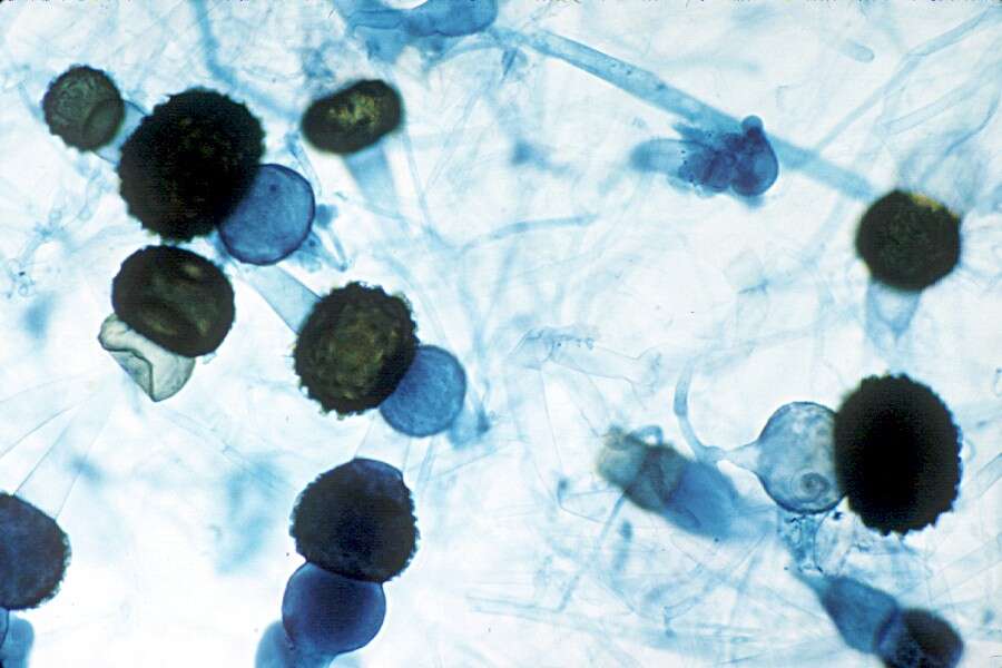
Description: English: Light microscopy of Rhizopus showing a mature zygosporangium with what is believed to be the sprouting sporangiophore that would release the meiospores. Scale bar = 0.1mm. Date: 21 May 2014, 09:52:35. Source: Jon Houseman and Matthew Ford. Author: Jon Houseman. Other versions: Labeled. : This is a retouched picture, which means that it has been digitally altered from its original version. Modifications: Balance (Color, brightness, and contrast) and adjust background color.
Description: English: Light microscopy of Rhizopus showing a mature zygosporangium with what is believed to be the sprouting sporangiophore that would release the meiospores. A=Mature zygosporangia, B=Sprouting sporangiophore, C=Suspensor cell. Scale bar = 0.1mm. Date: 26 May 2014, 11:15:16. Source: Jon Houseman and Matthew Ford. Author: Jon Houseman. Other versions: Original (unlabeled). : This is a retouched picture, which means that it has been digitally altered from its original version. Modifications: Balance (Color, brightness, and contrast) and adjust background color.
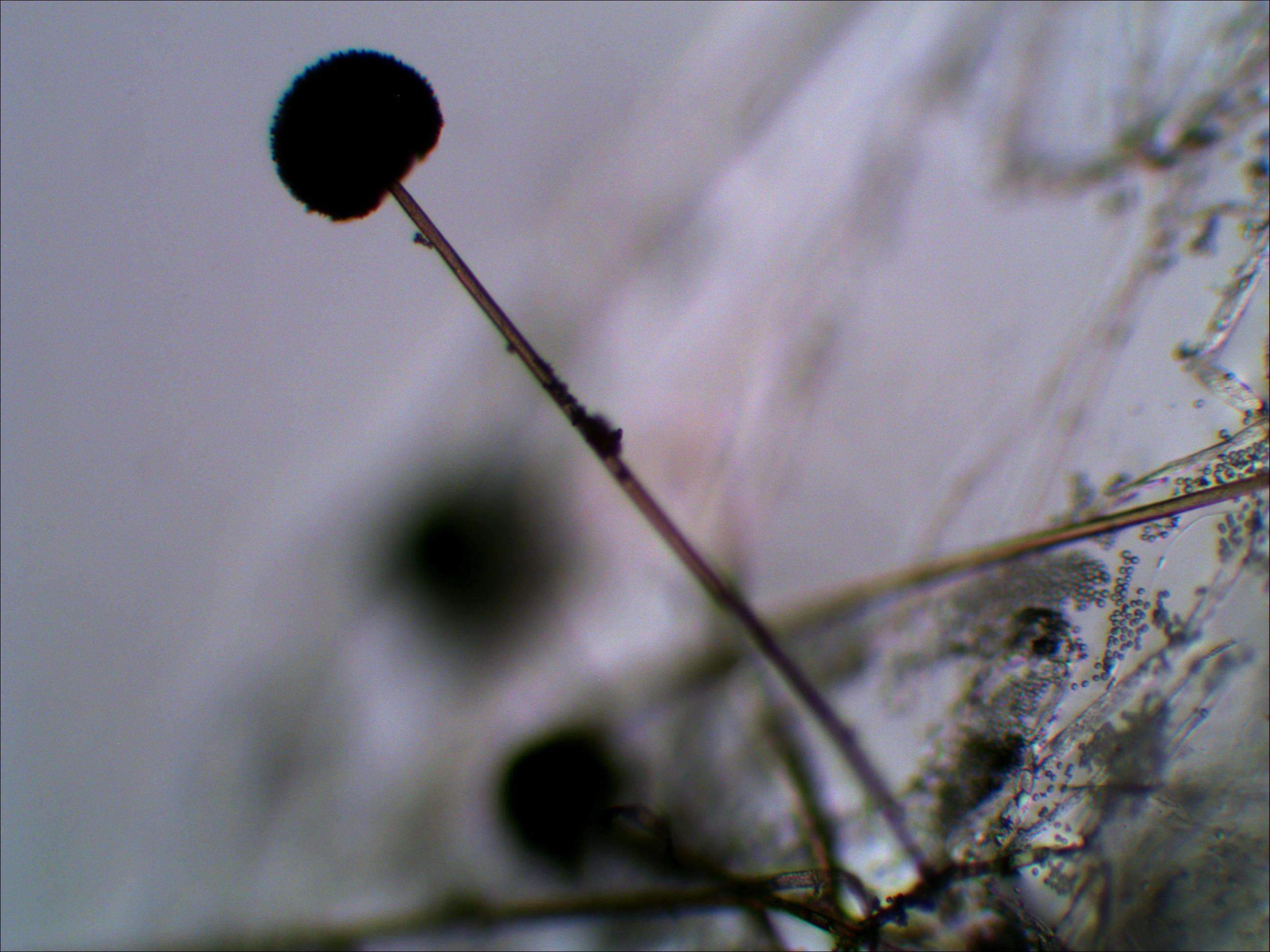
Description: فارسی: کپک نان که مدتی نزدیک به یک ماه در فضای باز رها شدهاست. Date: 5 April 2015, 15:59:11. Source: Own work. Author: Leyth. Camera location 35° 20′ 42.98″ N, 59° 13′ 44.35″ E: View all coordinates using: OpenStreetMap - Google Earth: 35.345272; 59.228986.
Description: فارسی: کپک نان که مدتی نزدیک به یک ماه در فضای باز رها شدهاست. Date: 5 April 2015, 15:59:24. Source: Own work. Author: Leyth. Camera location 35° 20′ 40.07″ N, 59° 13′ 45.53″ E: View all coordinates using: OpenStreetMap - Google Earth: 35.344464; 59.229314.
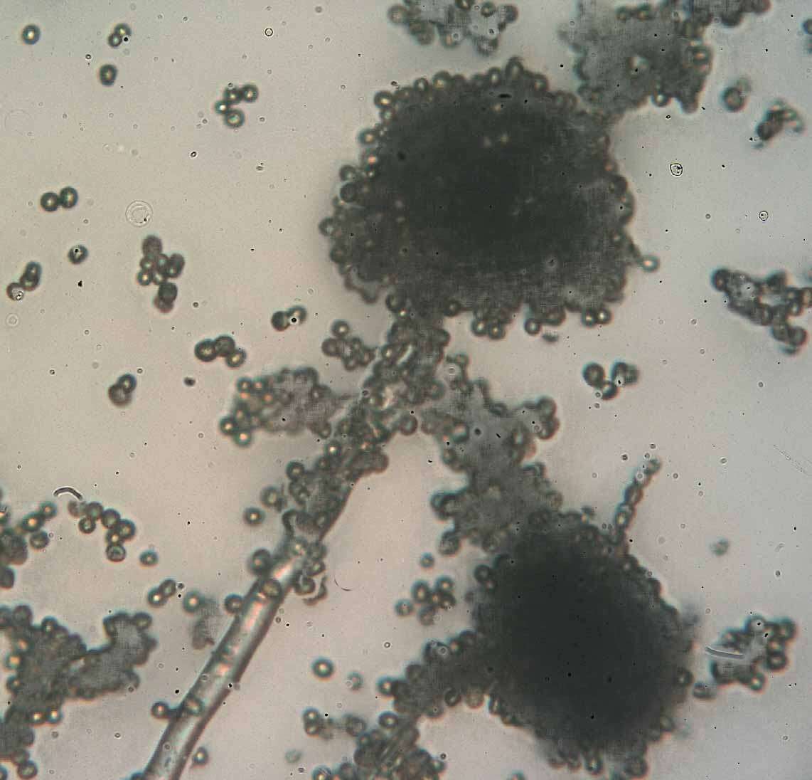
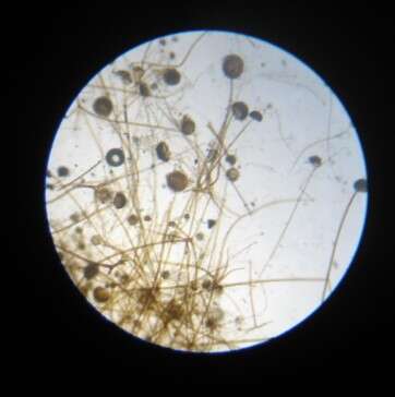
Description: English: A picture showing the true color of black mold (rhizopus sp) spores, using Light Microscope, Normal image without any coloring or processing By researcher El sayed Al gayar Faculty of Agriculture Damietta University. Date: 27 December 2017. Source: Own work. Author: Ramy algayar.
Description: English: A picture showing the true color of black mold (rhizopus sp) spores, using Light Microscope, Normal image without any coloring or processing By researcher El sayed Al gayar Faculty of Agriculture Damietta University. Date: 27 December 2017. Source: Own work. Author: Ramy algayar.
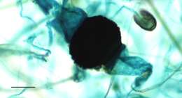
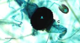
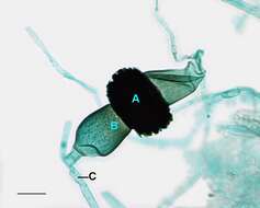
Description: English: Light microscopy of Rhizopus with an immature zygosporangium. A=Immature zygosporangium, B=Suspensor cell, C=Hypha. Scale bar = 0.1mm. Date: 26 May 2014, 11:15:17. Source: Jon Houseman and Matthew Ford. Author: Jon Houseman. Other versions: Original (unlabeled). : This is a retouched picture, which means that it has been digitally altered from its original version. Modifications: Balance (Color, brightness, and contrast) and adjust background color.
Description: English: Light microscopy of Rhizopus with an immature zygosporangium. Scale bar = 0.1mm. Date: 21 May 2014, 09:52:40. Source: Jon Houseman and Matthew Ford. Author: Jon Houseman. Other versions: Labeled. : This is a retouched picture, which means that it has been digitally altered from its original version. Modifications: Balance (Color, brightness, and contrast) and adjust background color.
Description: فارسی: کپک نان که مدتی نزدیک به یک ماه در فضای باز رها شدهاست. Date: 5 April 2015, 15:59:35. Source: Own work. Author: Leyth. Camera location 35° 20′ 40.07″ N, 59° 13′ 45.53″ E: View all coordinates using: OpenStreetMap - Google Earth: 35.344464; 59.229314.
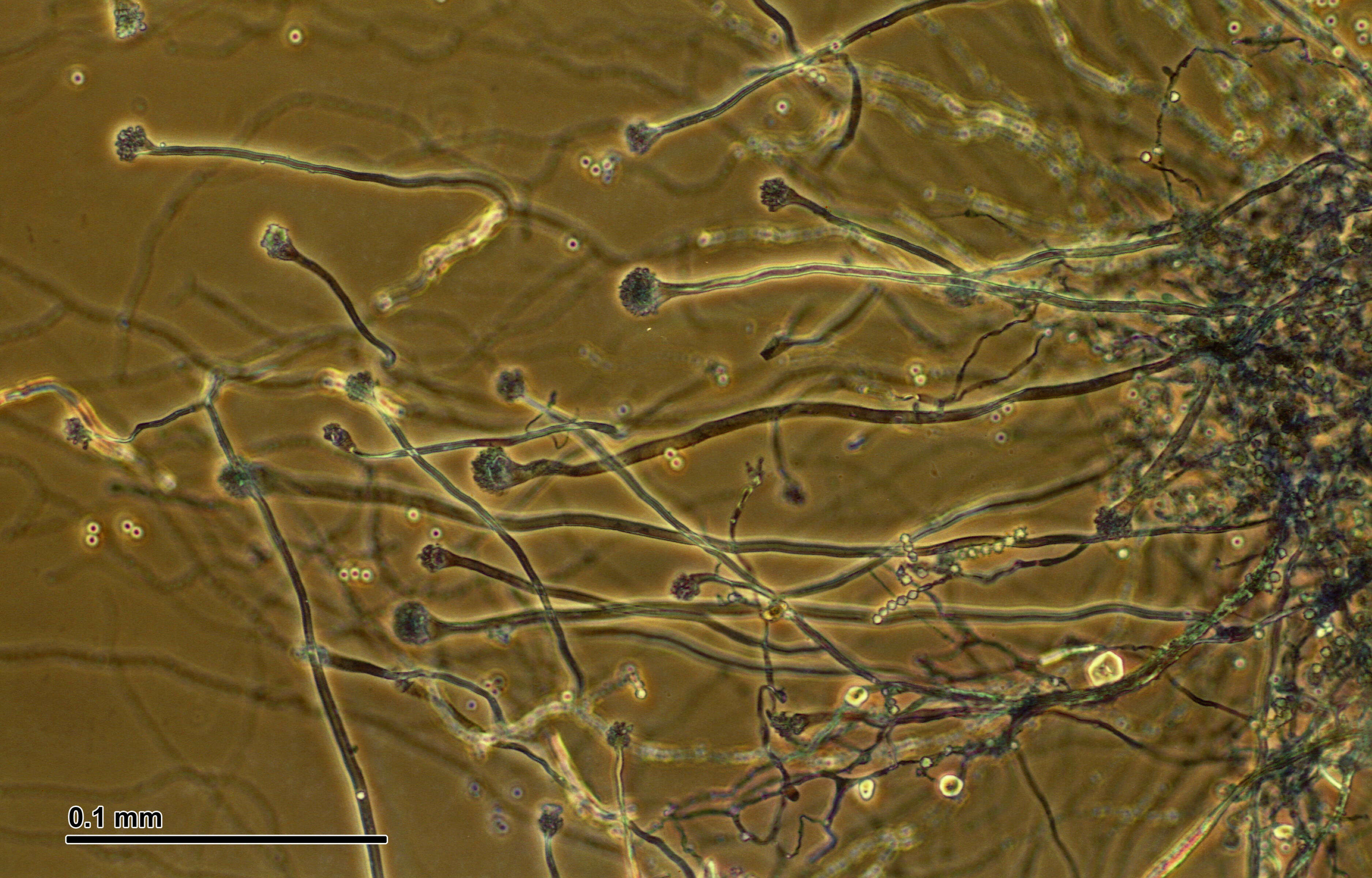
Description: English: Light microscopy of Rhizopus showing a close up view of two sporangium of Rhizopus where the differentiation between the spores and columella can be seen attached to the hypha. Scale bar = 0.1mm. Date: 13 May 2014, 11:16:53. Source: Jon houseman and Matthew Ford. Author: Jon Houseman. Other versions: Labeled. : This is a retouched picture, which means that it has been digitally altered from its original version. Modifications: Balance (Color, brightness, and contrast) and adjust background color.
Description: English: Light microscopy of Rhizopus showing a close up view of two sporangium of Rhizopus where the differentiation between the spores and columella can be seen attached to the hypha. A=Sporangium with spores, B=Columella, C=Hypha, D=Apophysis. Scale bar = 0.1mm. Date: 13 May 2014, 11:16:53. Source: Jon Houseman and Matthew Ford. Author: Jon Houseman. Other versions: Original (unlabeled). : This is a retouched picture, which means that it has been digitally altered from its original version. Modifications: Balance (Color, brightness, and contrast) and adjust background color.
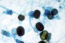
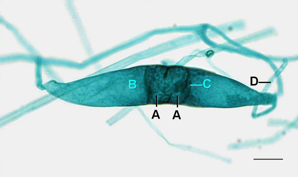
Description: English: Light microscopy of Rhizopus showing the early stages of plasmogamy when the immature zygosporangium begins to form. A=Gamatangia(n), B=Suspensor cell, C=Developing septal wall, D=Hypha. Scale bar = 0.1mm. Date: 26 May 2014, 11:15:25. Source: Jon Houseman and Matthew Ford. Author: Jon Houseman. Other versions: Original (unlabeled). : This is a retouched picture, which means that it has been digitally altered from its original version. Modifications: Balance (Color, brightness, and contrast) and adjust background color.
No machine-readable author provided. Timleo assumed (based on copyright claims).
Wikimedia Commons
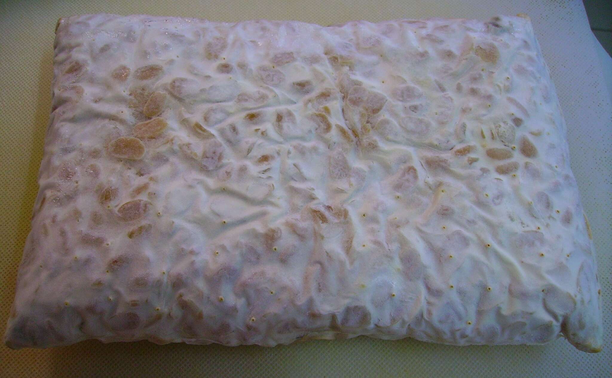
Description: A cake of tempeh made from dehulled soybeans and Rhizopus sp. 根黴屬真菌發酵脫皮大豆所得到的丹貝。. Date: 10 June 2006 (according to Exif data). Source: No machine-readable source provided. Own work assumed (based on copyright claims). Author: No machine-readable author provided. Timleo assumed (based on copyright claims).
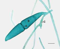
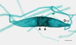
Description: English: Light microscopy of Rhizopus showing a mature zygosporangium with both of the suspensor cells and their hypha still attached. A=Mature zygosporangia, B=Suspensor cell, C=Hypha. Scale bar = 0.1mm. Date: 26 May 2014, 11:15:16. Source: Jon Houseman and Matthew Ford. Author: Jon Houseman. Other versions: Original (unlabeled). : This is a retouched picture, which means that it has been digitally altered from its original version. Modifications: Balance (Color, brightness, and contrast) and adjust background color.


