-
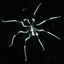
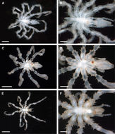
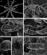
Figure 2.Ammotheidae 1; A, B: Achelia echinata, male, dorsal view; scales 500 µm and 250 µm, respectively; C, D: Achelia langi, male, dorsal view; scales 1 mm and 500 µm, respectively; E, F: Achelia vulgaris, male, dorsal view; scales 1 mm and 250 µm, respectively.
-
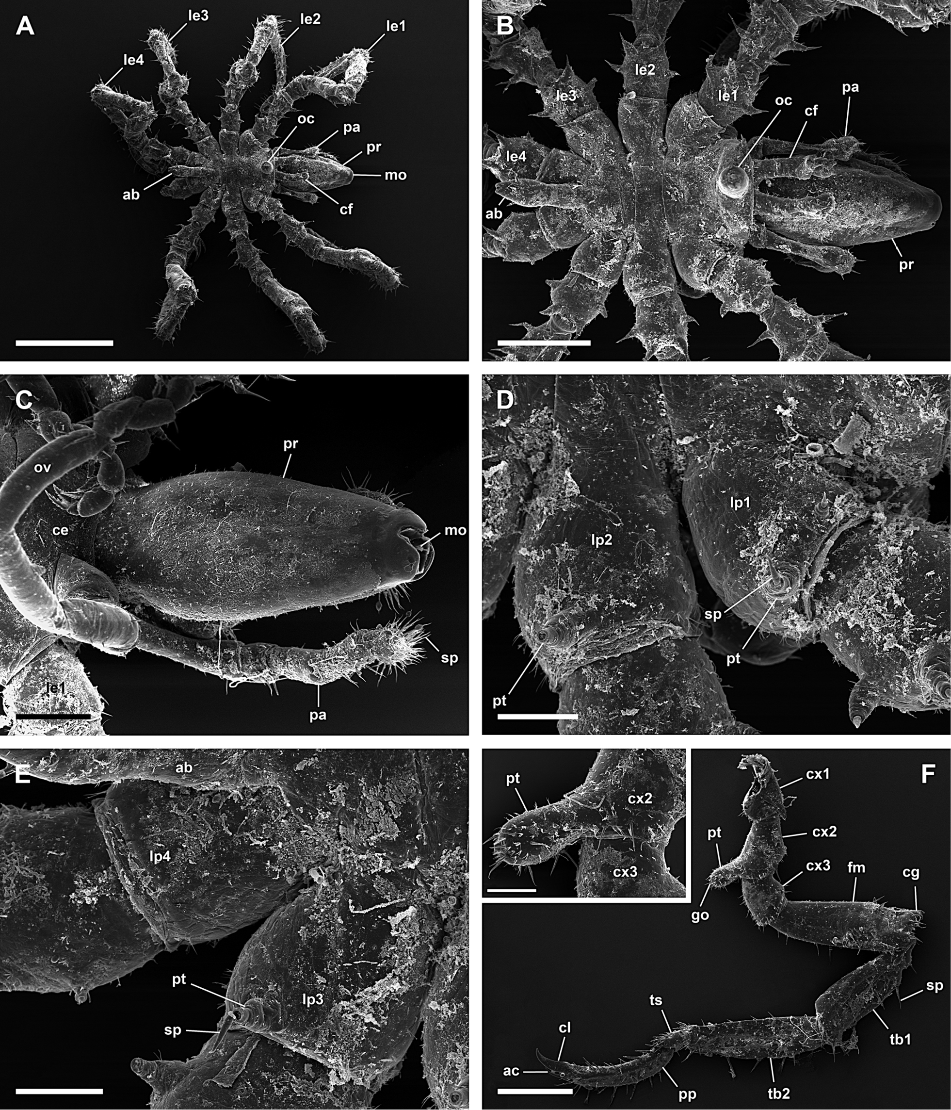
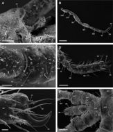
Figure 9.Achelia langi, male; A: Dorsal view; scale 1 mm; B: Dorsal view of trunk; scale 400 µm; C: Ventral view of proboscis; scale 200 µm; D: Dorsal view of right lateral processes 1 and 2, 2 protuberances with spine on lateral process 1 and 1 on lateral process 2; scale 100 µm; E: Dorsal view of right lateral processes 3 and 4, 1 protuberance with spine on lateral process 3 and lateral process 4 without protuberance or spine; scale 100 µm; F: Left 3rd leg; scale 400 µm; insert: Genital protuberance; scale 100 µm.
-
Figure 10.Achelia langi, female; A: Dorsal view of trunk; scale 400 µm; B: Dorsal view of left lateral processes, 1 protuberance with spine on lateral process 1-3, lateral process 4 without protuberance or spine; scale 200 µm; C: Right 3rd leg; scale 400 µm; D: Ventral view of coxa 2 with genital opening, distal is down (right 3rd leg); scale 100 µm; E: Genital opening; scale 20 µm; F: Tarsus, propodus, and claw, auxiliary claws about 2/3 of length of claw; scale 100 µm; G: Abdomen with anus; scale 40 µm.
-
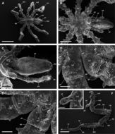
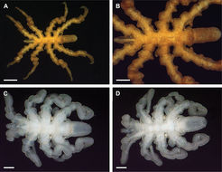
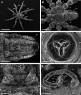
Figure 53.Pycnogonidae; A, B: Pycnogonum nodulosum, female, dorsal view; scales 1 mm and 500 µm, respectively; C, D: Pycnogonum (Retroviger) pusillum, male, dorsal and ventral view, respectively; scales 250 µm.
-
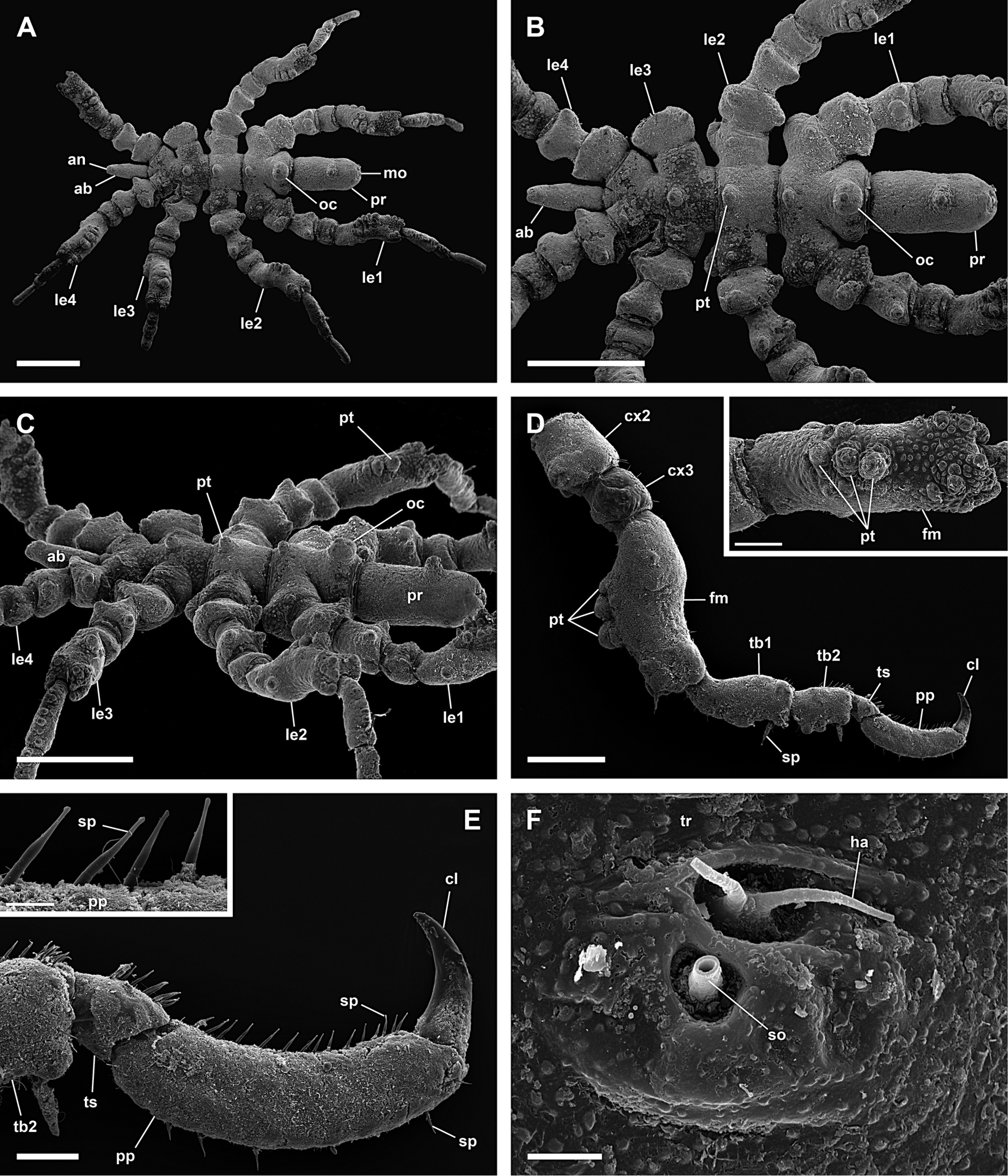
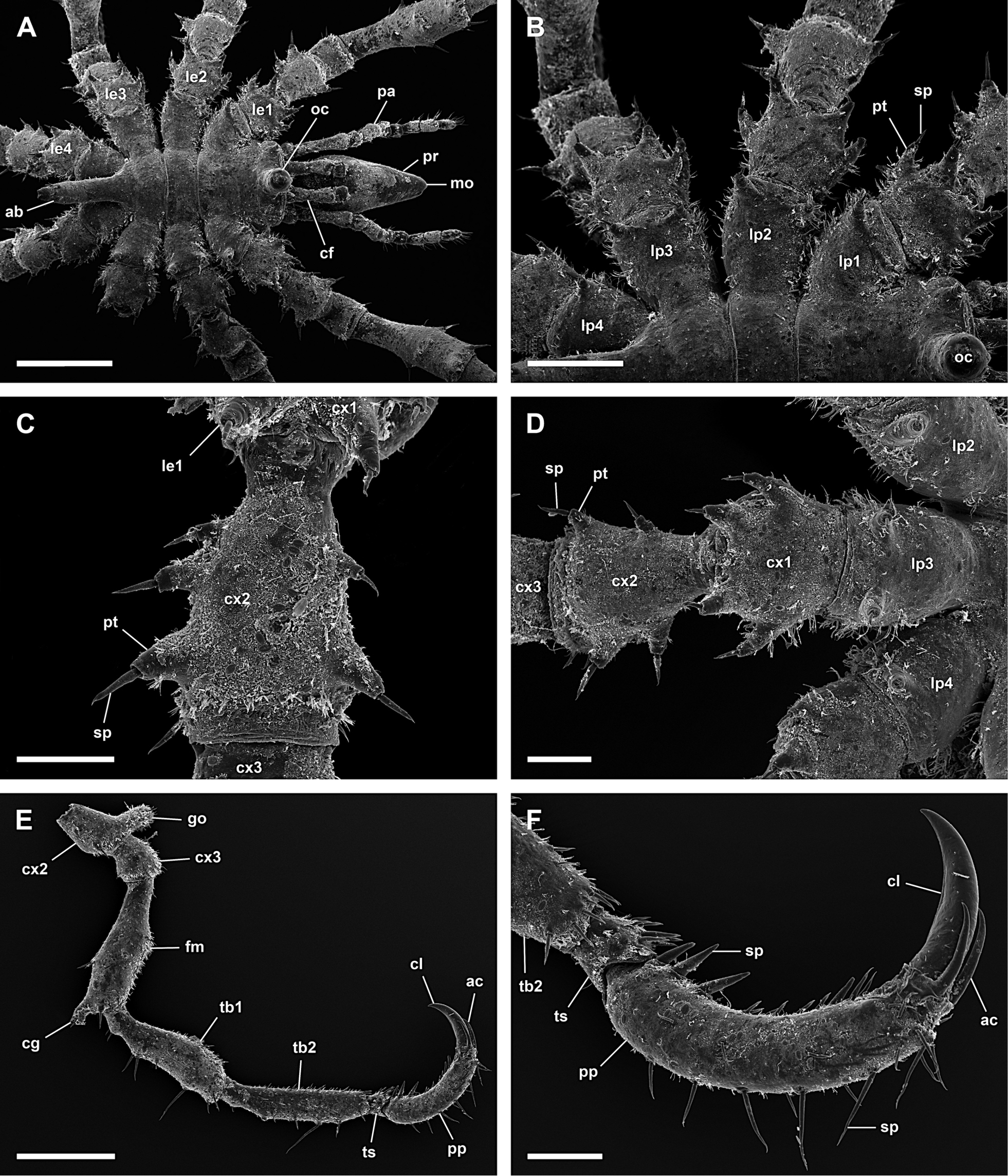
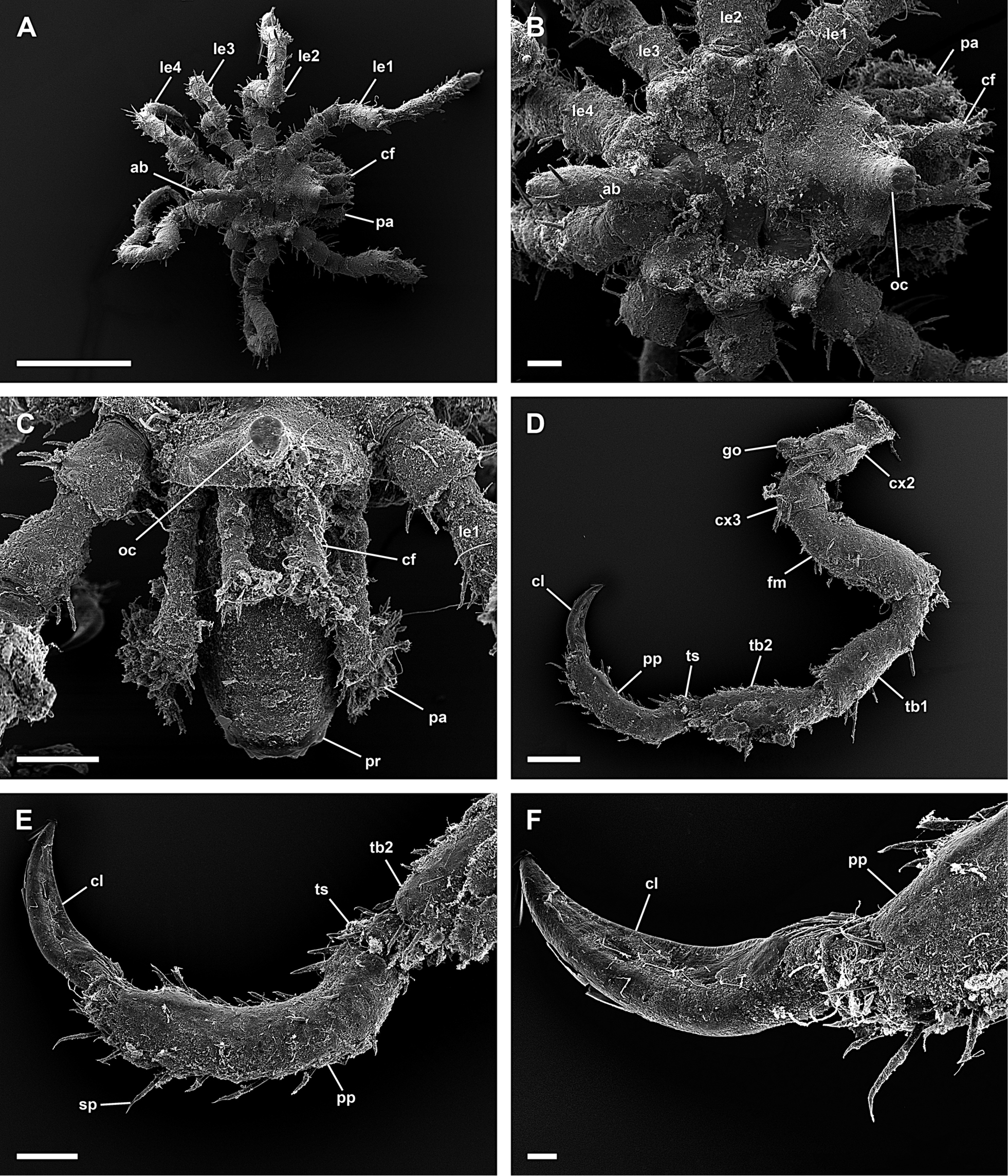
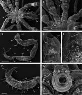
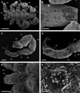
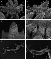
Figure 54.Pycnogonum nodulosum, female; A: Dorsal view; scale 1 mm; B: Dorsal view of trunk with protuberances on dorsal midline; scale 1 mm; C: Lateral view of trunk; scale 1 mm; D: Left 3rd leg with protuberances on femur; scale 400 µm; insert: Dorsal view of protuberances on femur; scale 200 µm; E: Tarsus, propodus, and claw without auxiliary claws (left 3rd leg); scale 100 µm; insert: Spines on propodus; scale 20 µm; F: Hair and slit organ on dorsal side of trunk; scale 5 µm.
-
Figure 53.Pycnogonidae; A, B: Pycnogonum nodulosum, female, dorsal view; scales 1 mm and 500 µm, respectively; C, D: Pycnogonum (Retroviger) pusillum, male, dorsal and ventral view, respectively; scales 250 µm.
-
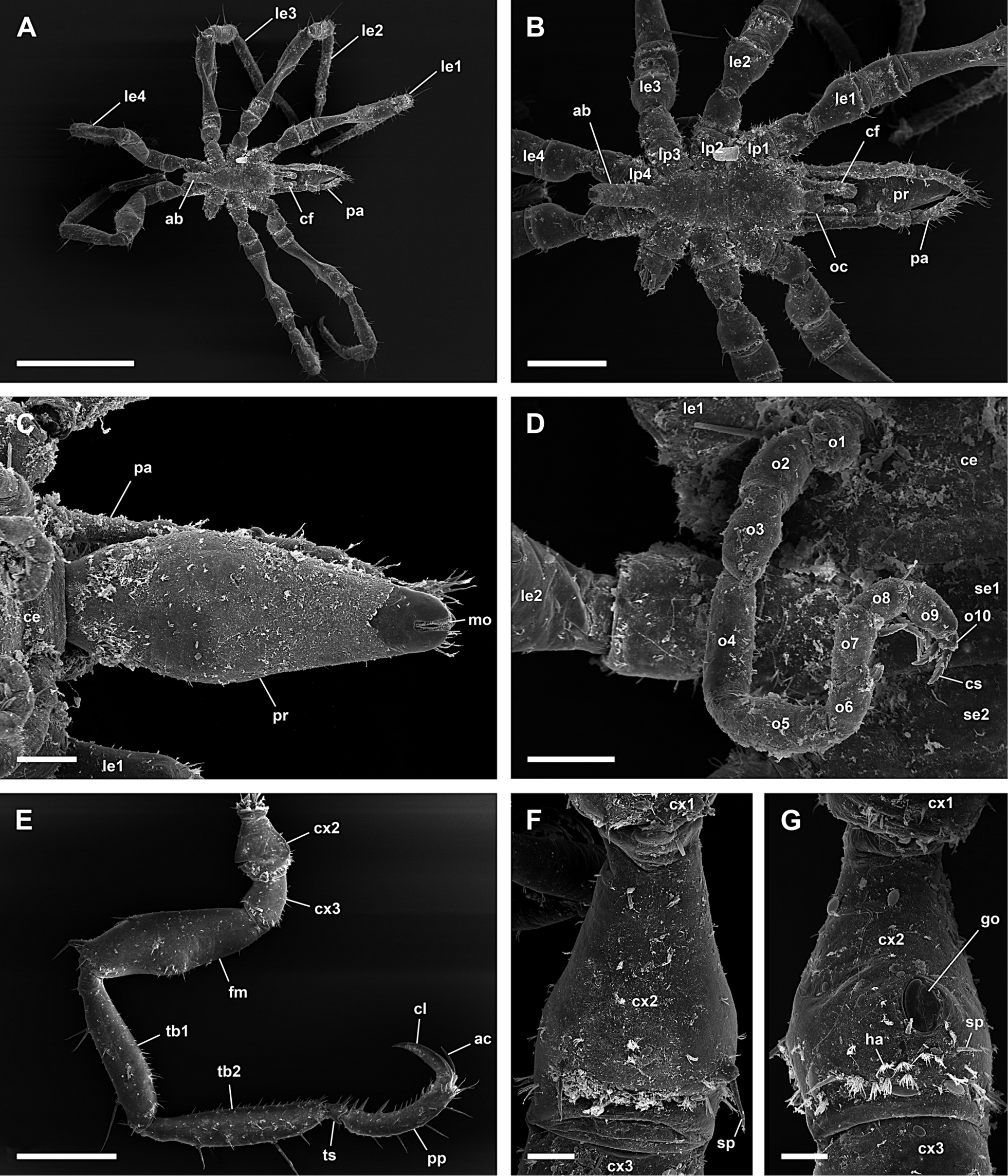
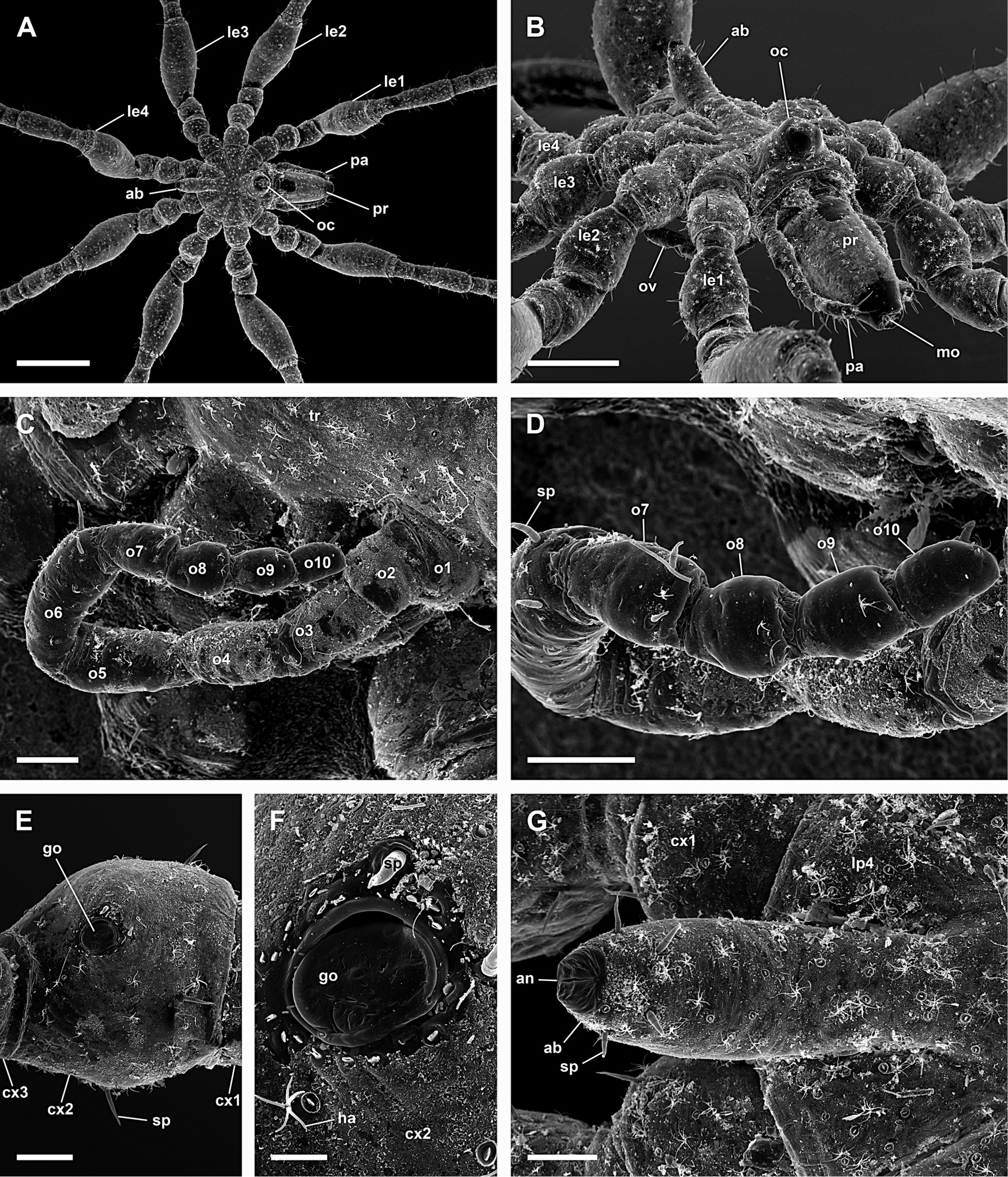
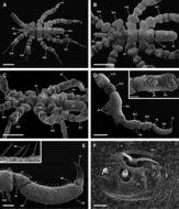
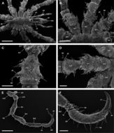
Figure 55.Pycnogonum (Retroviger) pusillum, male; A: Dorsal view; scale 400 µm; B: Dorsal view of proboscis; scale 100 µm; C: Right 3rd leg; scale 100 µm; D: Tarsus, propodus, and claw without auxiliary claws (right 3rd leg); scale 40 µm; E: Abdomen; scale 100 µm; F: Protuberance, scattered over trunk and legs; scale 5 µm.
-
Figure 2.Ammotheidae 1; A, B: Achelia echinata, male, dorsal view; scales 500 µm and 250 µm, respectively; C, D: Achelia langi, male, dorsal view; scales 1 mm and 500 µm, respectively; E, F: Achelia vulgaris, male, dorsal view; scales 1 mm and 250 µm, respectively.
-
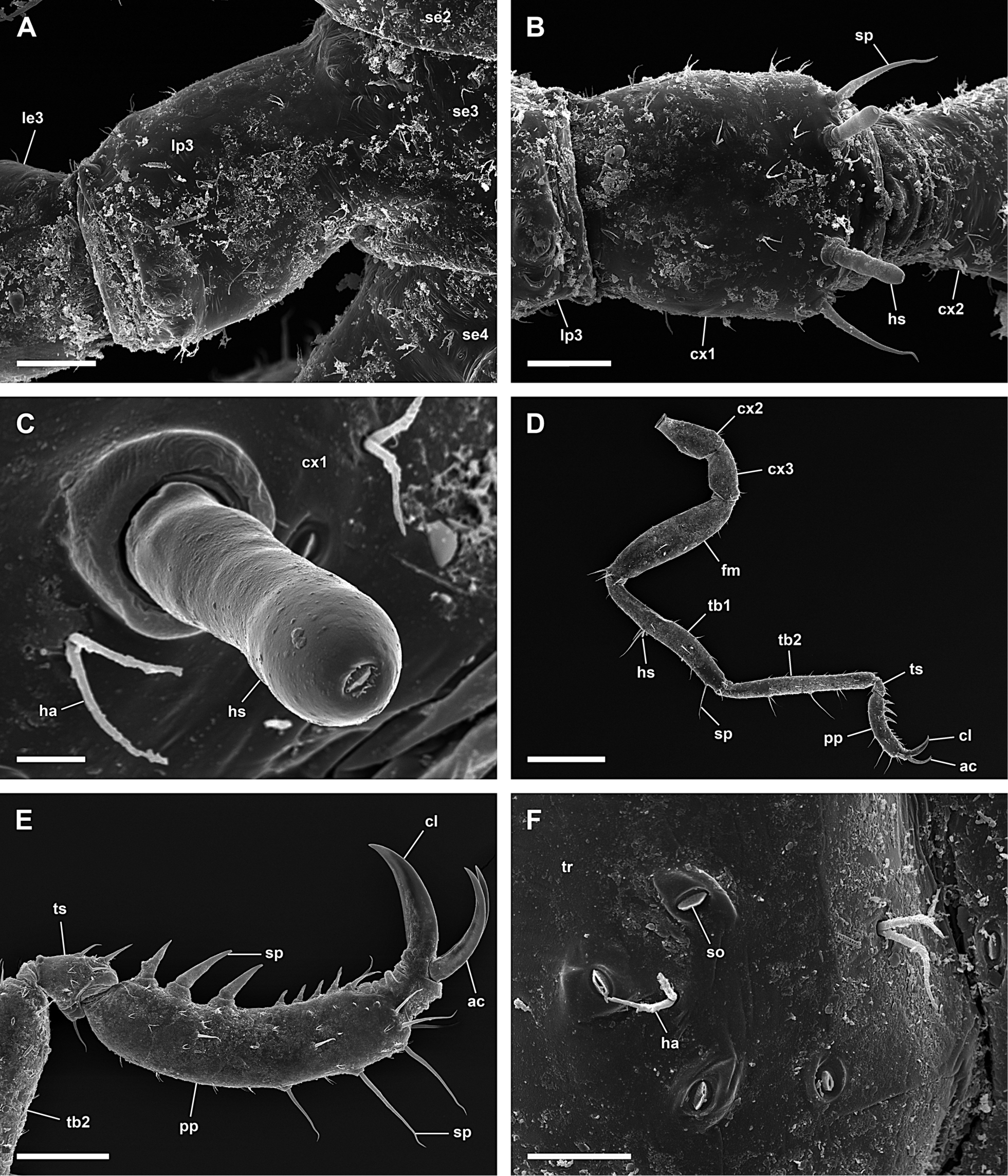
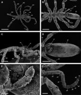
Figure 11.Achelia vulgaris, male; A: Dorsal view of trunk; scale 400 µm; B: Dorsal view of left lateral processes, lateral processes do not touch each other; scale 200 µm; C: 2 protuberances with spine on right side and 3 on left side of coxa 2, distal is down (right 1st leg); scale 100 µm; D: Lateral process, coxa 1 and 2, 2 protuberances with spine on each side of coxa 2 (left 3rd leg); scale 100 µm; E: Right 3rd leg; scale 400 µm; F: Tarsus, propodus, and claw, auxiliary claws about half as long as claw; scale 100 µm.
-
Figure 12.Achelia vulgaris, female; A: Dorsal view; scale 1 mm; B: Dorsal view of trunk, lateral processes do not touch each other; scale 300 µm; C: Ventral view of proboscis; scale 100 µm; D: Right 10-articled oviger; scale 100 µm; E: Right 3rd leg; scale 400 µm; F: Dorsal view of coxa 2 (left 3rd leg); scale 40 µm; G: Ventral view of coxa 2 with genital opening (left 3rd leg); scale 40 µm.
-
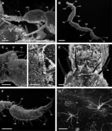
Figure 3.Ammotheidae 2; A, B: Ammothella appendiculata, female, dorsal view; scales 1 mm and 250 µm, respectively; C, D: Ammothella biunguiculata, male, dorsal view; scales 1 mm and 250 µm, respectively; E, F: Ammothella longipes, female, dorsal view; scales 1 mm and 250 µm, respectively.
-
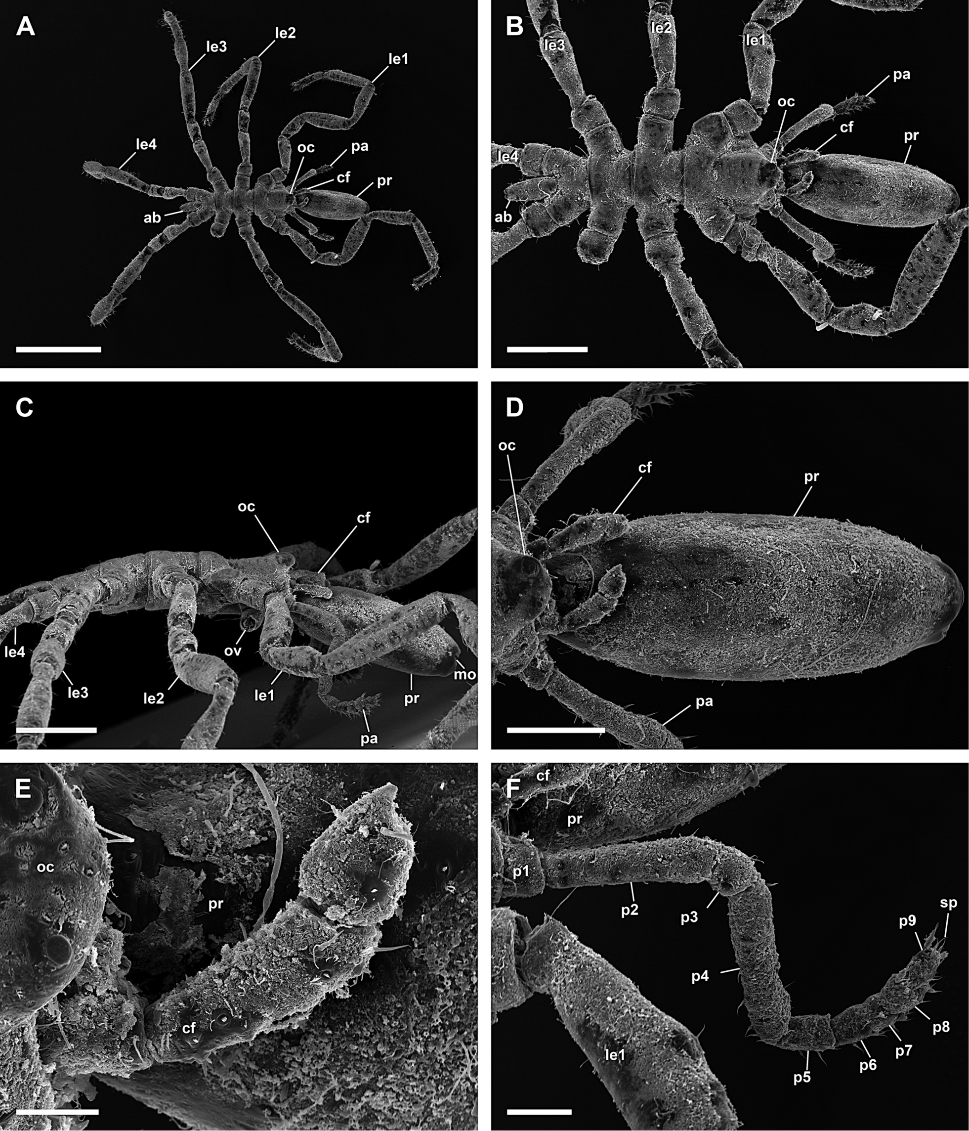
Figure 13.Ammothella appendiculata, female; A: Dorsal view of trunk; scale 400 µm; B: Lateral view of trunk; scale 200 µm; C: Ventral view of trunk; scale 200 µm; D: Mouth opening, dorsal is up; scale 20 µm; E: Right chelifore with reduced chela and right 9-articled palp; scale 100 µm; F: Ocular tubercle with lateral sense organ; scale 40 µm.
-
Figure 14.Ammothella appendiculata, female; A: Lateral process of left 3rd leg; scale 40 µm; B: Coxa 1 of right 3rd leg with hollow spines; scale 40 µm; C: Hollow spine; scale 5 µm; D: Right 4th leg; scale 400 µm; E: Tarsus, propodus, and claws (right 4th leg); scale 100 µm; F: Hairs and slit organs of dorsal side of trunk; scale 10 µm.
-
Figure 3.Ammotheidae 2; A, B: Ammothella appendiculata, female, dorsal view; scales 1 mm and 250 µm, respectively; C, D: Ammothella biunguiculata, male, dorsal view; scales 1 mm and 250 µm, respectively; E, F: Ammothella longipes, female, dorsal view; scales 1 mm and 250 µm, respectively.
-
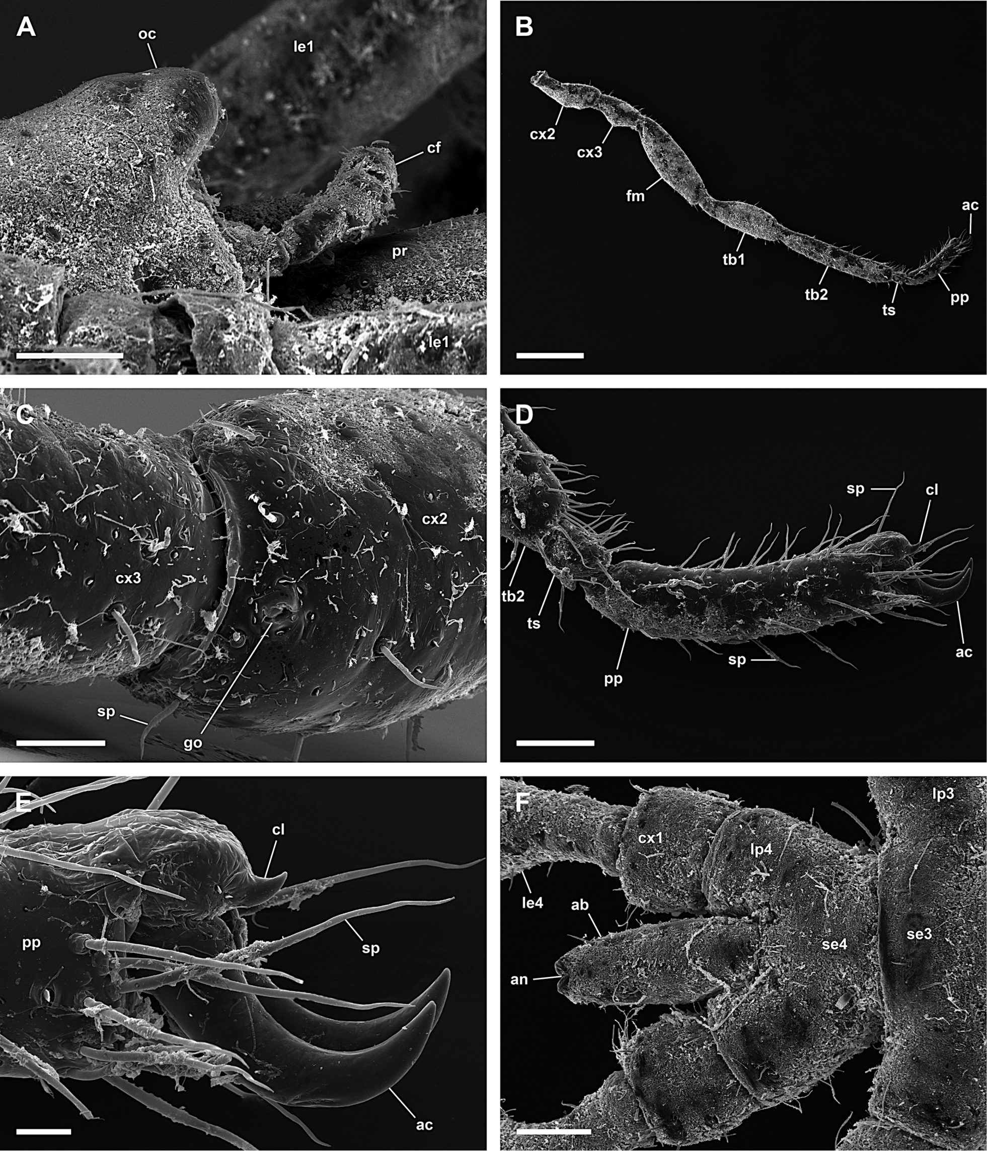
Figure 15.Ammothella biunguiculata, male; A: Dorsal view; scale 1 mm; B: Dorsal view of trunk; scale 400 µm; C: Lateral view of trunk, oviger dissected; scale 400 µm; D: Dorsal view of proboscis; scale 100 µm; E: Right chelifore with reduced chela; scale 40 µm; F: Right 9-articled palp; scale 100 µm.
-
Figure 16.Ammothella biunguiculata, male; A: Lateral view of ocular tubercle; scale 100 µm; B: Right 3rd leg; scale 400 µm; C: Ventral view of coxa 2 with genital opening, distal is left (right 3rd leg); scale 40 µm; D: Tarsus and propodus with reduced claw and dominating auxiliary claws; scale 100 µm; E: Reduced claw and dominating auxiliary claws; scale 20 µm; F: Dorsal view of trunk segment 4 and abdomen; scale 100 µm.
-
Figure 3.Ammotheidae 2; A, B: Ammothella appendiculata, female, dorsal view; scales 1 mm and 250 µm, respectively; C, D: Ammothella biunguiculata, male, dorsal view; scales 1 mm and 250 µm, respectively; E, F: Ammothella longipes, female, dorsal view; scales 1 mm and 250 µm, respectively.
-
Figure 17.Ammothella longipes, female; A: Dorsal view of trunk; scale 200 µm; B: Lateral view of trunk, one protuberance without spine aside on lateral processes; scale 200 µm; C: Ocular tubercle; scale 100 µm; D: Right lateral process; scale 100 µm; E: Left 3rd leg; scale 400 µm; F: Tarsus, propodus, and claws (left 3rd leg); scale 100 µm.
-
Figure 4.Ammotheidae 3; A, B: Ammothella uniunguiculata, male, dorsal view; scales 200 µm; C, D: Tanystylum conirostre, male, dorsal view; scales 1 mm and 250 µm, respectively; E, F: Trygaeus communis, male, dorsal view; scales 1 mm and 250 µm, respectively.
-
Figure 18.Ammothella uniunguiculata, male; A: Dorsal view; scale 1 mm; B: Dorsal view of trunk; scale 100 µm; C: Cephalon with proboscis, chelifore, and palps; scale 200 µm; D: Right 3rd leg; scale 200 µm; E: Tibia 2, tarsus, and propodus with claw (right 3rd leg); scale 100 µm; F: Propodus with claw; scale 20 µm.
-
Figure 4.Ammotheidae 3; A, B: Ammothella uniunguiculata, male, dorsal view; scales 200 µm; C, D: Tanystylum conirostre, male, dorsal view; scales 1 mm and 250 µm, respectively; E, F: Trygaeus communis, male, dorsal view; scales 1 mm and 250 µm, respectively.
-
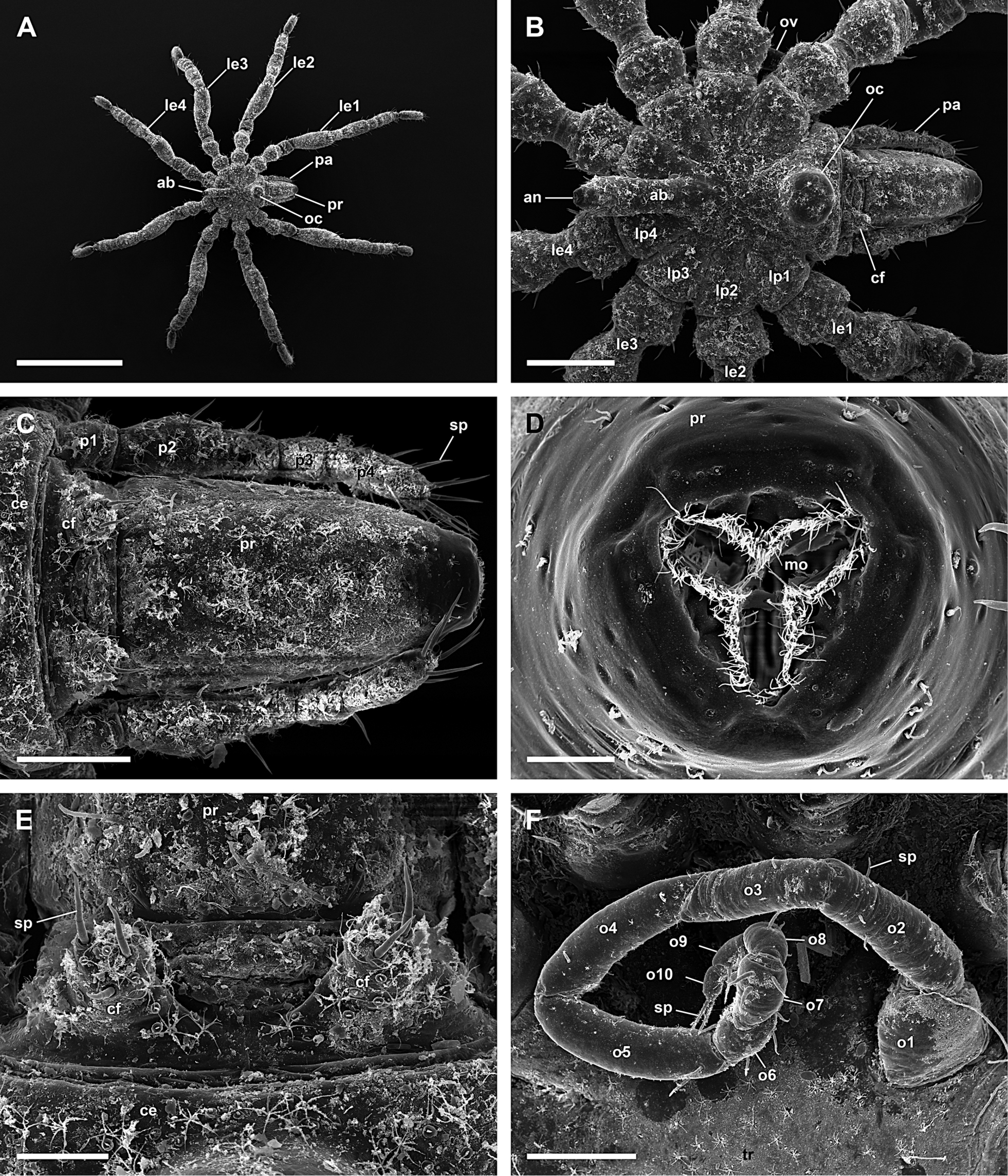
Figure 19.Tanystylum conirostre, male; A: Dorsal view; scale 1 mm; B: Dorsal view of trunk; scale 200 µm; C: Dorsal view of proboscis and 4-articled palp; scale 100 µm; D: Mouth opening; scale 20 µm; E: Reduced chelifores, frontal is up; scale 40 µm; F: Right 10-articled oviger; scale 100 µm.
-
Figure 20.Tanystylum conirostre, male; A: Distal articles of right oviger with spines on tip; scale 10 µm; B: Left 3rd leg; scale 100 µm; C: Lateral view of coxa 2 with genital opening on a protuberance, distal is right (left 4th leg); scale 40 µm; D: Genital opening; scale 10 µm; E: Cement gland on femur, distal is up (left 3rd leg); scale 20 µm; F: Tarsus, propodus, and claws (left 3rd leg); scale 100 µm; G: Hairs and slit organs on dorsal side of trunk; scale 10 µm.
-
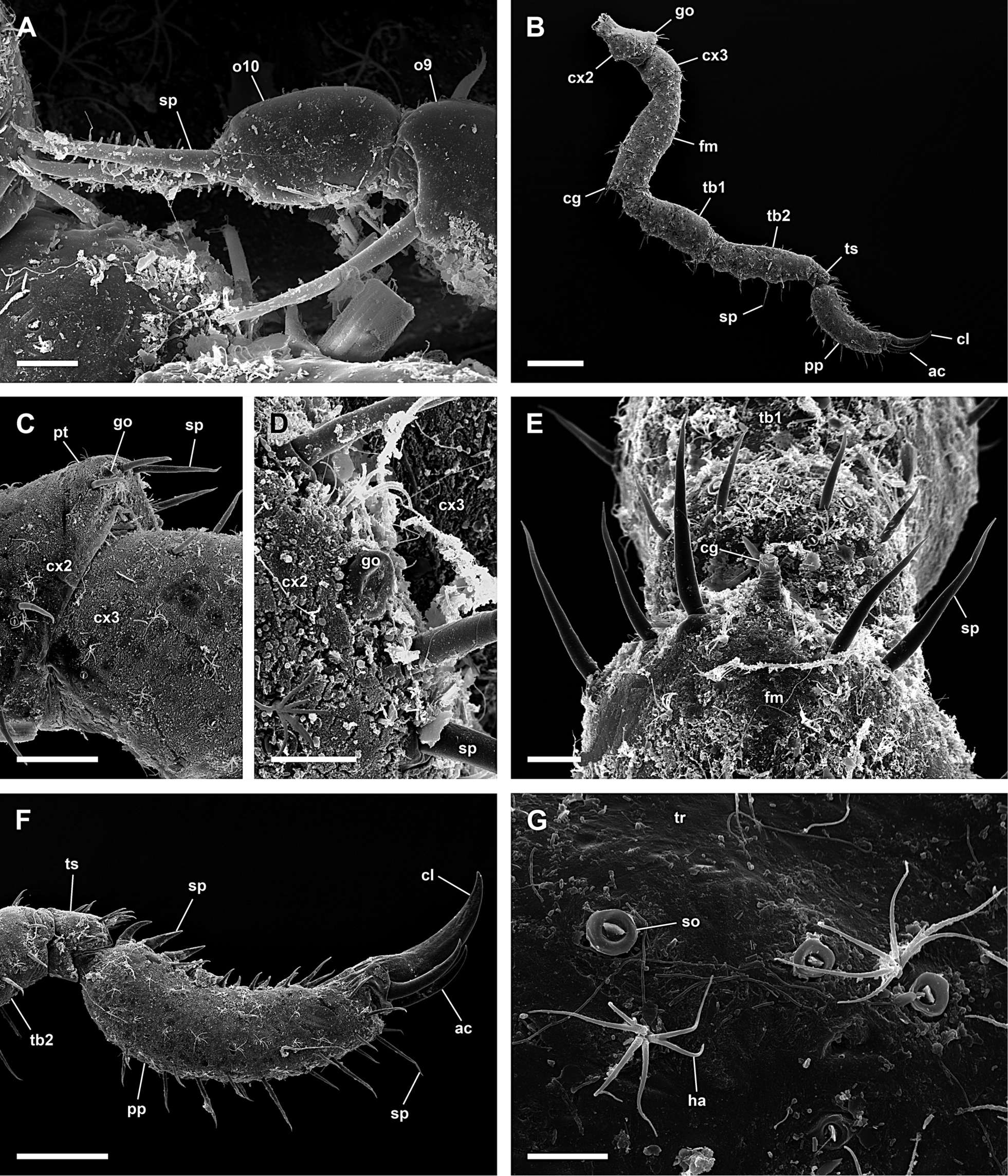
Figure 21.Tanystylum conirostre, female; A: Dorsal view of trunk; scale 400 µm; B: Frontal view of trunk; scale 200 µm; C: Left 10-articled oviger; scale 40 µm; D: Distal articles of left oviger; scale 40 µm; E: Ventral view of coxa 2 with genital opening, distal is right (right 4th leg); scale 40 µm; F: Genital opening; scale 10 µm; G: Abdomen; scale 40 µm.