Comprehensive Description
(
英語
)
由Smithsonian Contributions to Botany提供
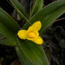
Barbacenia exscapa Martius
Barbacenia exscapa Martius, Nov. Gen. & Sp., 1:21, pl. 14, 1823.—Seubert in Martius, Fl. Bras., 3(1):69, 1847.—L. B. Smith, Contr. U.S. Nat. Herb., 35:280, 1962.—Ayensu, Smithsonian Contr. Bot., 15:63, pl. 46g, 1974.
Aylthonia exscapa (Martius) Menezes, Ciência e Cultura, 23(3):[421], 1971 [as “excapa”].
TYPE.—Among rocks of granular micaceous schist, summit of Itambé da Villa do Principe (Sêrro), Minas Gerais, Brazil, Martius s n (M, holotype; F, photo 18991).
DISTRIBUTION.—Brazil: Minas Gerais: Peak of Itambé, Sêrro.
64. Barbacenia rectifolia L. B. Smith & Ayensu, new species
A B. exscapa Martius, cui valde affinis, foliorum laminis rectis angustioribus laxe inconspicueque denticulatis differt.
Caudex simple, 2 to over 5 cm long. Leaf-sheaths completely covered; blades erect to reflexed, straight, flat, linear, filiform-attenuate, 16 cm long, 6–8 mm wide, laxly ciliate with minute bristle-tipped teeth, sulcate on both sides, otherwise smooth and glabrous.
Scape single, very short and completely hidden by the leaf-bases. Flower appearing sessile. Ovary turbinate, ca. 5 mm long, not distinct. Perianth-tube (whole) subcylindric, 40 mm long, 5 mm wide, very sparsely white-pubescent, green (!Anderson et al.). Tepals nearly uniform, elliptic, obtuse, 12 mm long, yellow (!Anderson et al.). Anthers 9 mm long, extending into the perianth-tube; coronoid appendages elliptic, 9 mm long, the 2 lobes short, subacute. Style subglobose at apex; stigmas linear, apically confluent.
LEAF ANATOMY (Anderson et al. 35674).—Surface View: Hairs: none observed. Epidermis: cells on both surfaces square to rectangular; walls thickened. Stomata: tetracytic; 24 × 21 μm; present in rows on abaxial surface.
Transverse Section of Lamina: Dorsiventral; widely V-shaped with recurved margins. Both surfaces gently undulating. Epidermis: cells on both surfaces rounded to dome-shaped; walls thickened. Subjacent to adaxial epidermis is 2 or 3 layers of rounded, thin-walled cells. Few sclerenchyma fibers present here. Adaxial epidermis and subjacent layers often large above midvein. Cuticle: thick and ridged on both surfaces. Stomata: present on abaxial surface only; stomata flush with surface; small substomatal chamber present. Mesophyll: three layers of elongated, palisade cells followed by 10 or 11 layers of rounded, compactly arranged cells. Vascular bundles: 25; commissural bundles observed. One or two large vessels present in each bundle. Two phloem units lying laterally in flanges of thin Y-shaped abaxial girder. Thin, inverted Y-shaped adaxial girder present. Abaxial girder extends to epidermis; adaxial girder extends to cells subjacent to epidermis. Each bundle surrounded by a prominent bundle sheath. Crystals: present in most mesophyll cells. Tannins: present.
TYPE.—Matted on sandstone rocks by river, southeastern drainage of Pico Itambé, about 5 km directly west and north of Santo Antonio de Itambé, Minas Gerais, Brazil, 950 m alt, 9 February 1972, Anderson, Stieber & Kirkbride 35674 (US, holotype; NY; UB, isotypes).
DISTRIBUTION.—Known only from the type-collection.
65. Barbacenia aurea L. B. Smith & Ayensu, new species
A B. sessiliflora L. B. Smith, cui affinis, foliis angustioribus albo-pubescentibus, floribus aureis, ovario vix distincto differt.
Caudex to 10 cm high, ca. 2 cm thick including old leaf-sheaths. Leaves sulcate on both sides; sheaths completely covered; blades linear-lanceolate, filiform-attenuate, 9 cm long, 10 mm wide, sub-densely white-pubescent throughout, finely setose-ciliate.
Scape not observed. Flower single, largely concealed by leaves and appearing sessile, densely white-pubescent, yellow-orange (!Anderson et al). Ovary 5 mm long, scarcely distinct. Perianth-tube (whole) cylindric, slightly flaring at apex, 32 mm long, 3 mm wide. Tepals oblong, obtuse, 10 mm long. Anthers 12 mm long, extending downward into the perianth-tube; coronoid appendages subovate, bidentate with short subacute lobes, 2.5 mm long. Style about equaling the anthers; stigmas not observed.
LEAF ANATOMY (Irwin et al. 29046).—Surface View: Hairs: long, multicellular, present on both surfaces. Epidermis: cells square to rectangular on both surfaces; thin-walled; cells in rows. Stomata: tetracytic; 21 × 18 μm; present on abaxial surface only.
Transverse Section of Lamina: Dorsiventral; widely V-shaped. Both surfaces gently undulating. Multicellular hairs present on both surfaces. Epidermis: cells on both surfaces rounded to dome-shaped and conical; thin-walled. Subjacent to epidermis occurs a layer of thin-walled parenchyma cells. Cuticle: thin and smooth over entire surface. Stomata: present on abaxial surface; fairly large substomatal chamber present; stomata flush with epidermal surface. Mesophyll: 2 or 3 layers of palisade cells followed by 5 or 6 layers of rounded spongy tissue. Two or three layers of translucent cells above midvein. Mesophyll cells thin-walled. Vascular bundles: 45; commissural bundles not observed. One or two large vessels present in each bundle. Two phloem units lying laterally in flanges of short U- or V-shaped abaxial girder extending to abaxial epidermis. Inverted Y-shaped adaxial girder present on each bundle; girder extends to adaxial surface and forms a T-shape beneath epidermis. Conspicuous bundle sheath completely surrounding each vascular bundle. Crystals: druses and styloids present in most of mesophyll. Tannins: few present in mesophyll.
TYPE.—On sandstone outcrops in cerrado, ca. 10 km west of Barão de Cocais, Minas Gerais, Brazil, 1500 m alt, 24 January 1971, Irwin, Harley & Omishi 29046 (US, holotype; NY; UB, isotypes).
DISTRIBUTION.—Known only from the type-collection.
66. Barbacenia coronata P. F. Ravenna, new species
A B. sessiliflora L. B. Smith, cui affinis, foliorum laminis obscure denticulatis, ovario vix distincto differt.
Caudex erect (?), to 15 cm high, simple or short-branched at apex, ca. 12 mm thick with leaf-bases. Leaves sulcate on both sides; sheaths completely hidden; blades linear, filiform-attenuate, 13 cm long, 4 mm wide, obscurely denticulate, glabrous.
Scapes solitary, to 20 mm long, very slender, completely hidden by the leaves. Flowers appearing sessile, yellow (!Hatschbach). Ovary slenderly fusiform, barely distinct, ca. 10 mm long. Perianth-tube slenderly subcylindric, 22 mm long, 2 mm wide, laxly vestite with minute pointed trichomes. Tepals oblong, broadly rounded, 11 mm long. Anthers 7 mm long, extending downward into the perianth-tube; coronoid appendages exceeding the anthers, suboblong with narrowly triangular obtuse lobes. Style about equaling the anthers; stigmas not observed.
LEAF ANATOMY (Hatschbach 30095).—Surface View: Hairs: none observed. Epidermis: cells on both surfaces square to rectangular; walls thickened. Stomata: paracytic and tetracytic; 24 × 18 μm; present on abaxial surface.
Transverse Section of Lamina: Dorsiventral; widely V-shaped with median adaxial groove. Surface only gently undulating. Epidermis: cells on both surfaces rounded to dome-shaped; few conical; walls thickened. One or two layers of rounded, thin-walled cells subjacent to adaxial epidermis; larger cells above midvein. Cuticle: thickened and ridged on abaxial surface; very thick and ridged on adaxial surface. Stomata: present on abaxial surface only; stomata flush with epidermal surface or slightly sunken; substomatal chamber present. Mesophyll: 3 or 4 layers of palisade cells followed by 6 or 7 layers of small, rounded, thin-walled cells. Vascular bundles: 15; commissural bundles observed. Two or three large vessels present in each bundle. Two phloem units lying laterally in flanges of Y-shaped abaxial girder. Long and slender inverted Y-shaped adaxial girder present on each bundle. Each vascular bundle completely surrounded by a bundle sheath. Crystals: druses present in mesophyll. Tannins: present.
TYPE.—On wet cliffs by small waterfall, road to Pico de Itambé, Mun. Santo Antonio de Itambé, Minas Gerais, Brazil, 1972, Hatschbach 27513 (US, holotype; MBM, isotype).
DISTRIBUTION.—Brazil: Minas Gerais: Type-locality, 9 August 1972, Hatschbach 30095 (MBM, US).
Note: Dr. P. F. Ravenna has kindly allowed us to publish his new species in our revision in order to relate it to others more immediately.
- 書目引用
- Smith, Lyman B. and Ayensu, Edward S. 1976. "A Revision of American Velloziaceae." Smithsonian Contributions to Botany. 1-172. https://doi.org/10.5479/si.0081024X.30
Comprehensive Description
(
英語
)
由Smithsonian Contributions to Botany提供
Barbacenia exscapa Martius
SPECIMENS EXAMINED.—Anderson et al. 35831.
SURFACE VIEW.—Hairs: present mostly on abaxial surface. Epidermis: adaxial cells mostly square to rectangular, few rounded; thin walled. Abaxial cells rectangular, thin walled. Stomata: paracytic, 27 × 21 μm; present on abaxial surface.
TRANSVERSE SECTION OF LAMINA.—Dorsiventral; V-shaped. Both adaxial and abaxial surfaces gently undulating. Epidermis: cells on adaxial and abaxial surfaces rounded to dome shaped, walls slightly thickened. Subjacent to adaxial epidermis are one to two layers of parenchyma cells; parenchyma layers in midvein region much larger. Cuticle: fairly thick and slightly ridged on adaxial and abaxial surfaces. Stomata: present on abaxial surface, small substomatal chamber observed; flush with epidermal surface. Mesophyll: three to four layers of palisade tissue occupying about one-half of the mesophyll. Abrupt change to spongy tissue. Elongated palisade cells not usually found in the midvein region. Vascular bundles: 25 to 27; of small sizes; few commissural bundles observed. Usually one large vessel present in each vascular bundle. Two phloem units lying laterally in flanges of Y-shaped abaxial girder. Inverted Y-shaped adaxial girder present on each vascular bundle. Each vascular bundle completely surrounded by a bundle sheath. Crystals: none observed. Tannins: few present in mesophyll.
NOTE: The adaxial and abaxial sclerenchyma girders are poorly developed. In some cases they are no more than lignified parenchyma cells.
- 書目引用
- Ayensu, Edward S. 1974. "Leaf Anatomy and Systematics of New World Velloziaceae." Smithsonian Contributions to Botany. 1-125. https://doi.org/10.5479/si.0081024X.15