NMNH Prorocentrum caribbaeum type specimen
描述:
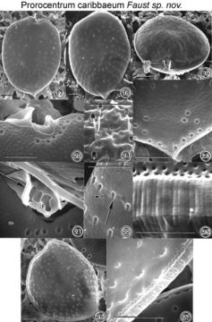
Figs. 17-27. Prorocentrum caribbaeum sp. nov. FIG.17. Cell shape is oval with a rounded anterior and a pointed posterior end. FIG.18. Valve surface is smooth with minute depressions. Radially arranged trichocyst pores are present on each valve. The two flagella are not shown. FIG.19. Cells are ovate to convex in side view. The periflagellar area is located in the right valve and is highly ornate. FIG.20. In valve view, the periflagellar area has a rectangular orientation and is composed of a curved apical collar (on the left) and a smaller protuberant apical plate (on the right). FIG.21. Curved apical collar (arrow) is the largest platelet situated adjacent to the auxiliary pore (A). The apical plate (arrowhead) is located next to the flagellar pore (F) and is separated by a rectangular platelet from the auxiliary pore. FIG.22. The trichocyst pores (arrow) are round with smooth edges and are similar in size. They are situated in furrowed depressions. Small, round pores (arrowhead) are also present, unevenly distributed on the valve surface. FIG.23. The posterior end is pointed and laced with trichocyst pores and small, round pores unique to this species. FIG.24.Ejected trichocysts are on valve surface. FIG.25. The intercalary band is transversally striated and sinous. FIG.6.Inner valve surface is smooth. The location of round trichocyst pores is illustrated, and a distinct striated intercalary band is present (arrowheads). FIG. 7. The inner face of the intercalary band is highly ornate and lacelike. Scale bars = 8 µm. EMu: Holotype SEM negative # 103001; SEM stub #103; Field # 358-90 Accession # 407164; Catalog # 49; Figure # 17.
來源資訊
- 許可
- cc-by-nc-sa-3.0
- 版權
- National Museum of Natural History, Smithsonian Institution
- 原始內容
- 原始媒體檔案
- 參訪來源
- 合作夥伴網站
- NMNH Marine Dinoflagellates
- ID