portrait
描述:
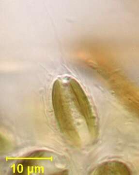
Individual cell detail of colonial chrysophyte, Synura. Species identification is based on ultrastructure of silica scales on the cell surface. Scales (seen in this image) originate in cytoplasmic vesicles and are extruded to the cell exterior. Several different types of scales occur on an individual cell. Cells have two subequal flagella, one hairy and the other smooth on electron microscopy (only one is seen in this image). Two yellow-brown chloroplasts flank a central nucleus. A posterior contractile vacuole is seen in the cell at the lower left of this image. From a freshwater pond near Boise, Idaho. Oblique illumination.