Astroviridae és una família de virus d'ARN monocatenari +. Va ser descoberta el 1975 en humans.[1] També s'han aïllat en nombrosos mamífers (classificats en el gènere Mammoastrovirus) i en aus (classificades en el gènere Aviastrovirus). Els astrovirus fan de 28 a 35 nm de diàmetre, i tenen càpside en forma d'icosaedre amb una superfície en forma d'estrella. No tenen embolcall.[2]
Causen gastroenteritis en infants i adults. El principal símptoma és la diarrea, seguida de nàusea, vòmits, febre, malestar i dolor abdominal. Els símptomes acostumen a durar de tres a quatre dies. Les infeccions no són normalment severes i només en casos rars porten a la deshidratació. No cal l'hospitalització.[3]
Al Regne Unit (1999) es va determinar una incidència de 3,8/1000 pacients l'any essent la quarta causa de gasteroenteritis viral.[4] Als Estats Units s'ha determinat que el 2-9% dels infants en presenten símptomes.[5]
No hi ha vacuna o tractament antiviral però la higiene personal en redueix la incidència.
Astroviridae és una família de virus d'ARN monocatenari +. Va ser descoberta el 1975 en humans. També s'han aïllat en nombrosos mamífers (classificats en el gènere Mammoastrovirus) i en aus (classificades en el gènere Aviastrovirus). Els astrovirus fan de 28 a 35 nm de diàmetre, i tenen càpside en forma d'icosaedre amb una superfície en forma d'estrella. No tenen embolcall.
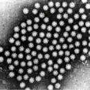
Astroviruses are a type of virus that was first discovered in 1975 using electron microscopes following an outbreak of diarrhea in humans.[1] In addition to humans, astroviruses have now been isolated from numerous mammalian animal species (and are classified as genus Mamastrovirus) and from avian species such as ducks, chickens, and turkey poults (classified as genus Avastrovirus). Astroviruses are 28–35 nm diameter, icosahedral viruses that have a characteristic five- or six-pointed star-like surface structure when viewed by electron microscopy. Along with the Picornaviridae and the Caliciviridae, the Astroviridae comprise a third family of nonenveloped viruses whose genome is composed of plus-sense, single-stranded RNA.[2] Astrovirus has a non-segmented, single stranded, positive sense RNA genome within a non-enveloped icosahedral capsid.[3] Human astroviruses have been shown in numerous studies to be an important cause of gastroenteritis in young children worldwide.[2] In animals, Astroviruses also cause infection of the gastrointestinal tract but may also result in encephalitis (humans and cattle), hepatitis (avian) and nephritis (avian).[4]
The International Committee on Taxonomy of Viruses (ICTV) established Astroviridae as a viral family in 1995.[5] There have been over 50 astroviruses reported, although the ICTV officially recognizes 22 species.[6] The genus Avastrovirus comprises three species; Chicken astrovirus (Avian nephritis virus types 1–3), Duck astrovirus (Duck astrovirus C-NGB), and Turkey astrovirus (Turkey astrovirus 1). The genus Mamastrovirus includes Bovine astroviruses 1 and 2, Human astrovirus (types 1-8), Feline astrovirus 1, Porcine astrovirus 1, Mink astrovirus 1 and Ovine astrovirus 1.[6]
Astroviruses have a star-like appearance with five or six points. Their name is derived from the Greek word "astron" meaning star. They are non-enveloped RNA viruses with cubic capsids, approximately 28–35 nm in diameter with T=3 symmetry.[7][8] Human astroviruses are part of the Mammastrovirus genus and contains 8 serotypes. The human astrovirus capsid spikes have a distinct structure. The spike domain in particular has a 3-layered beta-sandwiches fold and a core, 6-stranded beta-barrel structure. The beta-barrel has a hydrophobic core. The triple-layered beta-sandwich is packed outside the beta-barrel. The spike also forms a dimer. This unique structure was found to be similar to the protein projections found on the capsid of the hepatitis E virus. The projection domain of the human astrovirus contains a receptor binding site for polysaccharides. The amino acid sequence of the astrovirus capsid protein does not have similar homology to other known viral proteins, but the closest would be hepatitis E virus.[9]
Astroviruses infect birds and mammals through the fecal-oral route. They have a tissue tropism for enterocytes. Entry into the host cell is achieved by attachment to host receptors, which mediates endocytosis. Replication follows the positive-strand RNA virus replication model.[10] Astrovirus RNA is infectious and functions as a messenger RNA for ORF1a and ORF1b.[11] A frame-shifting mechanism between these two nonstructural polypeptides translates RNA-dependent RNA polymerase.[12] In replication complexes near intracellular membranes, ORF1a and ORF1b are cleaved to generate individual nonstructural proteins that are involved in replication. The resulting subgenomic RNA contains ORF2 and encodes precursor capsid protein (VP90). VP90 is proteolytically cleaved during packaging and produces immature capsids made of VP70. Following encapsidation, immature capsids are released from the cell without lysis.[5] Extracellular virions are cleaved by Trypsin and form mature infectious virions.[13]
Astroviruses have a genome composed of a single strand of positive sense RNA. The strand has a poly A tail at the 3' end, but no 5' cap. With the exclusion of polyadenylation at the 3' end, the genome is between 6.8–7.9 kb long. The genome is arranged into three open reading frames (ORFs), with an overlap of approximately 70 nucleotides between ORF1a and ORF1b. The remaining ORF is known as ORF2.[14] ORF2 encode the structural proteins, which are -at least- VP26, VP29 and VP32, the most antigenic and immunogenic of these being VP26. This protein is probably involved in the first steps of viral infection, being a key factor in the biological cycle of astroviruses.[15] The human astrovirus genome mutation rate has been estimated to be 3.7×10⁻³ nucleotide substitutions per site per year with the synonymous changes rate of 2.8×10⁻³ nucleotide substitutions per site per year.[16] The capability for genetic recombination appears to be present in type-3 and type-4 human astroviruses,[17][18] and in porcine astrovirus strains.[19]
The Astroviridae capsid is related to those of the Tymoviridae. The non-structural region is related to the Potyviridae. It appears that this group of viruses may have arise at some point in the past as a result of recombination event between two distinct viruses and that this even occurred at the junction of the structural and non-structural coding regions.[20]
Avastrovirus 1–3 are associated with enteric infections in turkeys, ducks, chicken and guinea fowl. In turkey poults 1–3 weeks of age, some symptoms of enteritis include diarrhea, listlessness, liver eating and nervousness. These symptoms are usually mild but in cases of poult enteritis and mortality syndrome (PEMS), which has dehydration, immune dysfunction and anorexia as symptoms, mortality is high.[21] Post mortem examination of the intestines of infected birds show fluid filled intestines. Hyperplasia of enterocytes is also observed in histopathology studies. However, in contrast to other enteric viruses, there isn't villous supply.[4]
Avastrovirus species often infect extraintestinal sites such as the kidney or liver resulting in hepatitis and nephritis.[4] Birds infected by avian nephritis virus typically die within 3 weeks of infection. The viral particles can be detected in fecal matter within 2 days and peak virus shedding occurs 4–5 days after infection.[22] The virus can be found in the kidney, jejunum, spleen, liver and bursa of infected birds. Symptoms of this disease include diarrhea and weight loss. Necropsies show swollen and discolored kidneys and there is evidence of death of the epithelial cells and lymphocytic interstitial nephritis.[4] Another extraintestinal avastrovirus is avian hepatitis virus which infects ducks. Hepatitis in ducks caused by this duck astrovirus (DAstV) is often fatal.[23]
In birds, Avastroviruses are detected by antigen-capture ELISA. In the absence of vaccines, sanitation is the prevalent way to prevent Avastrovirus infections.[4]
Mamastroviruses often cause gastroenteritis in infected mammals. In animals, gastroenteritis is usually undiagnosed because most astrovirus infections are asymptomatic. However, in mink and humans, astroviruses can cause diarrhea and can be fatal. The incubation period for Mamastrovirus is 1–4 days. When symptoms occur, the incubation period is followed by diarrhea for several days. In mink, symptoms include increased secretion from apocrine glands.[4] Human astroviruses are associated with gastroenteritis in children and immunocompromised adults.[24] 2–8% of acute non-bacterial gastroenteritis in children is associated with human astrovirus. These viral particles are usually detected in epithelial cells of the duodenum.[4] In sheep, ovine astroviruses were found in the villi of the small intestine.[25]
Mamastroviruses also cause diseases of the nervous system.[26] These diseases most commonly occur in cattle, mink and humans. In cattle, this occurs sporadically and infects individual animals. Symptoms of this infection include seizure, lateral recumbency and impaired coordination. Histological examinations showed neuronal necrosis and gliosis of the cerebral cortex, cerebellum, spinal cord and brainstem.[27]
Members of a relatively new virus family, the astroviridae, astroviruses are now recognised as a cause of gastroenteritis in children, whose immune systems are underdeveloped, and elderly adults, whose immune systems are generally somewhat compromised. Presence of viral particles in fecal matter and in epithelial intestinal cells indicate that the virus replicates in the gastrointestinal tract of humans.[28] The main symptoms are diarrhoea, followed by nausea, vomiting, fever, malaise and abdominal pain. Some research studies have shown that the incubation period of the disease is approximately three to four days. Astrovirus infection is not usually a severe situation and only in some rare cases leads to dehydration. The severity and variation in symptoms correlates with the region the case develops in. This could be due to climatic factors influencing the life cycle or transmission method for that particular strain of Astrovirus. Malnutrition and immunodeficiency tend to exacerbate the condition, leading to more severe cases or secondary conditions that could require hospital care.[29] Otherwise, infected people do not need hospitalization because symptoms will reduce by themselves, after 2 to 4 days.[30]
Electron microscopy, enzyme-immunoassay (ELISA), immunofluorescence, and polymerase chain reaction have all been used for detecting virus particle, antigens or viral nucleic acid in the stools of infected people.[31] A method using real-time RT-PCR, which can detect all human astrovirus genotypes, has been reported.[32] Some RT-qPCR techniques are able to simultaneously detect human astroviruses and other enteric viruses associated with gastroenteritis.[33] Microarrays are also used to differentiate between the eight different human astrovirus serotypes.[2]
Astroviruses cause gastroenteritis by causing destruction of the intestinal epithelium, leading to the inhibition of usual absorption mechanism, loss of secretory functions, and decrease in epithelial permeability in the intestines. Inflammatory responses were seen to not affect astrovirus pathogenesis.[34]
Astroviruses are associated with 5–9% of the cases of gastroenteritis in young children.[35] Humans of all ages are susceptible to astrovirus infection, but children, the elderly, and those that are immunocompromised are most prone. A study of intestinal disease in the UK, published in 1999, determined incidence as 3.8/1000 patient years in the community (95% CI, range 2.3–6.4), the fourth most common known cause of viral gastroenteritis.[36] Studies in the USA have detected astroviruses in the stools of 2–9% of children presenting symptoms; illness is most frequent in children younger than two years, although outbreaks among adults and the elderly have been reported. Early studies carried out in Glasgow demonstrated that a significant proportion of babies excreting virus particles did not exhibit gastrointestinal symptoms.[37] Seroprevalence studies carried out in the US have shown that 90% of children have antibody to HastV-1 by age 9, suggesting that (largely asymptomatic) infection is common. Looking at the pattern of disease, it suggests that antibodies provide protection through adult life until the antibody titre begins to decline later in life.[38][39]
The occurrence of astrovirus infections vary depending on the season. In temperate climates, infection is highest during winter months possibly due to lower temperatures which enhance the stability of the virus.[40] This is in contrast to tropical regions where prevalence is highest during the rainy season. The seasonal distribution in tropical climates can be explained by the effect of rain particularly on the breakdown of sanitation in developing countries.[37]
Human astroviruses are transmitted by the fecal–oral route. The main mode of astrovirus transmission is by contaminated food and water. Young children in childcare backgrounds or adults in military barracks are most likely to develop the disease. Human astroviruses may be released in large quantities in the stool of infected individuals and contaminate groundwater, fresh water and marine water due to inadequate wastewater treatment. Fruits and vegetables grown in such contaminated water may also act as sources of viral infection. Poor food handling practices, poor hand hygiene and contamination of inanimate objects are other factors that encourage enteric virus transmission.[41]
Astroviruses can also be transmitted to humans from other animal species. In comparison to individuals who had no contact with turkey, turkey abattoir workers were three times more likely to test positive for antibodies against turkey astroviruses.[42] Furthermore, some human, duck, chicken and turkey astroviruses are phylogenetically related and share genetic features.[43]
Human astroviruses can be prevented by detection and inactivation in contaminated food and water in addition to disinfection of contaminated fomites.[6]
In a study by Bjorkholm et al., a 78-year-old patient diagnosed with Waldenstrom's macroglobulinemia was given 0.4 g/kg of astrovirus immunoglobulin for four days, and the symptoms dissolved leading to a full recovery from astrovirus; however, further testing has yet to be completed.[44]
In a study by Tellez et al., extracts from a plant Achyrocline bogotensis was used to develop an antiviral therapy for both rotavirus and astrovirus. Achyrocline bogotensis was commonly used for skin and urinary infections. Drug testing methodology involved application of the extract to cell for pre-treatment (blocking), direct viral activity (evidence of killing the virus), and treatment (a decrease in the viral load after an infection is established). The extract demonstrated direct viral activity by killing astroviruses directly and treatment by leading to a decrease in the viral load after an established infection. A pre-treatment effect was not evident during the experiment.[45]
1975: Appleton and Higgins first discovered astrovirus in stool samples of children suffering from gastroenteritis by using electron microscopy (EM)
1975: Madeley and Cosgrove named the 20–30 nm viral particle Astrovirus based on the star-like EM (electron microscopy) appearance
1976-1992: Lee and Kurtz serotyped 291 astrovirus stool samples in Oxford; discovered serotypes 6 and 7
1981: Lee and Kurtz were able to grow astrovirus in tripsin-dependent tissue culture by using human embryo kidney cells (HEK)
1985: Lee and Kurtz discover two serotypes of astrovirus that are used to type 13 strains of community-acquired astrovirus
1987: Gray et al. discovered that a 22-day long gastroenteritis outbreak in an elderly home was caused by astrovirus type 1 and calicivirus
1988: Hermann and Hudson use antigen characterization of HEK grown astroviruses to develop monoclonal antibodies
1992: Cruz et al. analyzed 5,000 stool samples 7.5% of the diarrheal diseases found in Guatemalan ambulatory rural children were caused by astroviruses
1993: Jiang et al. sequence astrovirus RNA and determine the presence of three ORFs and ribosomal frameshifting
1993: Monroe et al. classify subgenomic data for astrovirus, providing support for astrovirus to be classified as a viral family
1994: Oishi et al. determine astrovirus as the main cause of gastroenteritis in schools in Katano City, Osaka, Japan
1995: Bjorkholm elt al. conducted a clinical study, and 78-year-old male Waldenström's macroglobulinemia patient with astrovirus-associated gastroenteritis was successfully treated with intravenous immunoglobulin
1995: Jonassen et al. uses PCR to detect all known serotypes (7) of astrovirus
1995: In their sixth report, ICTV establishes Astroviridae as a viral family
1996: Glass et al. states an epidemiological shift regarding astrovirus due to improvements in RT-PCT (reverse transcription PCR), monoclonal antibodies, and enzyme immunoassays (EIA); astroviruses are now considered one of the main causes of diarrheal disease worldwide
1996: Palombo and Bishop the epidemiology of astrovirus infections in children suffering from gastroenteritis in Melbourne, Australia (data collected include total incidence, genetic diversity, serotype characterization)
1998: Unicomb et al. conduct a clinical study in Bangladesh and conclude astrovirus infections involving nosocomial, acute, and persistent diarrheal diseases
1998: Gaggero et al. identify human astrovirus type 1 to be the main cause of acute gastroenteritis in Chilean children
1999: Bon et al. discover astrovirus in a gastroenteritis outbreak in Dijon, France
2001: Dennehy et al. collected stool samples from hospitalized children suffering from acute gastroenteritis; astrovirus was determined the second leading cause of gastroenteritis after rotavirus
2002: Guix et al. completes an epidemiological study on the presence of astrovirus in Barcelona, Spain; the total incidence of astrovirus in 2,347 samples was 4.95 with a peak in the number of cases in the winter
2003: Basu et al. discovered astrovirus in 2.7% of stool samples collected from 346 children suffering from gastroenteritis in Gaborone, Botswana
2009: Finkbeiner et al. used Sanger sequencing to discover a novel astrovirus in stool samples from children suffering from an acute gastroenteritis outbreak at a childcare center
2009: Using RT-PCR, Kapoor et al. discover novel astrovirus strains HMOAstV species A, B, C which are very similar to astroviruses found in mink and ovine species; this showed that the virus may have the ability to jump species
Astroviruses are a type of virus that was first discovered in 1975 using electron microscopes following an outbreak of diarrhea in humans. In addition to humans, astroviruses have now been isolated from numerous mammalian animal species (and are classified as genus Mamastrovirus) and from avian species such as ducks, chickens, and turkey poults (classified as genus Avastrovirus). Astroviruses are 28–35 nm diameter, icosahedral viruses that have a characteristic five- or six-pointed star-like surface structure when viewed by electron microscopy. Along with the Picornaviridae and the Caliciviridae, the Astroviridae comprise a third family of nonenveloped viruses whose genome is composed of plus-sense, single-stranded RNA. Astrovirus has a non-segmented, single stranded, positive sense RNA genome within a non-enveloped icosahedral capsid. Human astroviruses have been shown in numerous studies to be an important cause of gastroenteritis in young children worldwide. In animals, Astroviruses also cause infection of the gastrointestinal tract but may also result in encephalitis (humans and cattle), hepatitis (avian) and nephritis (avian).
Astroviridae es una familia de virus que infectan vertebrados. Tienen un genoma de ARN monocatenario positivo y por lo tanto se incluyen en el Grupo IV de la Clasificación de Baltimore.[1] Tienen una apariencia de estrella con 5 o 6 puntas y su nombre se deriva de la palabra griega "astron", que significa estrella. Estos virus fueron descubiertos en 1975 mediante el microscopio electrónico durante un brote de diarrea.[2] El genoma no es segmentado y la cápside es icosaédrica sin envoltura.[3] Cada partícula del virus tiene un diámetro de 28-30 nm.[4]
Los astrovirus humanos (HAstV) son una de las principales causas de gastroenteritis aguda (GEA) en todo el mundo, llegando a erigirse como la segunda causa principal de GEA infantil en algunas regiones.[5] Los síntomas principales son diarrea, seguida de náuseas, vómito, fiebre, malestar general, anorexia y dolor abdominal. Algunos estudios de investigación han mostrado que la duración de los síntomas es de aproximadamente tres a cuatro días. La infección no suele ser grave y sólo en algunos pocos casos conduce a la deshidratación. Las personas infectadas no necesitan hospitalización, debido a que los síntomas remiten por sí mismos después de un corto período.[6]
Los astrovirus pertenecen a la familia Astroviridae. Los miembros de esta familia de virus infectan mamíferos y aves. Tienen un genoma de ARN monocatenario positivo y por lo tanto se incluyen en el Grupo IV de la Clasificación de Baltimore. Tienen una apariencia de estrella con 5 o 6 puntas y su nombre se deriva de la palabra griega "astron", que significa estrella. Estos virus fueron descubiertos en 1975 mediante el microscopio electrónico durante un brote de diarrea.[2] El genoma no es segmentado y la cápside es icosaédrica sin envoltura.[3] Cada partícula del virus tiene un diámetro de 28-30 nm.[4]
La familia Astroviridae contiene dos géneros: Mamastrovirus que infecta a los humanos y Avastrovirus que infecta a aves. Dentro de cada género se conocen varias especies, cada una de las cuales se denomina en función del huésped que infecta. Además, cada especie es subclasificada en serotipos.[7]
Los astrovirus tienen un genoma compuesto por un solo filamento de ARN monocatenario de sentido positivo. La única cadena de ARN tiene una cola de poli A en el extremo 3', pero no 5' cap. Con la excepción de la poliadenilación en el extremo 3', el genoma tiene una longitud de 6,8-7,9 kb. El genoma está organizado en 3 marcos de lectura abierta (ORFs), con una superposición de aproximadamente 70 nucleótidos entre ORF1a y ORF1b. El ORF restante se conoce como ORF2.[8]
Los ORFs 1a y 1b codifican las proteínas no estructurales, mientras que el ORF2, codifica las proteínas estructurales. Estas últimas juegan un papel fundamental en la antigenicidad, inmunogenicidad y patogenia del virus. La traducción del ORF2 se produce a través de una poliproteína de aproximadamente 80 kDa que es posteriormente procesada intracelularmente hasta generar las tres proteínas que, principalmente, conforman la cápside viral.
El ORF2 codifica, al menos, 3 proteínas estructurales; VP26, VP29 y VP32. Estudios recientes llevados a cabo en España han determinado que la proteína estructural VP26 es la proteína más inmunogenica de todas, siendo la proteína que presenta una mayor antígenicidad frente a los anticuerpos neutralizantes, revelando que es la proteína implicada en el reconocimiento celular, uno de los pasos más importantes en el ciclo inefectivo del virus. Los estudios llevados a cabo con esta proteína revelan que podría ser un firme candidato para el abordaje de desarrollo de vacunas o como molécula para el desarrollo de algún tipo de molécula terapéutica.[1]
Astroviridae esta estrechamente con Potyviridae, una familia que infectan plantas y son de importancia agrícola ya que pueden dañar los cultivos. Estas dos familias son reunidas en la clase Stelpaviricetes.
Un estudio de la enfermedad intestinal en el Reino Unido, publicado en 1999, determinó una tasa de incidencia de 3,8/1000 pacientes-año en la comunidad (95% CI, range2.3-6.4), la cuarta causa más común de gastroenteritis viral.[9] Estudios en EE. UU. han detectado astrovirus en las heces del 2-9% de los niños que presentan síntomas, siendo la enfermedad más frecuente en los niños menores de dos años, aunque también se han detectado brotes entre los adultos y personas de edad avanzada. Los primeros estudios llevados a cabo en Glasgow han demostrado que un porcentaje significativo de los bebés excretan partículas del virus, el 12%, no presentan síntomas gastrointestinales. Los estudios de seroprevalencia realizados en EE. UU. han demostrado que el 90% de los niños tienen anticuerpos para HastV-1 a la edad de 9 años, lo que sugiere que la infección asintomática es común. Como en la mayoría de las causas de gastroenteritis viral, hay un pico de incidencia en el invierno.[10]
Los humanos de todas las edades son susceptibles a la infección por astrovirus, pero los niños, ancianos e inmunodeprimidos son más propensos. La mayoría de los niños han adquirido anticuerpos de astrovirus a la edad de 5 años y viendo el patrón de la enfermedad, esto sugiere que los anticuerpos brindan protección por toda la vida adulta, hasta que comienzan a descender más adelante en la vida.[11][12]
Los astrovirus circulan por todo el mundo y en determinadas regiones, son la segunda causa más importante tras los rotavirus como causa de diarrea infantil. La aparición de la infección por astrovirus varía en función de la estación. En climas templados la infección es más alta durante los meses de invierno. Esto contrasta con las regiones tropicales, donde la prevalencia es más alta durante la temporada de lluvias. Esta distribución estacional de la infección en los climas templados es bastante desconcertante. Sin embargo, la distribución estacional en los climas tropicales se explica por el impacto de la lluvia sobre los servicios de saneamiento en los países en desarrollo.[10]
El principal modo de transmisión de astrovirus es por alimentos y agua contaminados. Los niños pequeños en guarderías o los adultos en cuarteles militares tienen más probabilidades de desarrollar la enfermedad.
Los astrovirus son ahora reconocidos como una de las causas de gastroenteritis en niños y adultos. Los síntomas principales son diarrea, seguidos de náuseas, vómito, fiebre, malestar general, anorexia y dolor abdominal. Algunos estudios de investigación han mostrado que la duración de los síntomas es de aproximadamente tres a cuatro días. La infección no suele ser grave y sólo en algunos pocos casos conduce a la deshidratación. Las personas infectadas no necesitan hospitalización, debido a que los síntomas remiten por sí mismos después de un corto período de tiempo.[6]
El método clásico de diagnóstico ha sido mediante el uso del microscopía electrónica, sin embargo, debido a la laboriosidad de esta técnica, y su baja sensibilidad, este método prácticamente ha sido restringido a su uso en centros de referencia, como método de verificación. Con la aparición de metodología más sensible, como el enzimo inmuno ensayo (ELISA), el uso de la inmunofluorescencia ha sido drásticamente reducido. A pesar de la mayor sensibilidad de los métodos comerciales de ELISA, el uso de la técnica de PCR es el más extendido.[13] El uso de la técnica de PCR como posible método gold standard, se debe a que, no solo supera la sensibilidad de los enzimoinmunoensayos, sino que, además el un método mucho más rápido y te permite el posterior genotipado de las muestras analizadas.[5] Un equipo español desarrolló un método de RT-PCR en tiempo real, altamente sensible y específico para la detección molecular de todos los serotipos virales de astrovirus.[1]
No hay vacuna o tratamiento antiviral contra la infección por astrovirus, pero la higiene personal puede reducir la incidencia de la enfermedad.
Astroviridae es una familia de virus que infectan vertebrados. Tienen un genoma de ARN monocatenario positivo y por lo tanto se incluyen en el Grupo IV de la Clasificación de Baltimore. Tienen una apariencia de estrella con 5 o 6 puntas y su nombre se deriva de la palabra griega "astron", que significa estrella. Estos virus fueron descubiertos en 1975 mediante el microscopio electrónico durante un brote de diarrea. El genoma no es segmentado y la cápside es icosaédrica sin envoltura. Cada partícula del virus tiene un diámetro de 28-30 nm.
Astrovirukset (Astroviridae) ovat pieniä pyöreitä viruksia, jotka aiheuttavat gastroenteriittiä. Tyypillisimmin astrovirusinfektio ilmenee alle kouluikäisellä lapsella. Lisäksi sitä tavataan vanhuksilla ja niillä, joiden immuunijärjestelmä on heikentynyt. Itämisaika on yhdestä kahteen vuorokautta. Oireita ovat oksentelu, ripuli ja kuumeilu. Tauti voi olla myös oireeton. Tartunnan aiheuttama ripuli on tyypillisesti lieväoireinen.[2][3][4]
Astrovirukset löydettiin vuonna 1975.[5] Virukset aiheuttavat oireita ihmisten lisäksi myös useilla muilla lajeilla.[6]
Viruksen yleisin leviämistapa on kosketustartunta ihmisestä toiseen, harvemmin elintarvikkeiden tai veden värityksellä. Tartuntaa voidaan ehkäistä huolellisella käsihygienialla, pesemällä vihannekset huolellisesti ja kuumentamalla marjoja riittävästi. [4]
Astrovirukset (Astroviridae) ovat pieniä pyöreitä viruksia, jotka aiheuttavat gastroenteriittiä. Tyypillisimmin astrovirusinfektio ilmenee alle kouluikäisellä lapsella. Lisäksi sitä tavataan vanhuksilla ja niillä, joiden immuunijärjestelmä on heikentynyt. Itämisaika on yhdestä kahteen vuorokautta. Oireita ovat oksentelu, ripuli ja kuumeilu. Tauti voi olla myös oireeton. Tartunnan aiheuttama ripuli on tyypillisesti lieväoireinen.
Astrovirukset löydettiin vuonna 1975. Virukset aiheuttavat oireita ihmisten lisäksi myös useilla muilla lajeilla.
Viruksen yleisin leviämistapa on kosketustartunta ihmisestä toiseen, harvemmin elintarvikkeiden tai veden värityksellä. Tartuntaa voidaan ehkäistä huolellisella käsihygienialla, pesemällä vihannekset huolellisesti ja kuumentamalla marjoja riittävästi.
La famille des Astroviridae est une famille de virus appartenant au groupe IV des virus à ARN simple brin (à polarité positive). Cette famille de virus a été décrite pour la première fois en 1975.
Le nom d'astrovirus vient du mot grec astron qui signifie « étoile ».
Ils infectent les mammifères et les oiseaux et sont responsables principalement de troubles intestinaux de type gastro-entérite.
Ces virus ont peu été étudiés du fait des difficultés de leur culture.
Les Hommes de tout âge sont susceptibles d'être infectés, mais les enfants, les personnes âgées et les personnes immunodéprimés sont tout particulièrement sensibles.
Ces virus occasionnent souvent des infections sporadiques mais peuvent également être responsables d'épidémies de gastro-entérite, avec un pic d'incidence en hiver.
Ce sont de petits virus non enveloppés, icosaédriques, mesurant 28 à 30 nm. Ils ont une forme d'étoile à cinq ou six branches (d'où leur nom).
Leur génome est de type ARN simple brin. Le génome complet comprend entre 6800 et 7900 nucléotides.
À ce jour, sept sérotypes humains ont été décrits.
Les Astrovirus sont divisés en deux genres :
La transmission est orofécale. Elle se fait principalement par le biais de l'eau et des aliments.
La période d'incubation est de trois à quatre jours. Ils infectent, selon l'espèce, l'homme, les oiseaux, les chats, les chiens, les porcs, les moutons.
Ils peuvent être responsables de gastro-entérites (diarrhée, nausées, vomissements, fièvre, anorexie, douleurs abodominales).
La durée des symptômes est de trois à cinq jours.
Les infections à Astrovirus ne sont pas nécessairement graves ; seuls quelques cas vont jusqu'à la déshydratation, et sont cependant moins sévères que lors d'infections à Rotavirus.
Les espèces d’Astrovirus infectent les cellules matures de la muqueuse des villosités de l'intestin grêle, qui seront remplacées par des cellules épithéliales cuboïdes.
Le diagnostic peut être fait en microscopie électronique. Les techniques telles que ELISA, l'immunofluorescence et la RT-PCR peuvent être utilisées.
Sa culture est difficile car nécessite un milieu spécial (cellules fœtales).
Il n'y a pas de traitement spécifique, ni de vaccin. Le traitement est symptomatique, et s'appuie principalement sur la réhydratation.
La famille des Astroviridae est une famille de virus appartenant au groupe IV des virus à ARN simple brin (à polarité positive). Cette famille de virus a été décrite pour la première fois en 1975.
Le nom d'astrovirus vient du mot grec astron qui signifie « étoile ».
Ils infectent les mammifères et les oiseaux et sont responsables principalement de troubles intestinaux de type gastro-entérite.
Ces virus ont peu été étudiés du fait des difficultés de leur culture.
Astroviridae è una famiglia di virus scoperta nel 1975 mediante l'utilizzo della microscopia elettronica in seguito a un'epidemia di diarrea umana[1]. Oltre che negli esseri umani gli astrovirus sono stati isolati anche in numerose altre specie di mammiferi (classificati come Mamastrovirus) e uccelli (classificati come Avastrovirus).
Gli astrovirus sono virus di 28-35 nm di diametro con capside icosaedrico. Insieme ai Picornaviridae e ai Caliciviridae sono compresi tra i virus a RNA a singolo filamento positivo non dotati di envelope[2][3].
Gli astrovirus umani si sono dimostrati, in numerosi studi clinici, una delle cause mondiali più importanti di gastroenterite del bambino[2].
Astroviridae è una famiglia di virus scoperta nel 1975 mediante l'utilizzo della microscopia elettronica in seguito a un'epidemia di diarrea umana. Oltre che negli esseri umani gli astrovirus sono stati isolati anche in numerose altre specie di mammiferi (classificati come Mamastrovirus) e uccelli (classificati come Avastrovirus).
Gli astrovirus sono virus di 28-35 nm di diametro con capside icosaedrico. Insieme ai Picornaviridae e ai Caliciviridae sono compresi tra i virus a RNA a singolo filamento positivo non dotati di envelope.
Gli astrovirus umani si sono dimostrati, in numerosi studi clinici, una delle cause mondiali più importanti di gastroenterite del bambino.
Astrowirusy (łac. Astroviridae, z łac. astrum - gwiazda) – rodzina wirusów, charakteryzujących się następującymi cechami:
W obrębie tej rodziny wyróżnia się jeden rodzaj:
Astrowirusy ludzkie występują w postaci kilku serotypów i wywołują głównie trwające 2-3 dni choroby biegunkowe, przypominające łagodne choroby rotawirusowe.
Astrowirusy (łac. Astroviridae, z łac. astrum - gwiazda) – rodzina wirusów, charakteryzujących się następującymi cechami:
Symetria: ikosaedralna, charakterystyczny jest kształt wirionów, zbliżony do gwiazdki Otoczka lipidowa: brak Kwas nukleinowy: ssRNA(+), 6,8 - 7,9 tys. par zasad Replikacja: prawdopodobnie zachodzi w cytoplazmie zakażonej komórki, możliwe jednak, że pewne jej stadia zachodzą w jądrze komórkowym Wielkość: ok. 28 nm średnicy Gospodarz: kręgowce Cechy dodatkowe: prawdopodobnie bardzo szeroko rozpowszechnione w populacji ludzkiej, ale rzadko wywołują chorobyW obrębie tej rodziny wyróżnia się jeden rodzaj:
Rodzina: Astroviridae (Astrowirusy) Rodzaj: Astrovirus Human astrovirus (HastV), zwyczajowo ludzki astrowirusAstrowirusy ludzkie występują w postaci kilku serotypów i wywołują głównie trwające 2-3 dni choroby biegunkowe, przypominające łagodne choroby rotawirusowe.
Os Astrovirus são um grupo de vírus da família Astroviridae, de genoma de RNA simples de sentido positivo (usados diretamente na síntese de proteínas). Não possuem cápsula viral, o que lhes conferem resistência a agentes tensoativos. Devem o seu nome ao seu formato semelhante a uma estrela de cinco ou seis pontas, quando observados ao microscópio electrónico.[1] Causam gastroenterites limitadas com diarreia, vómitos, dor abdominal e náuseas. A transmissão se dá por via indireta mediante comida e água contaminada, porém também existe a transmissão pelo contato com pessoas e objetos contaminados com fezes em presença do vírus.[1] A prevenção se dá pelas medidas de higiene, no entanto em crianças é comum mesmo em ambientes higiénicos. O período de incubação do vírus é de três a quatro dias, e os sintomas geralmente permanecem de dois a três dias, podendo persistir até os doze dias, principalmente em indivíduos imunocomprometidos.
Os Astrovirus são um grupo de vírus da família Astroviridae, de genoma de RNA simples de sentido positivo (usados diretamente na síntese de proteínas). Não possuem cápsula viral, o que lhes conferem resistência a agentes tensoativos. Devem o seu nome ao seu formato semelhante a uma estrela de cinco ou seis pontas, quando observados ao microscópio electrónico. Causam gastroenterites limitadas com diarreia, vómitos, dor abdominal e náuseas. A transmissão se dá por via indireta mediante comida e água contaminada, porém também existe a transmissão pelo contato com pessoas e objetos contaminados com fezes em presença do vírus. A prevenção se dá pelas medidas de higiene, no entanto em crianças é comum mesmo em ambientes higiénicos. O período de incubação do vírus é de três a quatro dias, e os sintomas geralmente permanecem de dois a três dias, podendo persistir até os doze dias, principalmente em indivíduos imunocomprometidos.
Astrovirus là một loại vi rút được phát hiện lần đầu tiên vào năm 1975 bằng kính hiển vi điện tử sau một đợt bùng phát tiêu chảy ở người.[1] Ngoài con người, astroviruses hiện đã được phân lập từ nhiều loài động vật có vú (và được phân loại là chi Mammoastrovirus) và từ các loài chim như vịt, gà và gà tây (được phân loại là chi Avastrovirus). Astrovirus có đường kính 28-35 nm, vi rút icosahedral có cấu trúc bề mặt giống như năm hoặc sáu điểm đặc trưng khi được xem bằng kính hiển vi điện tử. Cùng với Picornaviridae và Caliciviridae, Astroviridae bao gồm một họ thứ ba của virus nonenveloped mà bộ gen của nó bao gồm RNA đơn cộng, mạch đơn.[2] Astrovirus có một chuỗi không phân đoạn, đơn lẻ, dương tính không có bộ gen RNA trong một capsid icosahedral không bao bọc.[3] Astrovirus ở con người đã chứng minh trong nhiều nghiên cứu là nguyên nhân quan trọng của viêm dạ dày ruột ở trẻ nhỏ trên toàn thế giới.
Thành viên của một gia đình virus tương đối mới, astroviridae, astroviruses được công nhận là nguyên nhân gây viêm dạ dày ruột ở trẻ em, có hệ thống miễn dịch kém phát triển, và ở người lớn tuổi, có hệ thống miễn dịch nói chung bị tổn hại. Sự hiện diện của các hạt virus trong phân phân và trong các tế bào ruột biểu mô chỉ ra rằng virus sao chép trong đường tiêu hóa của con người.[4] Các triệu chứng chính là tiêu chảy, sau đó là buồn nôn, nôn mửa, sốt, mệt mỏi và đau bụng. Một số nghiên cứu đã chỉ ra rằng thời gian ủ bệnh là khoảng 3-4 ngày. Nhiễm Astrovirus thường không phải là tình trạng nghiêm trọng và chỉ trong một số trường hợp hiếm hoi dẫn đến mất nước. Mức độ nghiêm trọng và biến đổi trong các triệu chứng tương quan với các khu vực với sự phát triển của ca bệnh. Đây có thể là do các yếu tố khí hậu ảnh hưởng đến vòng đời hoặc phương thức lây truyền vi rút Astrovirus. Suy dinh dưỡng và suy giảm miễn dịch có xu hướng làm trầm trọng thêm tình trạng, dẫn đến các trường hợp nặng hơn hoặc các tình trạng thứ cấp nên được chăm sóc tại bệnh viện.[5] Nếu không, người bị nhiễm bệnh không cần nhập viện vì các triệu chứng sẽ tự giảm, sau 2 đến 4 ngày[6]
Vắc-xin bất hoạt đang được sử dụng cho một số chủng nhất định của Chicken Astrovirus (CastV).
Astrovirus là một loại vi rút được phát hiện lần đầu tiên vào năm 1975 bằng kính hiển vi điện tử sau một đợt bùng phát tiêu chảy ở người. Ngoài con người, astroviruses hiện đã được phân lập từ nhiều loài động vật có vú (và được phân loại là chi Mammoastrovirus) và từ các loài chim như vịt, gà và gà tây (được phân loại là chi Avastrovirus). Astrovirus có đường kính 28-35 nm, vi rút icosahedral có cấu trúc bề mặt giống như năm hoặc sáu điểm đặc trưng khi được xem bằng kính hiển vi điện tử. Cùng với Picornaviridae và Caliciviridae, Astroviridae bao gồm một họ thứ ba của virus nonenveloped mà bộ gen của nó bao gồm RNA đơn cộng, mạch đơn. Astrovirus có một chuỗi không phân đoạn, đơn lẻ, dương tính không có bộ gen RNA trong một capsid icosahedral không bao bọc. Astrovirus ở con người đã chứng minh trong nhiều nghiên cứu là nguyên nhân quan trọng của viêm dạ dày ruột ở trẻ nhỏ trên toàn thế giới.
Astroviridae
Роды Группа по БалтиморуIV: (+)оцРНК-вирусы
Астровирусы[2] (лат. Astroviridae) — семейство вирусов, обнаруженных в 1975 году при помощи электронной микроскопии в стуле человека[3].
Наряду с Picornaviridae и Caliciviridae[en], астровирусы являются третьим семейством безоболочечных вирусов с одноцепочечной геномной (+)РНК[4][5]. Астровирусы человека являются одной из причин гастроэнтеритов у детей раннего периода[4].
У детей и взрослых вызывают гастроэнтериты. Ключевые симптомы — диарея, тошнота, рвота, недомогание и боли в животе. Инфицированные не нуждаются в госпитализации так как симптомы заболевания проходят сами собой в течение короткого периода[6].
Электронная микроскопия, ИФА, иммунофлуоресценция, ПЦР позволяют выявить вирусные частицы, антигены или нуклеиновые кислоты в фекалиях инфицированных людей[7].
Метод ПЦР в реальном времени позволяет обнаружить все генотипы астровирусов человека[8].
Не существует вакцины или противовирусных средств против астровирусной инфекции. Личная гигиена снижает вероятность заболевания.
Вирионы астровирусов имеют размер около 28—35 нм в диаметре, икосаэдрическую форму, а также звездчатую поверхность, с пятью или шестью отростками. Вирусы не содержат оболочки, геном представлен РНК[9].
Семейство астровирусов включает два рода: Mamastrovirus (19 видов), инфицирующие млекопитающих, и Avastrovirus (3 вида), инфицирующие птиц. Виды также разделяются на серовары[10].
|month= (справка); |access-date= требует |url= (справка) |month= (справка)
Астровирусы (лат. Astroviridae) — семейство вирусов, обнаруженных в 1975 году при помощи электронной микроскопии в стуле человека.
Наряду с Picornaviridae и Caliciviridae[en], астровирусы являются третьим семейством безоболочечных вирусов с одноцепочечной геномной (+)РНК. Астровирусы человека являются одной из причин гастроэнтеритов у детей раннего периода.
星狀病毒科(Astroviridae) 星狀病毒科是一種感染哺乳類及鳥類的病毒,它的基因體是不分節,正股的RNA,它沒有套膜且蛋白殼體為正20面體,星狀病毒科為比較新發現之病毒科,主要造成腹瀉、下腹痛等症狀,星狀病毒科主要是藉由水及飲食傳播。
2012国际病毒分类委员会ICTV最新报告将哺乳动物星状病毒属分为19个种,Mamastrovirus 1--19;禽星状病毒属分为3个种,Avastrovirus 1--3。
星狀病毒科(Astroviridae) 星狀病毒科是一種感染哺乳類及鳥類的病毒,它的基因體是不分節,正股的RNA,它沒有套膜且蛋白殼體為正20面體,星狀病毒科為比較新發現之病毒科,主要造成腹瀉、下腹痛等症狀,星狀病毒科主要是藉由水及飲食傳播。
アストロウイルス科(Family Astroviridae)とはウイルスの分類における1科。そのビリオンは直径約30nmの球形粒子でありエンベロープを欠く。一本鎖プラスRNAをゲノムとする。
아스트로바이러스(Astroviruses)는 1975년 설사 집단발병 사태 도중에 전자현미경을 통해 처음 발견된 바이러스이다.[1] 지금까지 사람뿐만 아니라 다른 동물들에게서도 발견되었는데, 포유류에게서 발견된 종들을 묶어 마모아스트로바이러스(Mamoastrovirus)로, 오리, 닭, 칠면조 등의 가금류에서 발견된 종들을 묶어 아바스트로바이러스(Avastrovirus)로 각각 별개의 속을 만들어 분류한다. 아스트로바이러스는 28~35 nm 직경의 정이십면체 캡시드를 가지는데, 전자현미경 아래에서는 마치 오각형이나 육각형의 별처럼 나타난다. 따라서 그리스어로 별이라는 의미의 아스트로(고대 그리스어: ἀστήρ)를 따 아스트로바이러스로 명명한 것이다. 바이러스 외피가 없으며, 양성 단일가닥 RNA 바이러스를 유전체로 가지는데,[2] 이런 성질을 가진 바이러스들 중에서 피코르나바이러스과, 칼리시바이러스과와 함께 세 번째로 발견된 것이다.[3] 사람 아스트로바이러스는 전세계의 어린이들에게 장염을 유발한다는 것이 알려져 있다.[3]
몇몇 아르보바이러스들은 아직 면역계가 약한 어린이들과 청소년, 노인들에게 장염을 일으키는 원인으로 인식되고 있다. 대변과 장 상피세포에서 바이러스 입자가 발견되어 위장관에서 바이러스 복제가 발생함을 시사한다.[4] 주요 증상으로는 설사가 있고, 메스꺼움, 구토, 발열, 불편감, 복통 등이 잇따라 발생한다. 잠복기는 대략 3~4일 정도인 것으로 나타났다. 심각한 질병을 일으키지는 않으며, 드물게 탈수를 일으키기도 한다. 증상의 종류와 심각성은 발생한 지역에 따라 달라지는데, 이는 기후적 요인이 바이러스의 생활사나 전염력에 영향을 주기 때문으로 보인다. 영양실조와 면역결핍 환자의 경우 심각한 증상이나 합병증이 발생할 수 있다.[5] 이들을 제외하고서는 대부분 2일에서 4일 정도가 지나면 증상이 저절로 줄어들기 때문에 굳이 입원할 필요는 없다.[6]
진단은 전자현미경, 효소결합면역흡착검사(ELISA), 면역 형광법, 중합효소 연쇄 반응 등을 통해 대변에서 항원이나 바이러스의 유전체 등을 발견함으로써 이루어진다.[7] 실시간 중합효소연쇄반응(RT-PCR)로 아스트로바이러스의 모든 유전자형을 검사할 수 있다.[8]
아스트로바이러스는 장내 상피세포를 파괴함으로써 대장의 본래 기능인 흡수 기작을 억제하고 장액 분비를 멈추며, 장 상피조직의 투과성을 낮춤으로써 장염을 일으킨다. 염증반응은 관측되지 않는다.[9]
왈덴스트룀 마크로클로불린혈증에 걸린 78세 노인에게 4일간 아스트로바이러스 항체를 0.4g/kg만큼 투여하여 아스트로바이러스 감염증을 완치한 사례가 있다.[10]
아스트로바이러스과는 포유류를 감염시키는 마모아스트로바이러스속(Mamoastrovirus)과 조류를 감염시키는 아바스트로바이러스속(Avastrovirus)으로 나뉜다. 각 종의 이름은 그 종이 감염시키는 숙주에 따라 명명되었는데, 일부 종의 경우 여러 가지 혈청형이 존재하는 것이 드러났다.[11] 소 아스트로바이러스는 최소 3개의 유전자형이 존재한다.[12]
별과 같은 외형을 가지고 있어, 그리스어로 별을 의미하는 단어로부터 그 이름이 지어졌다. 외피가 없는 RNA 바이러스로, 정육면체의 캡시드를 가지고 있다. 직경은 28~35 nm이다.[13]
사람 아스트로바이러스는 마모아스트로바이러스속의 한 종으로, 8개의 혈청형을 가지고 있으며 캡시드에는 뚜렷한 돌기 구조가 나타난다. 돌기의 도메인은 특히 3겹으로 된 베타-샌드위치 폴드(beta-sandwich fold)와, 6개의 폴리펩타이드 가닥으로 이루어진 베타-배럴(beta-barrel) 구조를 보인다. E형 간염바이러스도 이와 유사한 구조를 보이는 것으로 나타났다. 따라서 이 두 바이러스가 계통적으로 가까이 위치할 것이라는 추측이 존재한다.[14]
아스트로바이러스(Astroviruses)는 1975년 설사 집단발병 사태 도중에 전자현미경을 통해 처음 발견된 바이러스이다. 지금까지 사람뿐만 아니라 다른 동물들에게서도 발견되었는데, 포유류에게서 발견된 종들을 묶어 마모아스트로바이러스(Mamoastrovirus)로, 오리, 닭, 칠면조 등의 가금류에서 발견된 종들을 묶어 아바스트로바이러스(Avastrovirus)로 각각 별개의 속을 만들어 분류한다. 아스트로바이러스는 28~35 nm 직경의 정이십면체 캡시드를 가지는데, 전자현미경 아래에서는 마치 오각형이나 육각형의 별처럼 나타난다. 따라서 그리스어로 별이라는 의미의 아스트로(고대 그리스어: ἀστήρ)를 따 아스트로바이러스로 명명한 것이다. 바이러스 외피가 없으며, 양성 단일가닥 RNA 바이러스를 유전체로 가지는데, 이런 성질을 가진 바이러스들 중에서 피코르나바이러스과, 칼리시바이러스과와 함께 세 번째로 발견된 것이다. 사람 아스트로바이러스는 전세계의 어린이들에게 장염을 유발한다는 것이 알려져 있다.


