-
-

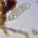
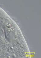
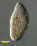
Portrait of Ophryoglena atra (Lieberkühn,1856), a histophagous hymenostome ciliate. some species of which are parasitic. Hosts include the nymphs of dragonflies and mayflies. The life cycle of parasitic forms is polymorphic, this portrait shows what is probably the theront or free-swimming hunter stage. The body is oval to elongate. The somatic ciliature consists of dense longitudinal kineties with a preoral suture. The oral aperture is located in the anterior quarter of the cell and has the outline of a right human ear. The buccal cavity spirals deep into the body and has an undulating membrane and three membranelles. Adjacent to the buccal cavity is the refringent organelle of Lieberkühn also referred to as the "watchglass organelle".This structure is thought to play a role in phototaxis which is positive in some phases of the life cycle and negative in others. O. atra is distinguished from other Ophryoglena species by its vermiform macronucleus (seen here). There is one contractile vacuole which empties through several pores. Collected from a freshwater pond populated by dragonfly and mayfly nymphs near Boise, Idaho. DIC.
-
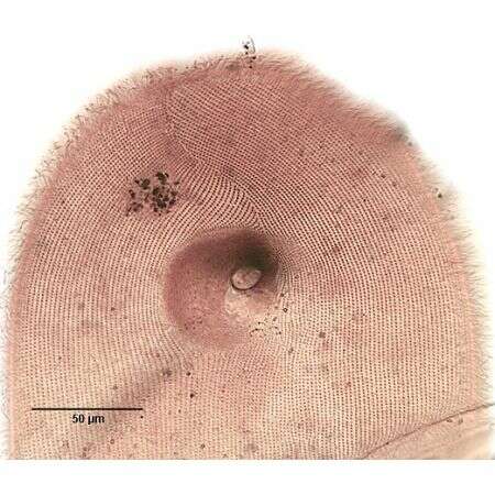
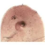
Infraciliature (ventral, anterior) of the ciliate, Ophryoglena flava (Ehrenberg, 1833). The numerous closely spaced longitudinal kineties on the right and left of the oral aperture converge at a sigmoid preoral suture. At least 15 postoral kineties terminate at the oral aperture. The dense inner ciliary rows of the buccal opening are seen here as a brown crescent in the concavity of which the refractile "watchglass organelle" (Lieberkühn's organelle) is located. Collected from a freshwater pond near Boise, Idaho. Silver carbonate method (Foissner, W.Europ. J. Protistol.27,313-330;1991). Brightfield.
-
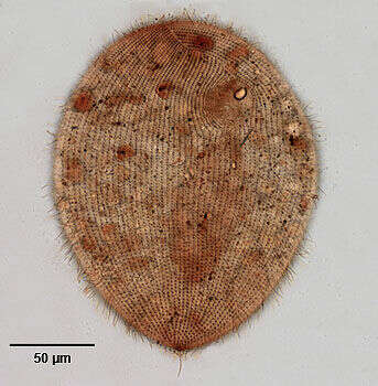
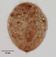
Infraciliature (ventral) of the ciliate, Ophryoglena utriculariae (Kahl, 1831). The numerous closely spaced longitudinal kineties on the right and left of the oral aperture converge at a sigmoid preoral suture. At least 15 postoral kineties terminate at the oral aperture. There is a single long caudal cilium. The dense inner ciliary rows of the buccal opening are seen here as a brown crescent in the concavity of which the refractile "watchglass organelle" (Lieberkühn's organelle) is located (seen well here).There is one right lateral contractile vacuole (not seen here). The large ellipsoid macronucleus is seen here with its anterior end at the posterior margin of the oral aperture. Collected from a freshwater pond near Boise, Idaho. Silver carbonate method (see Foissner, W.Europ. J. Protistol.27,313-330;1991). Brightfield.