-
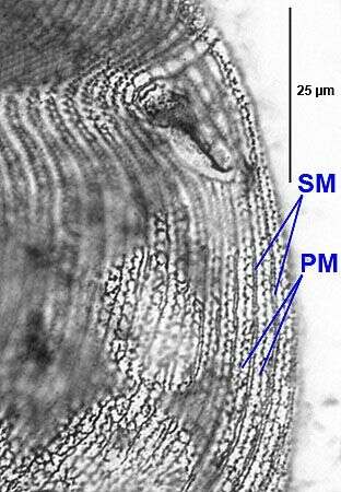
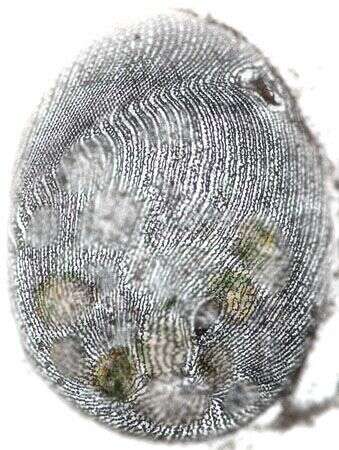
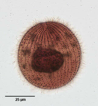
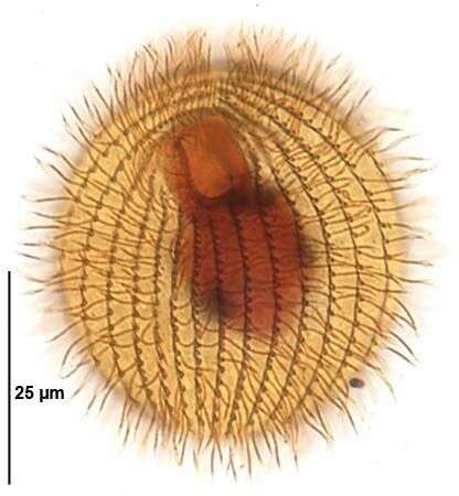
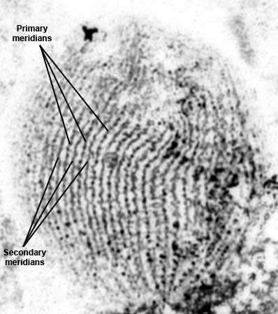
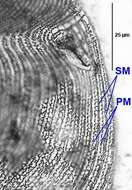
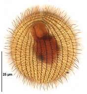
Silverline system of Colpidium colpoda (LOSANA,1829) STEIN,1860.2 secondary meridians (SM) run between pairs of primary meridians (PM). This pattern occurs sporadically in the same cell with some areas showing a single branched secondary meridian between pairs of primary meridians (similar to the pattern in C. kleini). From a putrefying raw culture from a freshwater pond near Boise, Idaho.October 2007. Stained by the dry silver nitrate technique (see Foissner, W. Europ. J. Protistol., 27:313-330;1991).Brightfield.
-
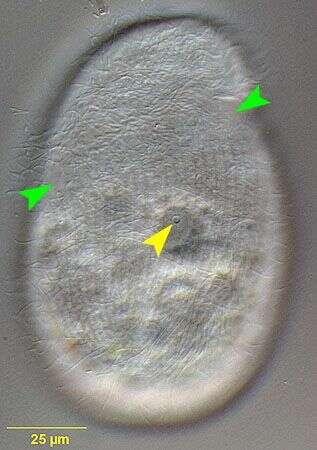
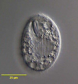
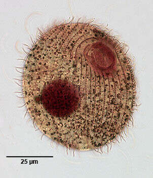
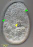
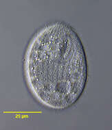
Right lateral view of the infraciliature of Colpidium colpoda (LOSANA,1829) STEIN,1860.The green arrowheads mark the oblique furrow that extends from the oral aperture accross the right side of the cell to the center of the dorsal surface.The somatic kineties are more closely spaced and bend strongly to the left in this depression.In the living cell this area appears as a more densely ciliated region on the right.The single pore of the contractile vacuole (yellow arrowhead) is located on the right dorsolateral surface. From a putrefying raw culture from a freshwater pond near Boise, Idaho.October 2007. DIC.
-
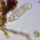
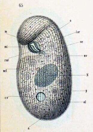
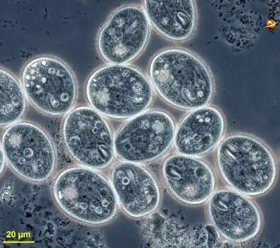
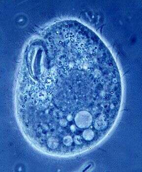
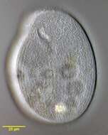
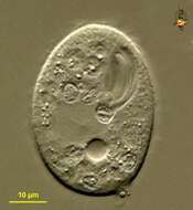
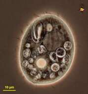
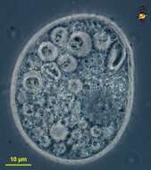
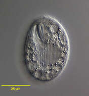
in vivo portrait (ventral view) of Colpidium colpoda (LOSANA,1829) STEIN,1860.From a putrefying raw culture from a freshwater pond near Boise, Idaho.October 2007. DIC.
-
Silverline system of Colpidium colpoda (LOSANA,1829) STEIN,1860.2 secondary meridians run between pairs of primary meridians. This pattern occurs sporadically in the same cell with some areas showing a single branched secondary meridian between pairs of primary meridians (similar to the pattern in C. kleini). From a putrefying raw culture from a freshwater pond near Boise, Idaho.October 2007. Stained by the dry silver nitrate technique (see Foissner, W. Europ. J. Protistol., 27:313-330;1991).Brightfield.
-
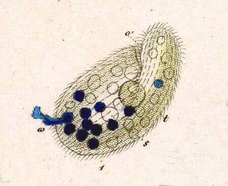
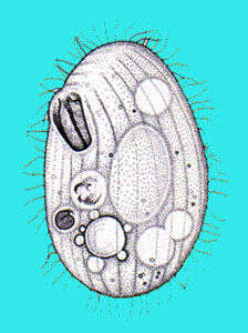
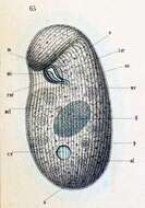
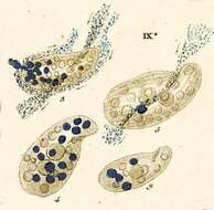
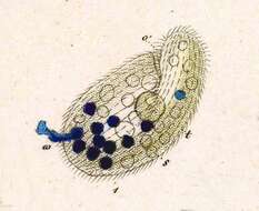
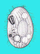
Key to Schewiakoff's abbrevations: a -- Anus al -- Pellicular alveoli cv -- Contractile vacuole l.or -- Left edge of mouth m -- Undulating membrane ni -- Inner undulating membrane N -- Macronucleus ncl -- Micronucleus o -- Mouth nv -- Food vacuole r.or --Right edge of mouth
-
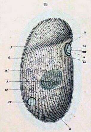
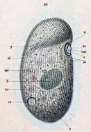
a -- Anus Key to Schewiakoff's abbreviations: al -- Pellicular alveoli cv -- Contractile vacuole m -- Undulating membrane mi -- Inner undulating membrane N -- Macronucleus ncl -- Micronuclues nv -- Food vacuole o -- Mouth oe -- Throat p -- Pellicle r.mr -- Right edge of mouth
-
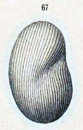
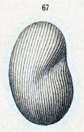
Dorsal view.
-
Described by Ehrenberg under the name Paramecium kolpoda.
-
Originally described by Ehrenberg under the name Paramecium colpoda.
-
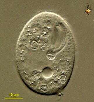
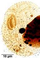
Glaucoma (glaw-comb-a) is a tetrahymenine ciliate (very closely related to Tetrahymena). It is flattened, but is made distinctive because the three membranelles in the mouth are very strongly developed. Eats bacteria which are brought into the oral cavity by the cilia in the mouth, and then packaged into food vacuoles at the cytostome - at the posterior end of the oral cavity. Large homogeneous region to the left is the macronucleus. Differential interference contrast.
-
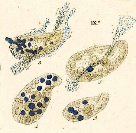
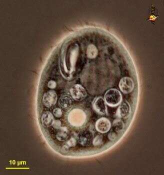
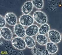
Glaucoma (glaw-comb-a) is a tetrahymenine ciliate (very closely related to Tetrahymena). It is flattened, but is made distinctive because the three membranelles in the mouth are very strongly developed. Eats bacteria, very obvious here in the food vacuoles. Large homogeneous region to the upper right is the macronucleus. Phase contrast.
-
Glaucoma (glaw-comb-a) is a tetrahymenine ciliate (very closely related to Tetrahymena). It is flattened, but is made distinctive because the three membranelles in the mouth are very strongly developed. Eats bacteria, very obvious here in the food vacuoles. Large homogeneous region to the lower right is the macronucleus. Phase contrast.
-
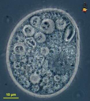
Glaucoma (glaw-comb-a) is a tetrahymenine ciliate (very closely related to Tetrahymena). It is flattened, but is made distinctive because the three membranelles in the mouth are very strongly developed. Eats bacteria. This shows variation among individuals within a population. Phase contrast.
-
-
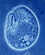
Phase contrast micrograph of lving cell showing well developed oral ciliature.
-
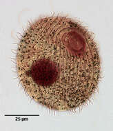
Portrait (ventral view) of the hymenostome ciliate, Glaucoma scintillans (Ehrenberg, 1830).
-
Portrait (dorsal view)of the hymenostome ciliate, Glaucoma scintillans (Ehrenberg, 1830).
-
Infraciliature (ventral view) of the hymenostome ciliate, Glaucoma scintillans (Ehrenberg, 1830). Stained by the silver carbonate technic (see Foissner, W. Europ. J. Protistol. 27:313-330;1991). Brightfield
-
Infraciliature (dorsal view) of the hymenostome ciliate, Glaucoma scintillans (Ehrenberg, 1830). Stained by the silver carbonate technic (see Foissner, W. Europ. J. Protistol. 27:313-330;1991). Brightfield
-
Oral infraciliature of the common hymenostome ciliate, Glaucoma scintillans (EHRENBERG,1830). Stained by the silver carbonate technique (Foissner,W. Europ. J. Protistol.27:313-330;1991).Brightfield.
-
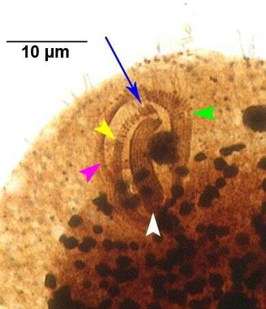
Oral infraciliature of the common hymenostome ciliate, Glaucoma scintillans (EHRENBERG,1830). The three adoral membranelles (M1,M2 and M3) are indicated by the green, white and yellow arrowheads respectively. The "X-body", a rhomboidal group of kinetids just anterior to the end of M2, is indicated by the blue arrow.The undulating membrane on the right margin of the oral aperture is indicated by the pink arrowhead.Stained by the silver carbonate technique (Foissner,W. Europ. J. Protistol.27:313-330;1991).Brightfield.
-
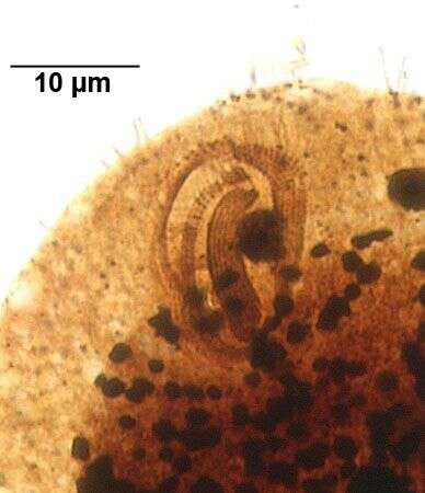
Oral infraciliature of the common hymenostome ciliate, Glaucoma scintillans (EHRENBERG,1830). There are three adoral membranelles (M1,M2 and M3) The "X-body" is a rhomboidal group of kinetids just anterior to the end of M2.Stained by the silver carbonate technique (Foissner,W. Europ. J. Protistol.27:313-330;1991).Brightfield.
-
Infraciliature (ventral view) of the hymenostome ciliate, Glaucoma scintillans (Ehrenberg, 1830). Stained by the silver carbonate technic (see Foissner, W. Europ. J. Protistol. 27:313-330;1991). Brightfield.
-
Silverline system of the hymenostome ciliate, Glaucoma scintillans (Ehrenberg, 1830). Stained by the dry silver nitrate technic (see Foissner, W. Europ. J. Protistol. 27:313-330;1991). Brightfield.