Melibe arianeae
(
İngilizce
)
wikipedia EN tarafından sağlandı
Melibe arianeae is a species of sea slug, a nudibranch, a marine gastropod mollusc in the family Tethydidae.[1]
Description
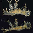
The overall shape of Melibe arianeae is elongate and limaciform. Anterolaterally, it is compressed to a small degree. It tapers posteriorly to form a long and conical shape to the end of the foot.[2][3][4]
The surface of the body of this species is almost transparent. It and the cerata and rhinophoral sheaths are completely covered with multiple, tiny tubercles that are white and opaque. The surface has blotches that are white and opaque as well as multiple orange flecks. The internal organs appear orange-brownish.
The cerata are mainly warty in appearance due to complete coverage of small tubercules. They are oval and are inflated with their distal ends being bifurcate or trifurcate, or, simple. Within the cerata, the branches of the digestive gland are visible due to the cerata being transparent. This gland appears as an axis brownish in colour.[5][6]
Distribution
This species is found in the tropical western Atlantic, the type material, so far the only material known, was found in Florida.[7]
References

- lisans
- cc-by-sa-3.0
- telif hakkı
- Wikipedia authors and editors
Melibe arianeae: Brief Summary
(
İngilizce
)
wikipedia EN tarafından sağlandı
- lisans
- cc-by-sa-3.0
- telif hakkı
- Wikipedia authors and editors
Description
(
İngilizce
)
Zookeys tarafından sağlandı
The living animals are nearly transparent, with numerous orange flecks and opaque white blotches all over its surface, and orange-brownish colored internal organs (Fig. 2). The body is limaciform and elongate, somewhat compressed anterolaterally, tapering posteriorly into a long, conical posterior end of the foot. The entire body surface, including cerata and rhinophoral sheaths are covered by numerous minute, opaque white tubercles. In the center of the dorsum of the holotype there are several (8) transparent tentacular papillae of different sizes, also covered with small white tubercles and having opaque white apices. The foot sole is wider anteriorly, it is covered with orange flecks and opaque white blotches as the dorsal surface, but it also has a faint white rim. The circular oral hood is small compared to the rest of the body. The margin of the hood is entire (with no indentations) and bears two rows of elongate papillae. There are papillae on the dorsal surface of the hood, generally resembling those on the body surface, and more concentrated towards the anterior margin. The rhinophores emerge from the posterior end of the oral hood. The rhinophores have 3–4 perfoliations.The rhinophoral sheaths are somewhat inflated and cylindrical, lacking a leaf-like posterior process. The sheaths have 2–3 posterior papillae. The cerata are inflated, oval, completely covered with small tubercles that give it a broadly warty look. Their distal ends of the cerata are either simple, bifurcate or trifurcate, independently of their size. The cerata are transparent, and the branches of the digestive gland within them are visible as a brownish axis. There are seven cerata alternating on each side of the dorsal midline of the holotype. The anus is located dorsol-lateraly, midway between the first and second anterior cerata. The position of the nephroproct could not be determined. The gonopore is lateral, anterior to the anteriormost right ceras. There are no papillae associated with the gonopore.
The buccal mass is devoid of a radula but contains a pair of simple, chitinous jaws. The jaws (not illustrated) have smooth borders and lack denticles on the masticatory border. The short esophagus emerges from the posterior end of the buccal mass and expands into a muscular stomach (Fig. 3B). Two small salivary glands are located laterally, one on each side of the buccal mass. The posterior portion of the stomach contains 18 elongate, thick and robust chitinous plates of various sizes (Fig. 4). The reproductive system is triaulic and contains a series of four spherical, well-separated ovotestis bodies, connected to a large ampulla. The ampulla connects into the female gland complex (Fig. 3A) near the point where the prostate emerges. The prostate is a short and wide glandular structure connected to a long, and convoluted deferent duct that expands distally into the penial sac. The vagina is short and wide and connects directly into a large bursa copulatrix. The narrow and straight uterine duct connects to the female gland complex. A serial seminal receptacle (present in other species of Melibe) was not observed. The central nervous system (Fig. 3B) is located above the esophagus and contains a fused pair of cerebral and pleural ganglia, as well as a pair of pedal ganglia. The buccal ganglia are located at the proximal end of the buccal mass.
- lisans
- cc-by-3.0
- telif hakkı
- Erika Espinoza, Anne DuPont, Ángel Valdés
- bibliyografik atıf
- Espinoza E, DuPont A, Valdés (2013) A tropical Atlantic species of Melibe Rang, 1829 (Mollusca, Nudibranchia, Tethyiidae) ZooKeys 316: 55–66
- yazar
- Erika Espinoza
- yazar
- Anne DuPont
- yazar
- Ángel Valdés
_-_ZooKeys-316-055-g002B.jpeg)