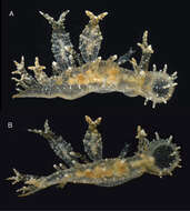
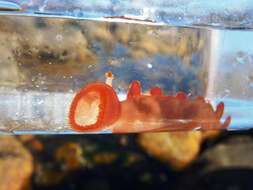
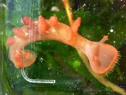
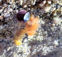
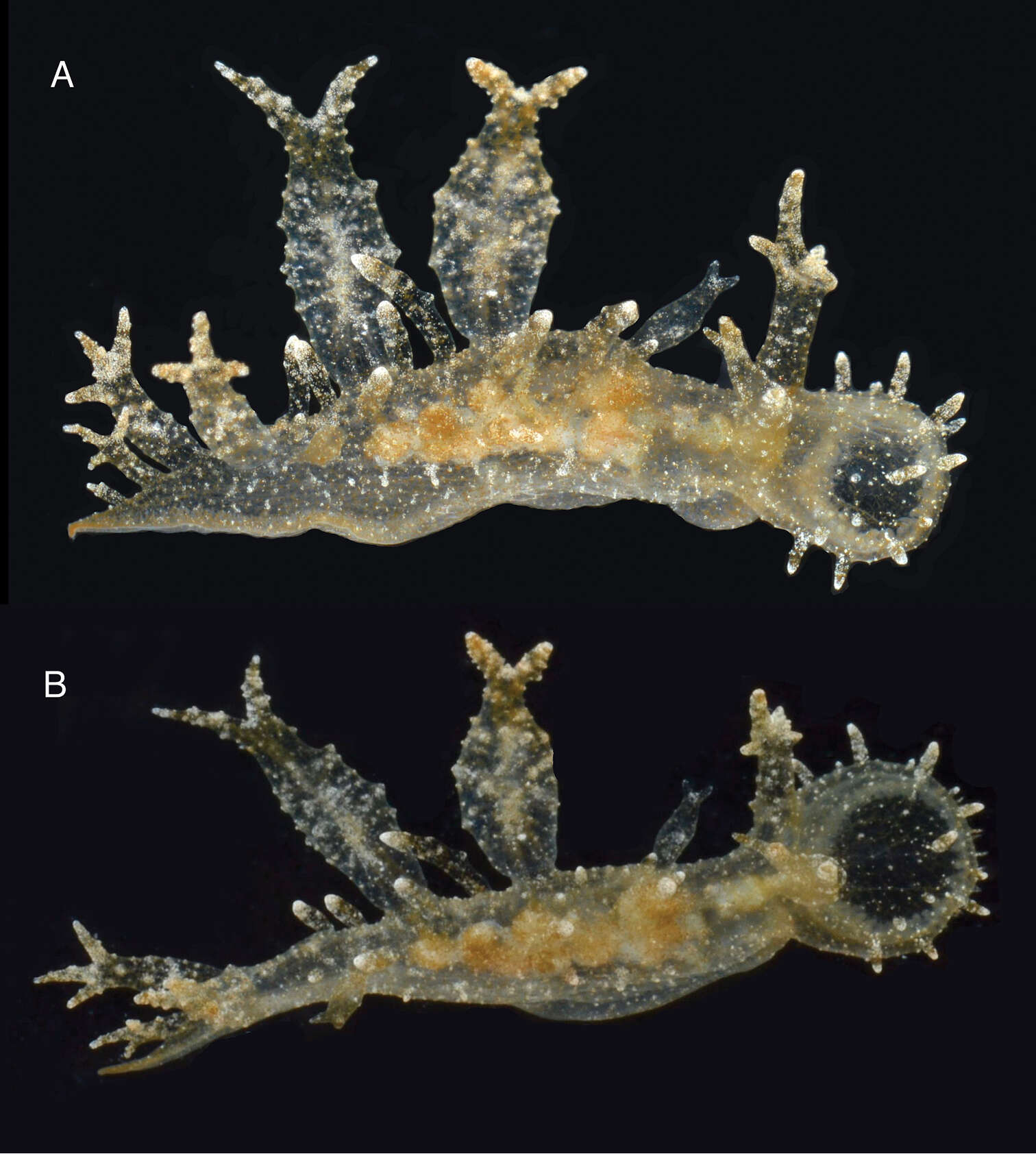
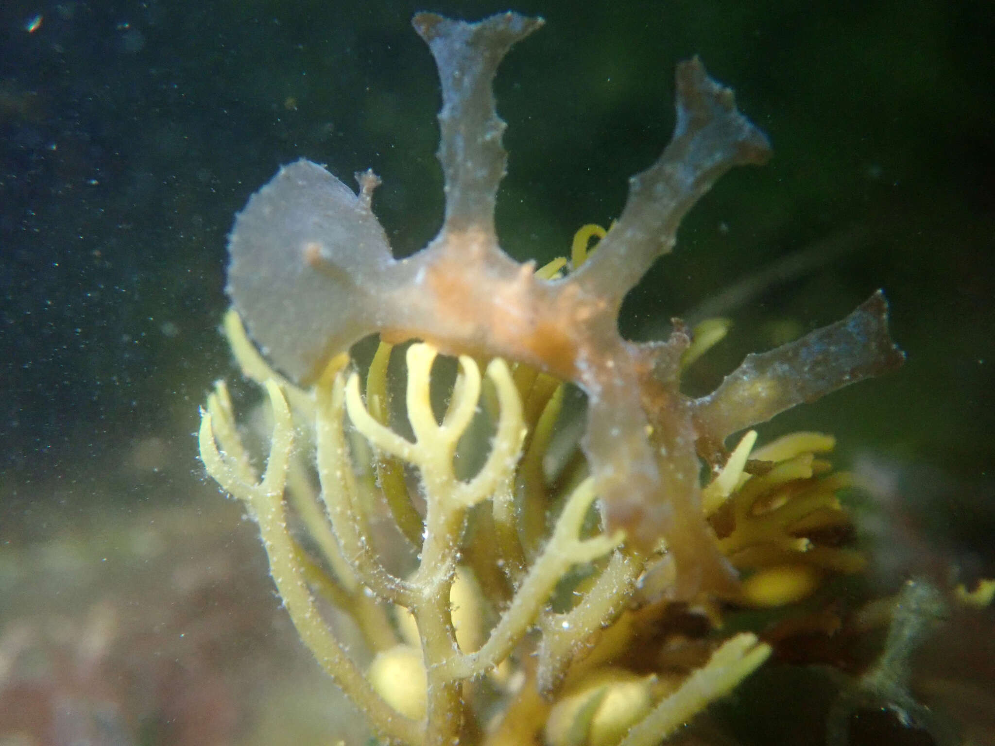
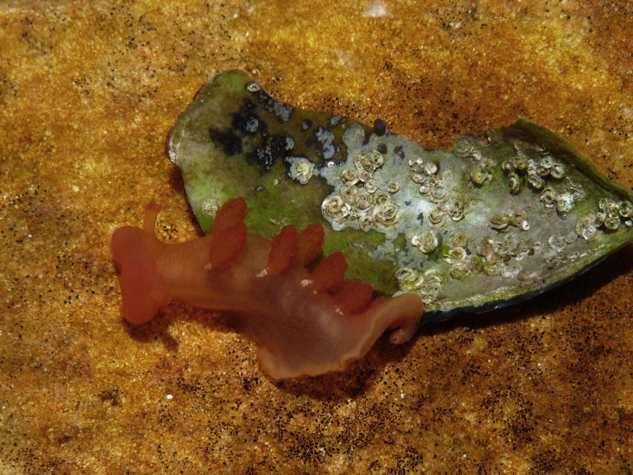
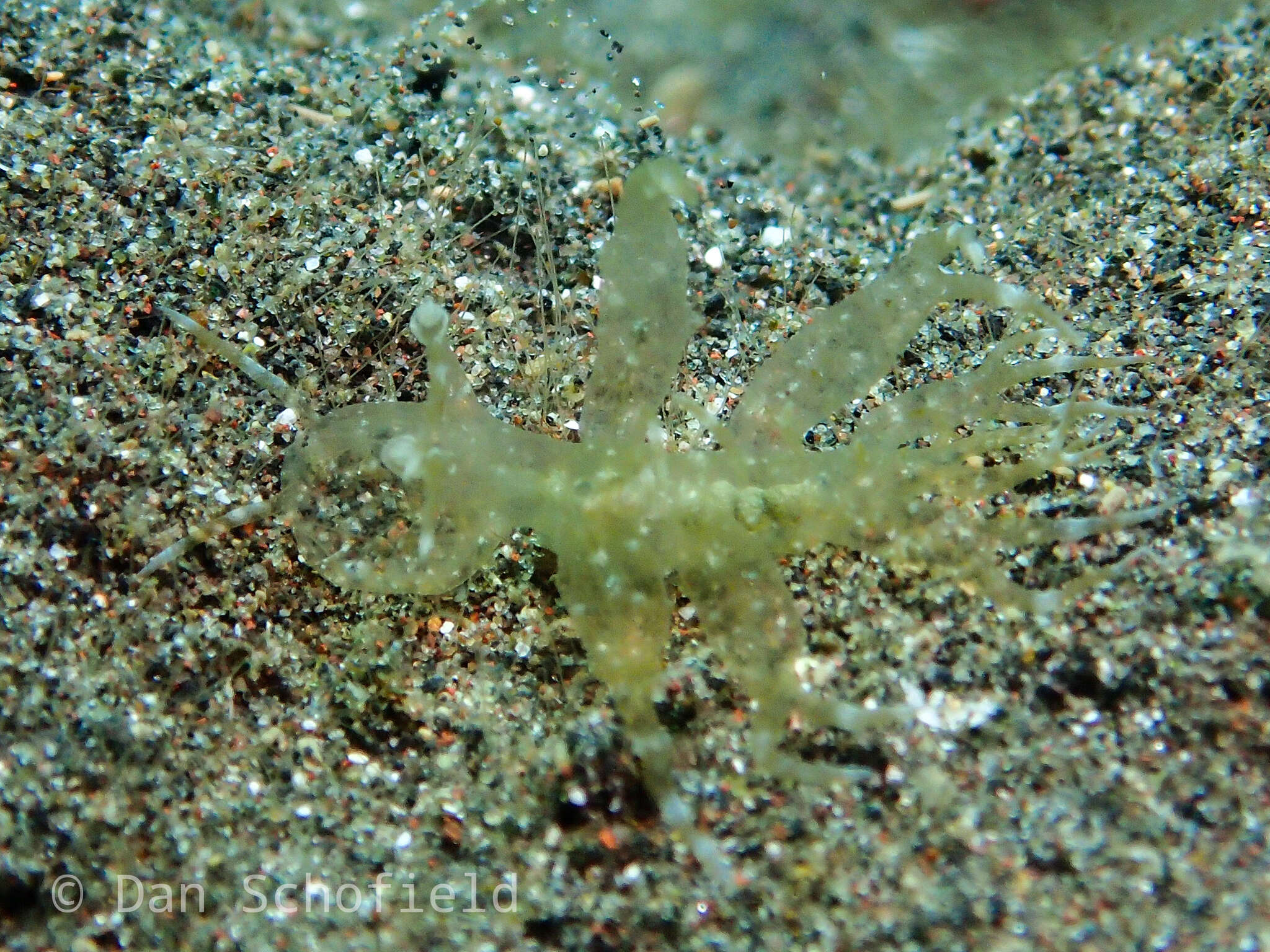
Figure 2.Two views of the holotype of Melibe arianeae sp. n. (LACM 3258). A. Dorsolateral view showing the right side of the animal. B. Dorsal view showing the oral hood border through the semi-transparent skin.
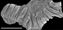
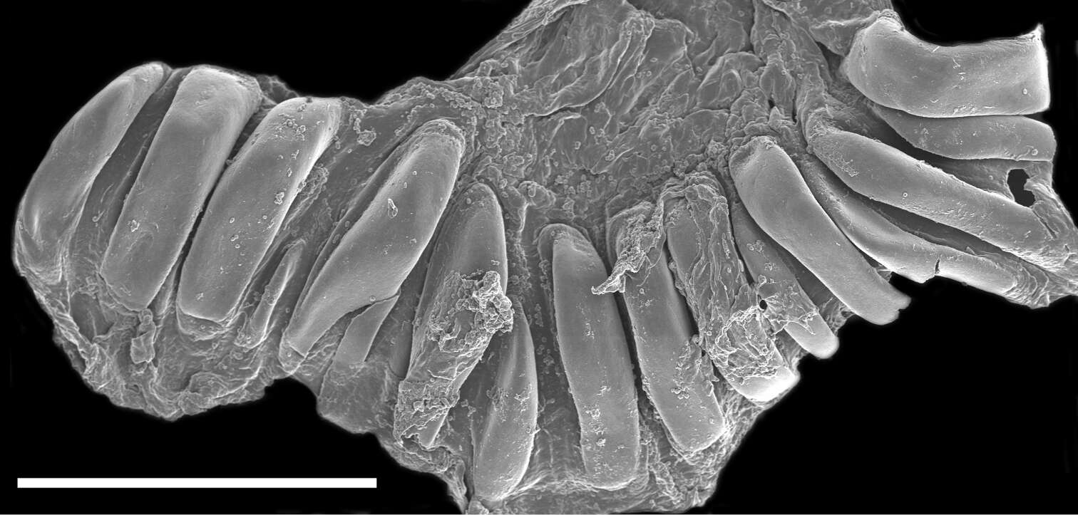
Figure 4.Scanning electron micrograph of the dissected stomach of the paratype of Melibe arianeae sp. n. (LACM 3259) showing the stomach plates. Scale bar = 500 µm.