-
All Biocode files are based on field identifications to the best of the researcher’s ability at the time.
-
2005 California Academy of Sciences
CalPhotos
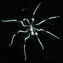
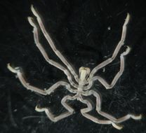
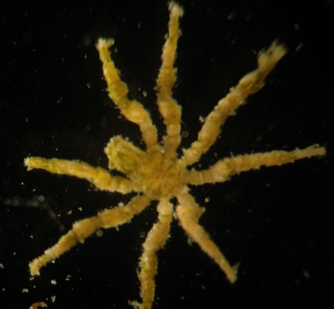
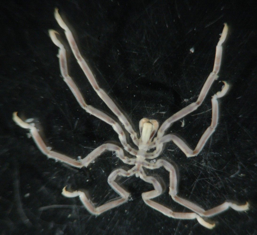
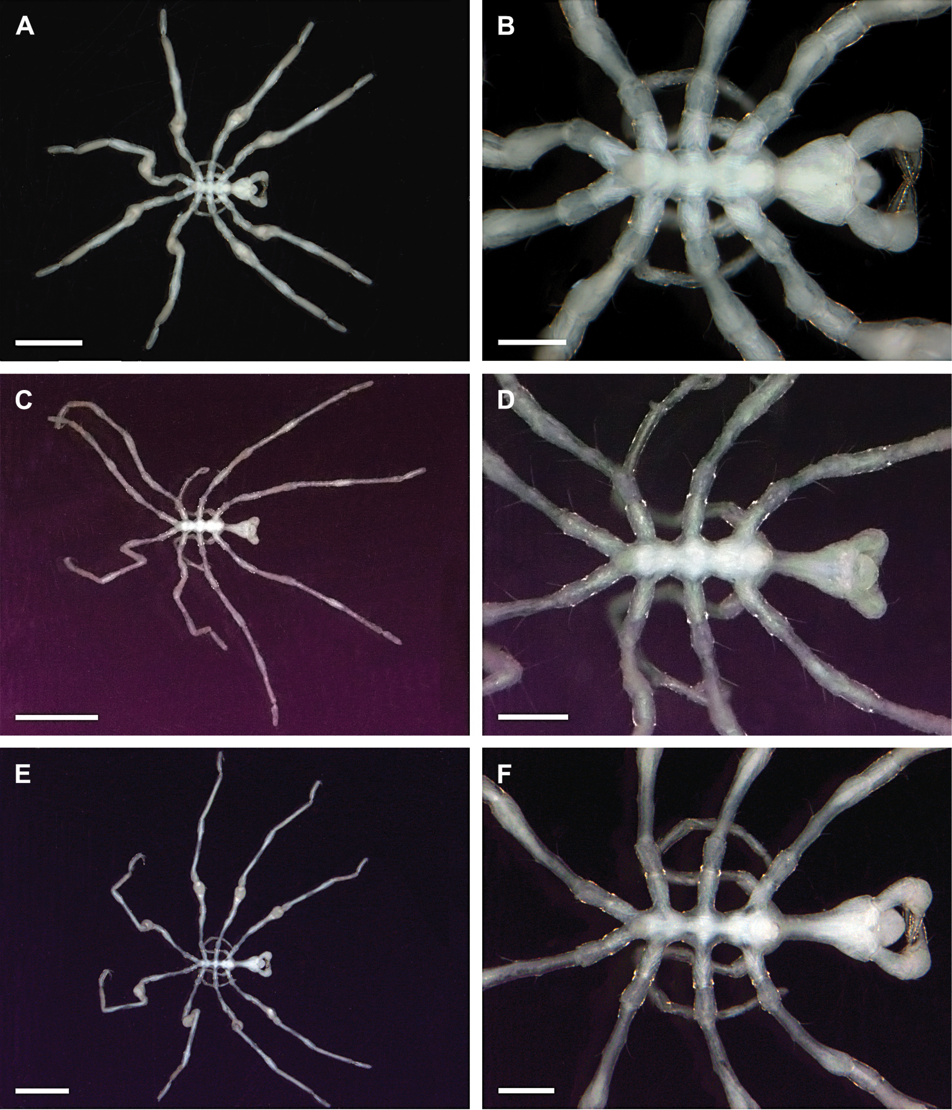
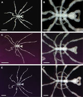
Female sea spider on hydroid Aglaophenia latirostris, 8 mm across. Moves very deliberately. Feeds on the hydroid. This individual has lost one leg, but it will not be replaced as these arthropods do not molt.
-
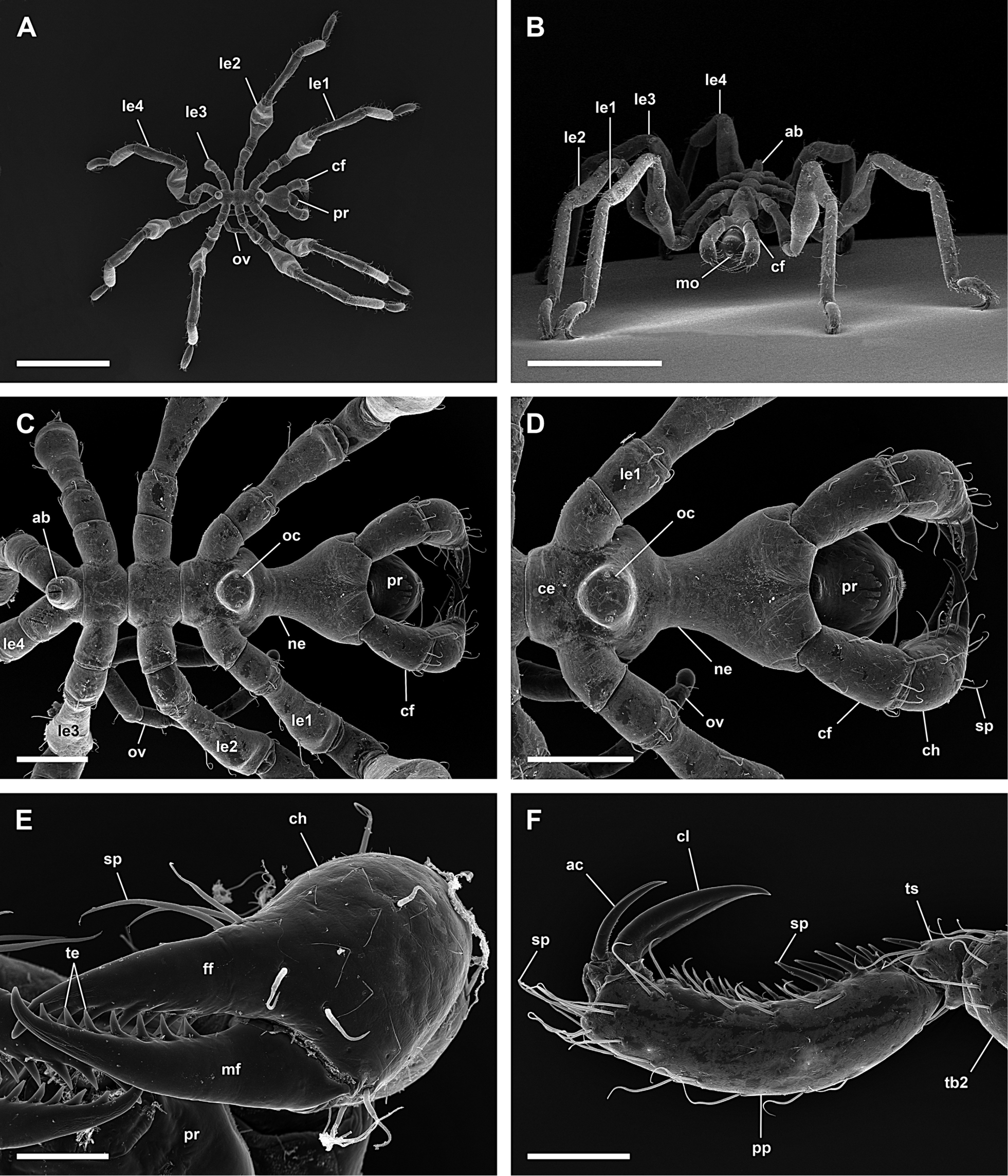
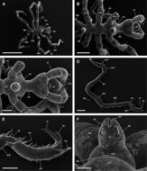
Figure 2.Ammotheidae 1; A, B: Achelia echinata, male, dorsal view; scales 500 µm and 250 µm, respectively; C, D: Achelia langi, male, dorsal view; scales 1 mm and 500 µm, respectively; E, F: Achelia vulgaris, male, dorsal view; scales 1 mm and 250 µm, respectively.
-
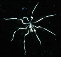
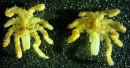
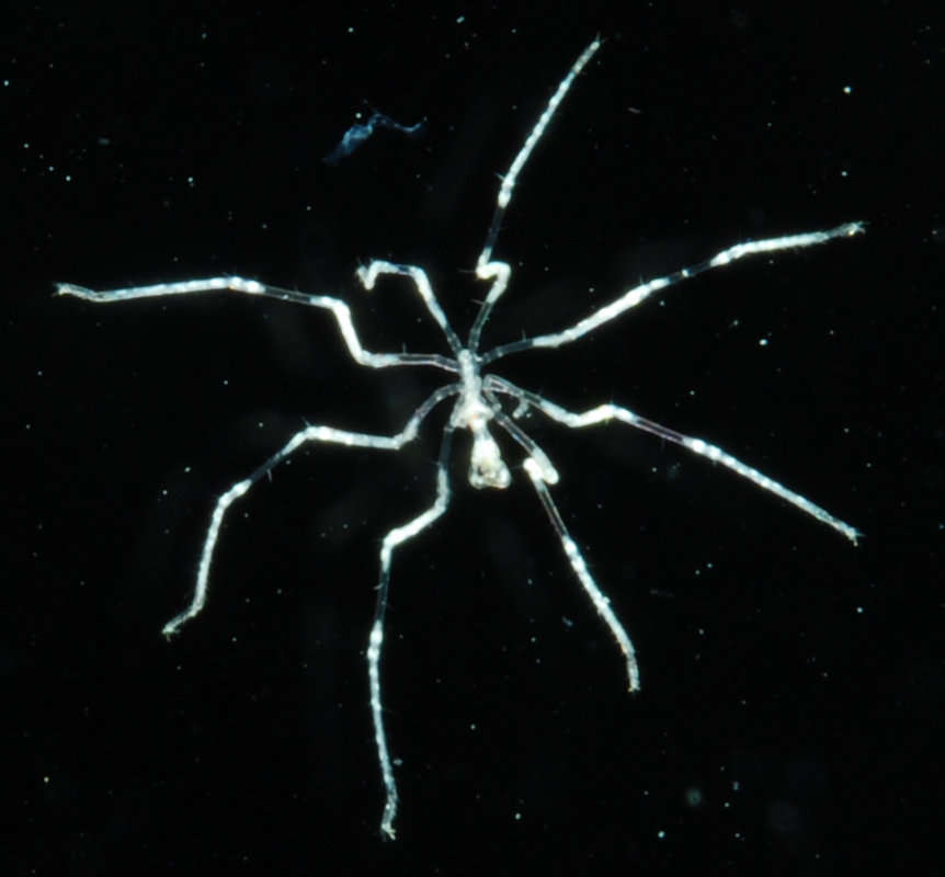
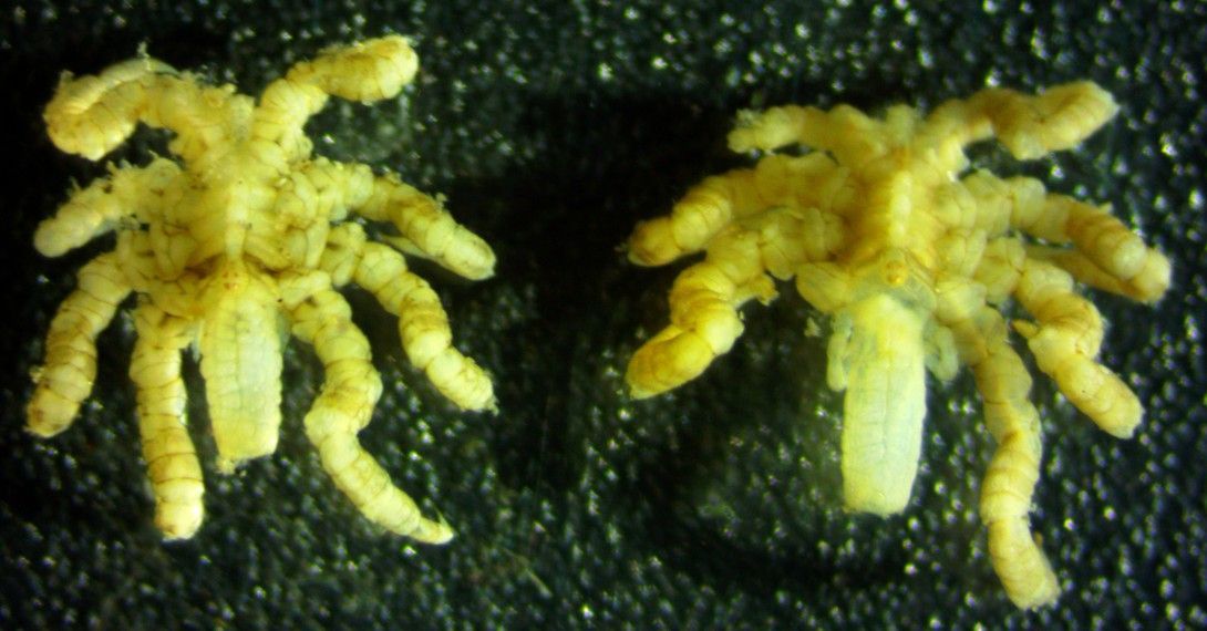
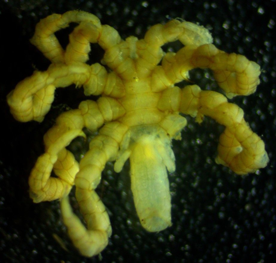
This pair of individuals, which includes the individual shown at the top of the page, were both on the same anenome. Though I did not check thoroughly, I did not see ovigerous legs on either individual suggesting they are both female.
-
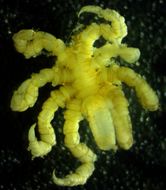
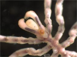
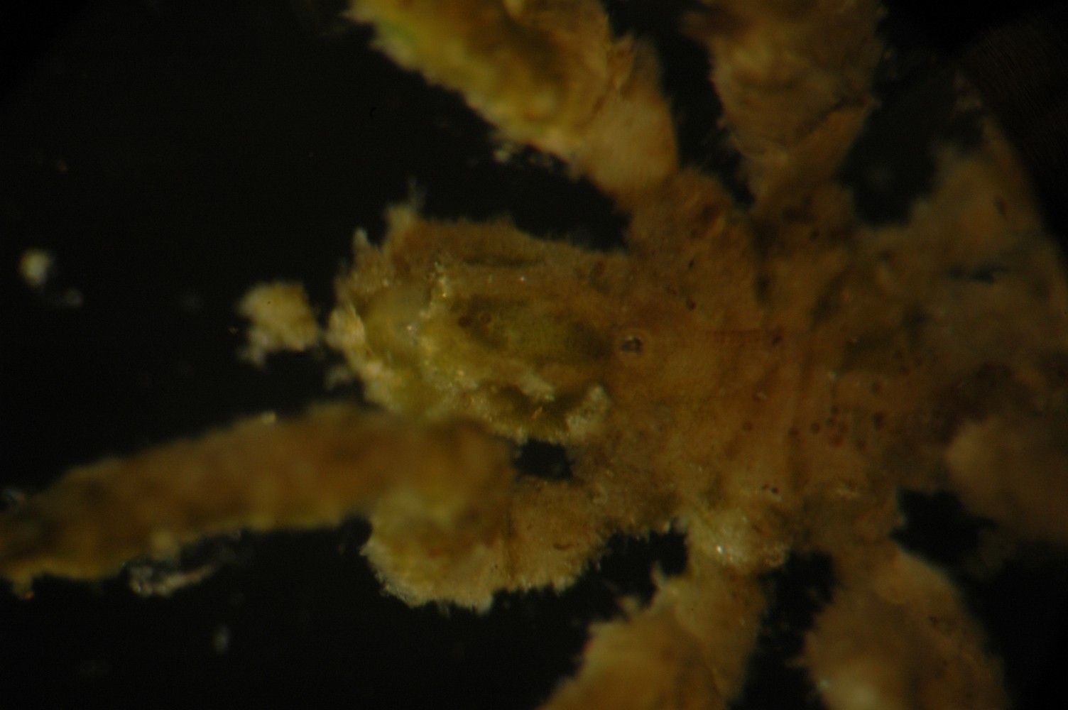
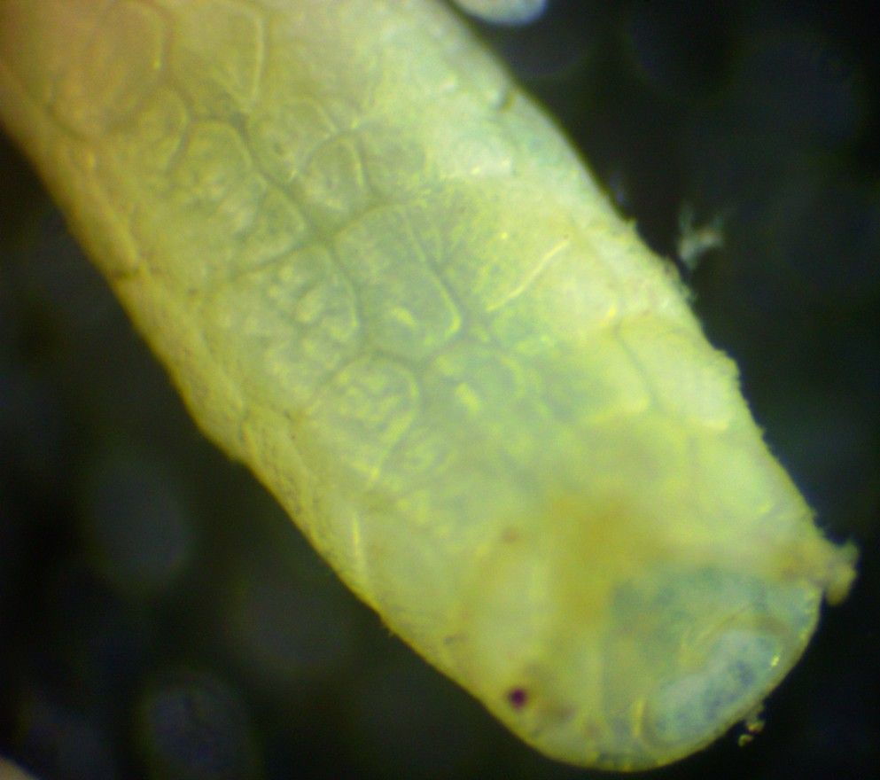
In this closeup view one can see the tiny turretlike head with 4 eyes (near the left end of the body, between the left (front) pair of legs). The proboscis is much larger than the head and extends to the left of the body.
-
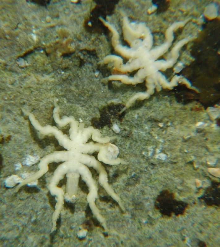
Pycnogonum stearnsi from Little Corona del Mar, CA. Total leg span about 1 cm. Animal was in a tidepool next to the base of an Anthopleura sola anemone. The head with its large proboscis is facing left (there is a bubble at the tip of the proboscis) (Photo by: Dave Cowles, November 2006)
-
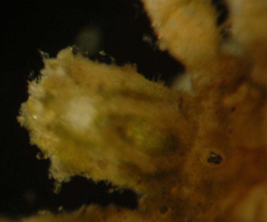
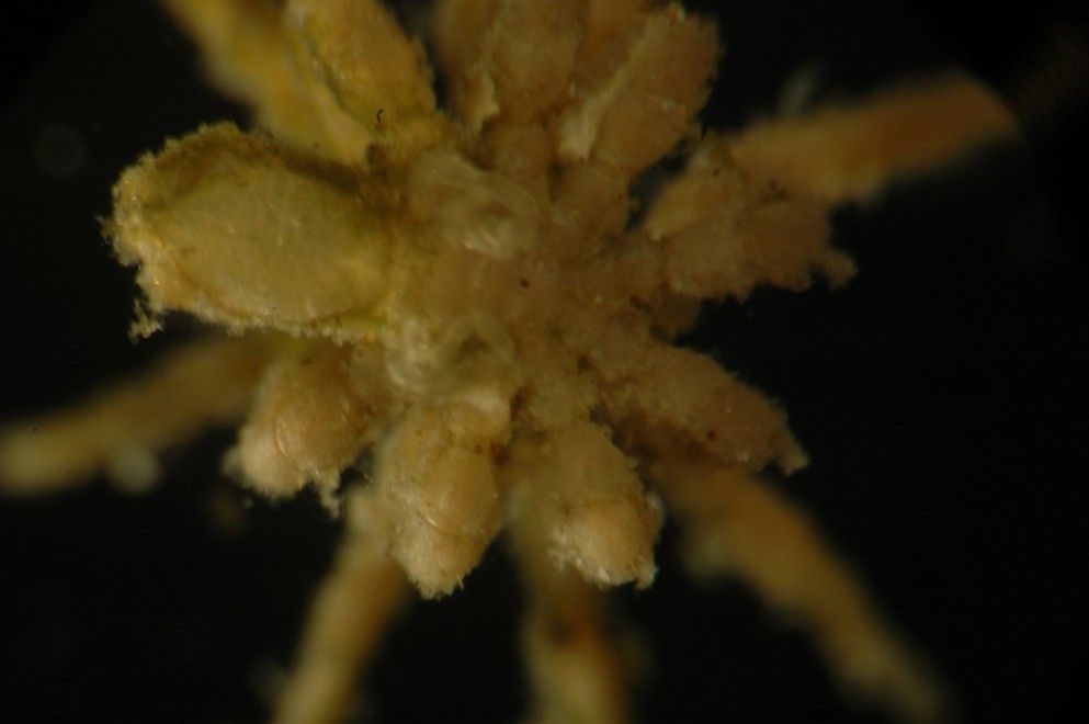
Here is an even closer view of the turret-like head, which appears as a small tubercle covered with 4 eyes, and also of the proboscis
-
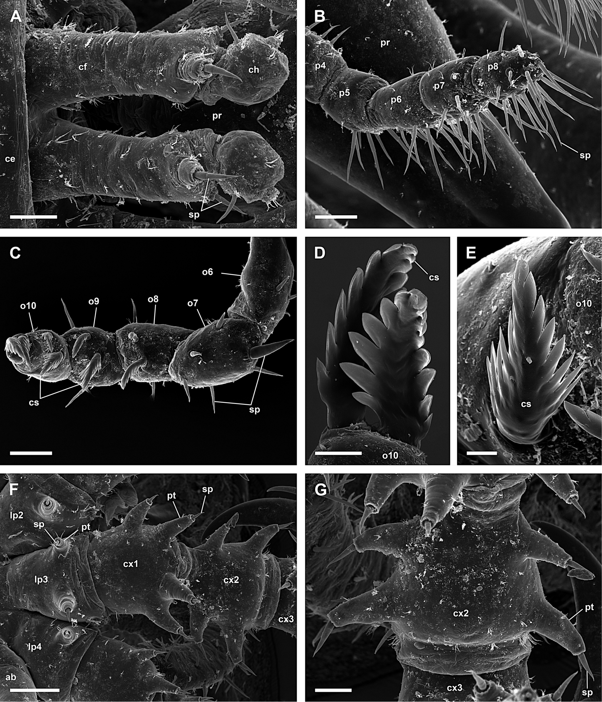
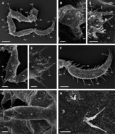
Figure 6.Achelia echinata, male; A: Chelifores with reduced chela; scale 40 µm; B: Distal articles of right 8-articled palp; scale 40 µm; C: Distal articles of 10-articled oviger; scale 40 µm; D, E: Compound spines on last oviger-article; scales 10 µm and 5 µm, respectively; F: Lateral process, coxa 1 and 2 of right 3rd leg, 2 protuberances with spine on each side of coxa 2; scale 100 µm; G: Coxa 2 with 2 protuberances with spine on each side (right 3rd leg); scale 40 µm.
-
-
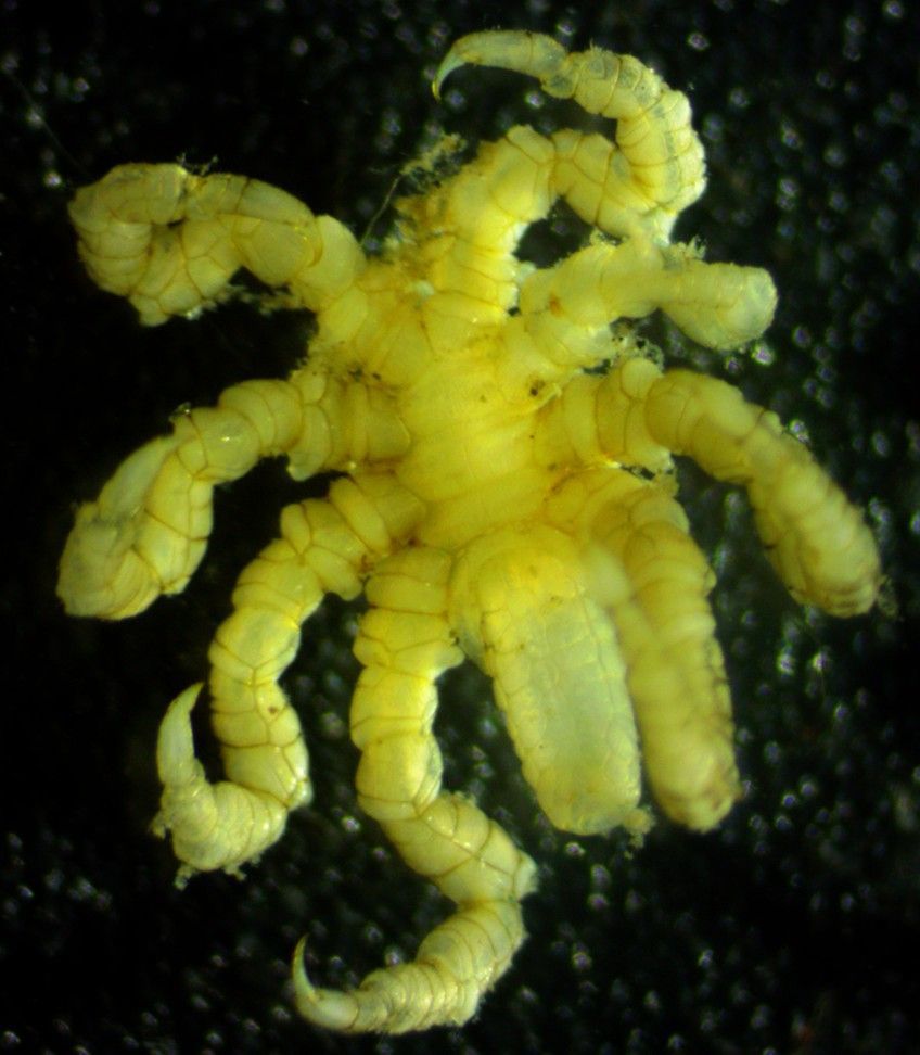
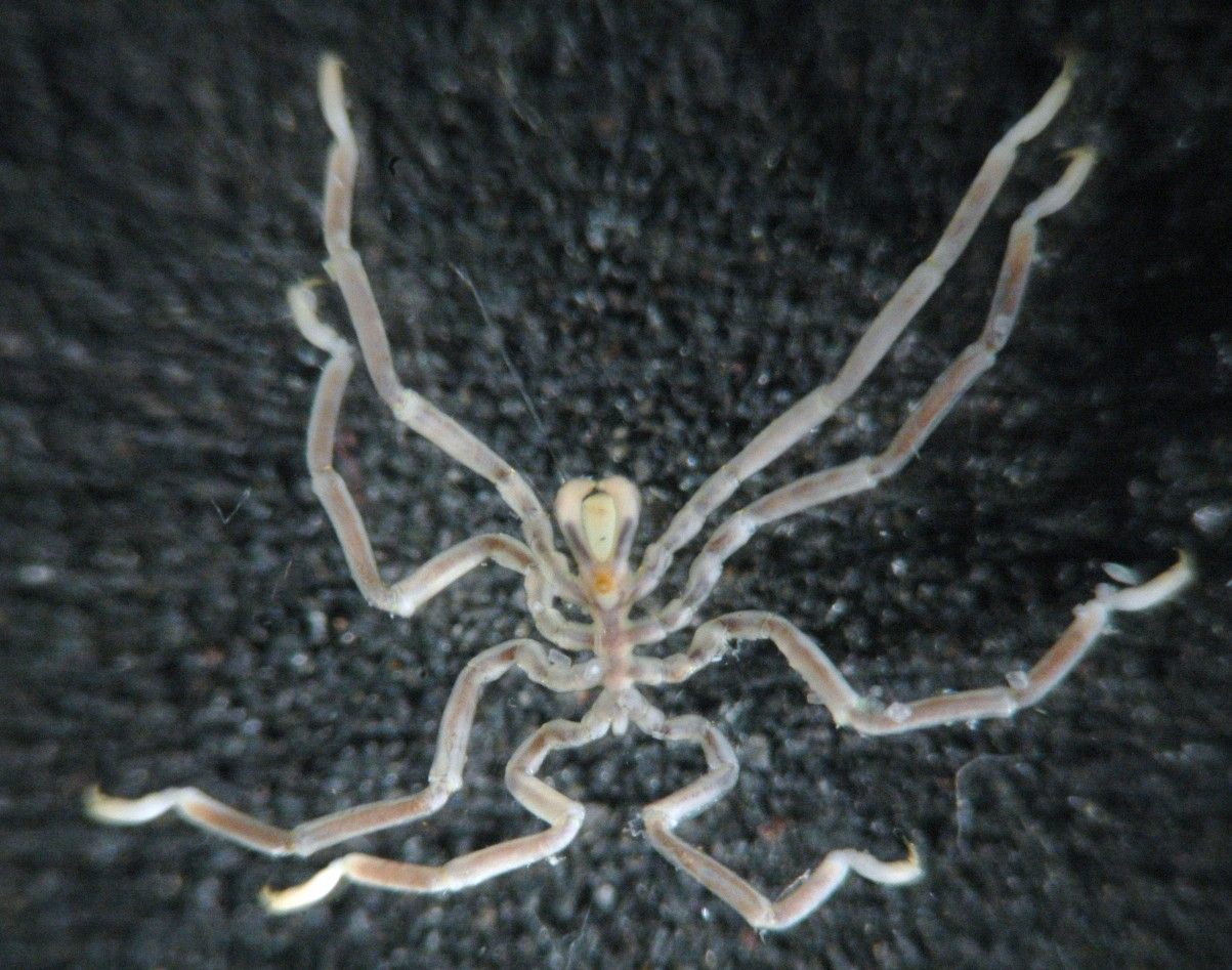
In this ventral view, the small ovigerous legs can be seen on the underside of the first body segment (left side, just to the right of the proboscis). Here the animal is extending its other, walking legs upward and away from the camera. The proboscis is to the left. Females of some families lack ovigerous legs and in other families they are reduced in females.
-
Figure 7.Achelia echinata, male; A: Left 3rd leg; scale 200 µm; B: Lateral view of coxa 2 with genital protuberance (right 4th leg); scale 40 µm; C: Genital opening (right 3rd leg); scale 20 µm; D: Lateral view of femur with cement gland on distal part (left 3rd leg); scale 100 µm; E: Cement gland (right 4th leg); scale 40 µm; F: Tarsus, propodus, and claw, auxiliary claws about half as long as claw (left 3rd leg); scale 100 µm; G: Abdomen; scale 40 µm; H: Hair and slit organ on dorsal side of trunk; scale 5 µm.
-
-
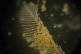
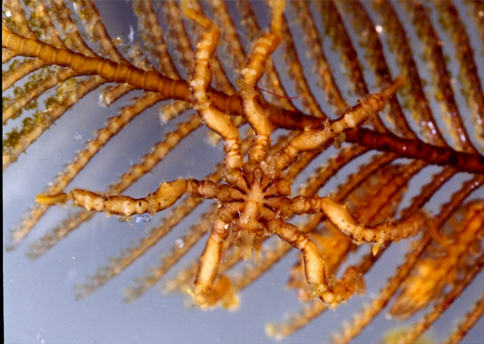
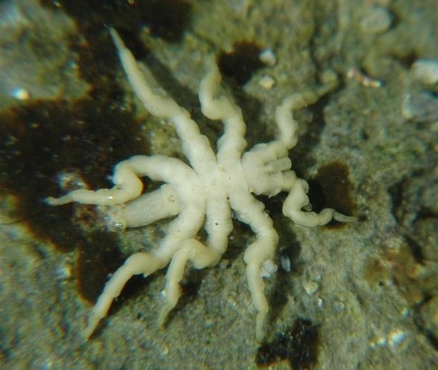
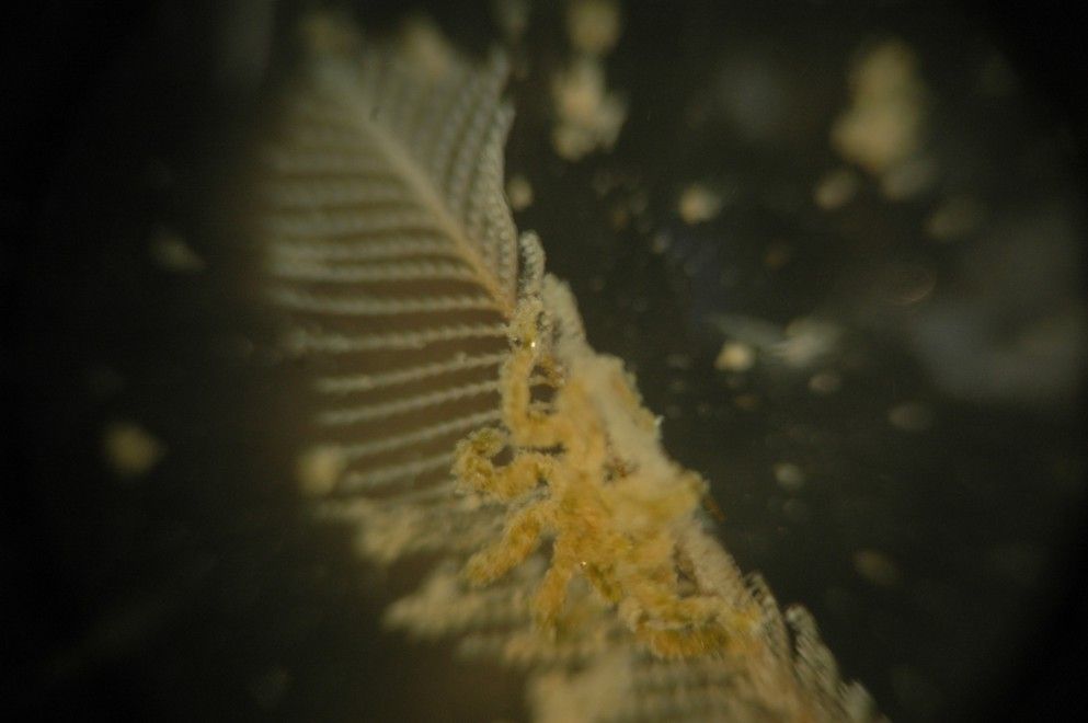
Pycnogonids are frequently found clinging to hydroids, as here on the hydroid Aglaophenia sp.
-
Figure 8.Achelia echinata, female; A: Dorsal view; scale 400 µm; B: Ventral view of proboscis; scale 100 µm; C: Left 10-articled oviger; scale 100 µm; D: Lateral process, coxa 1 and 2 of right 3rd leg, lateral processes touch each other; scale 100 µm; E: Left 3rd leg; scale 200 µm; F: Ventral view of coxa 2 with genital opening, distal is right (left 3rd leg); scale 40 µm; G: Genital opening; scale 20 µm.
-
-
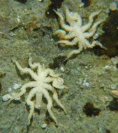
Tanystylum occidentalis from under an intertidal rock near the S end of Lopez Island. Total leg span about 8 mm. (Photo by: Dave Cowles, July 2006)
-
Figure 26.Callipallenidae 1; A, B: Callipallene emaciata, female, dorsal view; scales 1 mm and 250 µm, respectively; C, D: Callipallene phantoma, male, dorsal view; scales 1 mm and 250 µm, respectively; E, F: Callipallene producta, female, dorsal view; scales 1 mm and 250 µm, respectively.
-
-
Figure 28.Callipallene emaciata, male; A: Dorsal view; scale 1 mm; B: Dorsal view of trunk; scale 200 µm; C: Dorsal view of cephalon with rather slender neck; scale 100 µm; D: Right 3rd leg; scale 100 µm; E: Tarsus, strongly curved propodus, and claw, auxiliary claws about half as long as claw (right 3rd leg); scale 100 µm; F: Segment 3, 4 and abdomen; scale 40 µm.
-
This is a view of the underside of the whole animal
-
Figure 29.Callipallene emaciata, female; A: Dorsal view; scale 1 mm; B: Frontal view; scale 1 mm; C: Dorsal view of trunk; scale 200 µm; D: Dorsal view of cephalon with rather slender neck; scale 200 µm; E: Left chela, dorsal is up; scale 40 µm; F: Tarsus, strongly curved propodus, and claw, auxiliary claws about half as long as claw (left 3rd leg); scale 100 µm.
-
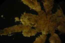
Phoxichilidium femoratum found by Joanna Cowles on an Epiactis ritteri anemone in a sea cave at Cape Flattery. Leg span 1.5 cm. Since no ovigerous legs are present, this must be a female. (Photo by: Dave Cowles, July 2009)
-
Figure 26.Callipallenidae 1; A, B: Callipallene emaciata, female, dorsal view; scales 1 mm and 250 µm, respectively; C, D: Callipallene phantoma, male, dorsal view; scales 1 mm and 250 µm, respectively; E, F: Callipallene producta, female, dorsal view; scales 1 mm and 250 µm, respectively.
-