-
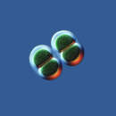
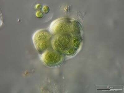
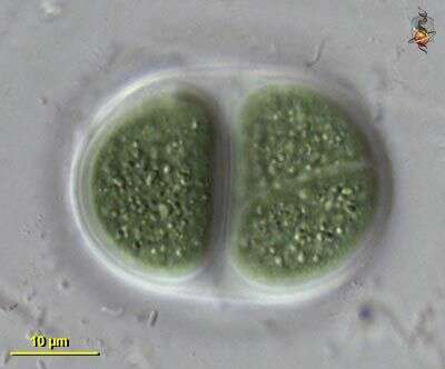
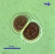
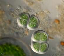
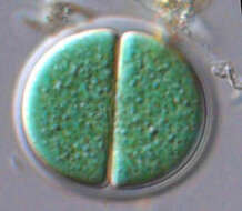
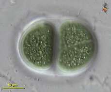
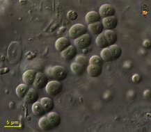
Chroococcus turgidus, (Cyanobacteria, Chroococales), from Lake Kinneret pelagic waters, April 2006, showing 2 daughter cells after division by simple binary fission â as characteristic for most Chroococcales species. This species is common in the plankton of Lake Kinneret throughout the year. Usually there are 2 â 8 cells in a colony. Clearly delimited colorless mucilaginous envelopes surround the individual cells following their contours, and the entire colony. Cell diameter: 8 â 11 µm.
-
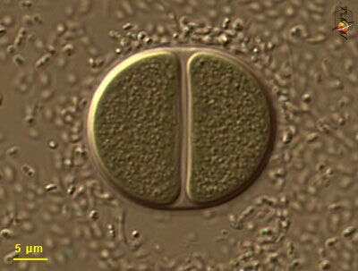
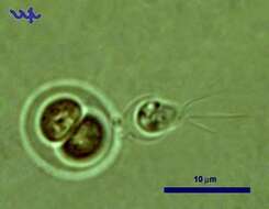
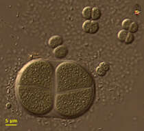
Chroococcus turgidus (Cyanobacteria, Chroococcales) is common in Lake Kinneret in recent years. In this unusual photograph a choanoflagellate is attached to its outer shell.
-
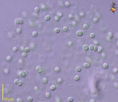
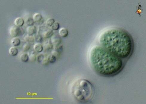
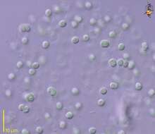
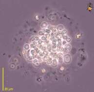
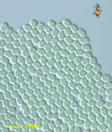
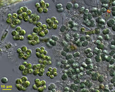
Anacystis (a-na-cyst-is) is a cyanobacterium in which globular cells are located within a gelatinous matrix. Some of the cyanobacteria with this form can be toxic. Differential interference contrast.
-
Anacystis (a-na-cyst-is), a cyanobacterium or blue green algae, in which the cells are clumped together within a mucus material. The clusters have to be compressed so that the individual cells can be observed. This genus adopts a variety of forms. Some Anacystis species are known to produce toxins. Phase contrast.
-
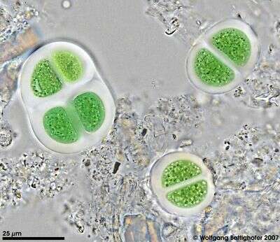
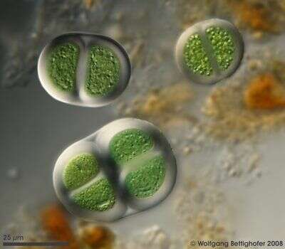
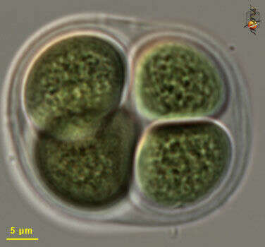
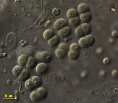
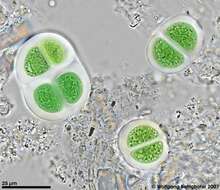
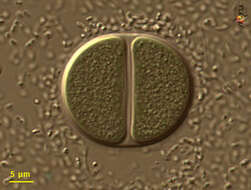
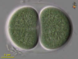
High resolution photo of Chroococcus turgidus using Planapo 63/1.4. Sample from sphagnum pond situated in the northern alpine region of Austria near Salzburg. Images were taken using Zeiss Universal with Olympus C7070 CCD camera.
-
High resolution photo of Chroococcus turgidus using Planapo 63/1.4 with DIC. Sample from sphagnum pond situated in the northern alpine region of Austria near Salzburg. Images were taken using Zeiss Universal with Olympus C7070 CCD camera.
-
High resolution photo of Chroococcus turgidus using Planapo 63/1.4 with DIC. Sample from sphagnum pond situated in the northern alpine region of Austria near Salzburg. Images were taken using Zeiss Universal with Olympus C7070 CCD camera.
-
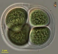
Scale bar indicates 25 µm. Sample from sphagnum pond situated in the northern alpine region of Austria near Salzburg. Images were taken using Zeiss Universal with Olympus C7070 CCD camera.
-
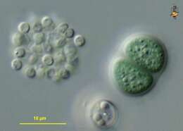
Chroococcus (crow-o-cock-us), large cyanobacterium, typically two (but sometimes one) cells enclosed within a mucus sheath. Photosynthetic pigment distributed through cytoplasm, which may have a granular texture, but does not have subcompartments (organelles). Differential interference contrast.
-
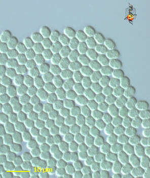
Chroococcus (crow-owe-cock-us) coccoid cyanobacteria, adhering to each other to form extensive flat sheets, no evident mucus sheath or heterocysts. Differential Interference Contrast.
-
Chroococcus (crow-o-cock-us), large cyanobacterium, typically two (but sometimes one) cells enclosed within a mucus sheath. Photosynthetic pigment distributed through cytoplasm, which may have a granular texture, but does not have subcompartments (organelles). Differential interference contrast.
-
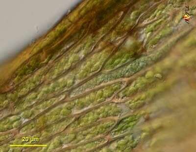
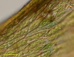
Chroococcus (crow-owe-cock-us) tentative identification. Coccoid blue green algal cells. Found as one of several cyanobacterial epibionts on the leaves of the moss Hygrohypnum, a site which seems to be a focus for nitrogen fixation. In this case the cyanobacterial cells have occupied one of the cortical cells of the plant. Differential interference contrast.
-
Chroococcus (crow-owe-cock-us) tentative identification. Coccoid blue green algal cells. Found as one of several cyanobacterial epibionts on the leaves of the moss Hygrohypnum, a site which seems to be a focus for nitrogen fixation. Differential interference contrast.
-
Chroomonas. Cell observed in freshwater sediments in the vicinity of Broome, Western Australia in September 2003. This image was taken using differential interference contrast optics. Â Â This work was supported by the Australian Biological Resources Study.
-
Variously sized individuals - some species have been reported with at least a five fold size range - so these may be of a single species. Nomarksi, diferential interference contrast optics.
-
Large blue-green algal (cyanobacterial) cells in mucus sheath. Differential interference contrast optics.
-
Blue green algae in a mucus sheath. Differential interference contrast optics.
-
Most Chroococcus cells are quite large. Some empty capsules can be seen to the right of the living cells. Differential interefence contrast optics.
-
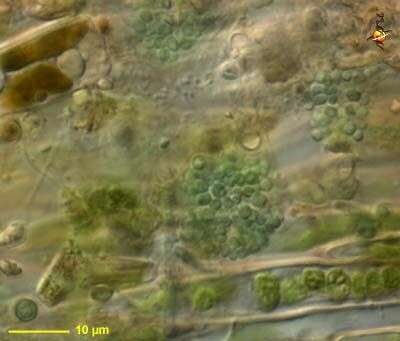
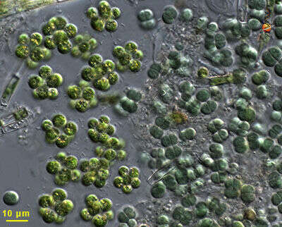
The chroococcus cells are to the right, and the cells of a similar size but a brighter green colour to the left are cells of an unidentified chlorophycean green alga (a eukaryote). This image illustrates the blue-green colour from which the blue-green algae get their name. Differential interference contrast optics.
-
Typical arrangement with paired cells inside a mucus sheath. Differential interference contrast optics.