We report an invasive mucormycosis caused byActinomucor elegansin a patient with refractory aplastic anemia. The organism was isolated from a necrotic skin lesion on the patient's left arm and demonstrated angioinvasive features on histopathology examination. In contrast to three cases described previously, we describe the first case ofA. elegansinvasive fungal infection in an immunocompromised patient. This report, along with the three previously reported cases, is convincing evidence thatA. elegansis an emerging fungal pathogen capable of causing invasive mucormycosis in humans. (Journal of Clincal Microbiology 2015)
Actinomucor elegans has been found on a wide array of substrates found around the world varying from air and soil to dung and also on soya fermenting products and even found in hop and wine fields (Marachisio and Mosca 1982)(Phalip et al 2006).It has been cultured from decomposing conifer litter in Idaho (Brandsberg 1969). It has even been isolated in harsh growing conditions as in some desert‘crusts’ (Grishkan et al 2006).It has even been isolated from a sandbox in Italy, though the authors attribute this to pigeon fecal origins (Marachisio and Mosca 1982).
Actinomucor elegans is best known for fermentation product of sufu (Chinese cheese soya product), but has recently been shown in 4 cases for pathogenicity in human, some resulting in mortality.
Overall Biology and Relevance for Humans
Sufu production; Sufu translates to ‘moulded milk’, and is found throughout the far east.A long tradition in Chinese food where the first documentation of written evidence from the Wei Dynasty (220 AD) but suspected much earlier implementation before it was documented (Han et al 2001).Sufu is made by solid state fungal fermentation in either a salty brine solution or in alcohol (Han et al 2001).Actinomucor, Mucor, and Rhizopus are all used in manufacture of Sufu with over 300 metric tons are made annually in China alone(Han et al 2001).
Actinomucor elegans has also been shown to have mild growth deterrents of other organisms when growing on Soya products (Wang et al 1972).
Actinomucor elegans has shown to be pathogenic in humans and mice, especially immune-compromised patients. While there are not many documented cases, rapid progression and difficulty in diagnosis often results in a higher mortality rates than from other fungi.There is also significant genetic diversity between suspected pathogenic pathovars and the holotype (Khan et al 2008).It has been noted that there is some variance in observed temperature conditions and previously reported optimal conditions (Khan et al 2008) (Tully et al 2009) and that this is not a good way to distinguish species (Tully et al 2009).It has been suggested for further identification based on Carbon usage analysis (Khan et al 2008). Documented cases from around the world show that this trait is as wide spread as the Actinomucor elegans habitat around the world.See Table 1 (Mahmud et. al 2012). .While a significant number of the cases are from immune-compromised patients (Khan et al 2008) (Tully et al 2009) the case of the child in Argentina suggests that it may have stronger pathogenicity than as an opportunistic pathogen though further cases are needed due to lack of information.The documented case of the Argentinean girl, age 11, differed from the other 3 documented cases in length of time and lower virulence (Daval et al 2001), confirmed by observation alone.However (Khan et al 2008) did isolate the strain, and through Koch’s postulate showed mortality in mice infected with the Actinomucor elegans var. Kuwaitensis.Further information should be conducted on antibiotic and fungal treatments as (Tully et al 2009) showed a higher mortality rate under some drugs (Khan et al 2008).
Actinomucor elegans has been suggested for usage in degradation of petroleum products relating to oil spills and other remediation/clean up events from petroleum spills. (Thirumalachar & Narasimhan 1983).Due to its widespread habitat, and survivability in harsh conditions, this may make Actinomucor elegans a strong candidate for remediation around the world.
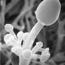
Actinomucor elegans is a facultative anaerobic fungi (Kurakov et al 2008) with white cotton-cobwebby mycelia growth on SMA and PDA agar that will age slightly brown but never black. A very detailed description can be found in Benjamin & Hesseltine (1957), who analyzed several strains in determination of a neotype.This hyphal growth spreads rapidly and will cover the petri dish even reaching up to the lid (Benjamin &Hesseltine 1957).The fungus will give off a slightly acrid and yeasty odor. The rhizoids that developed are well defined and branched, having sporangiophores on the opposite rhizoids.Whorls of short branches with sporangia with some that have broken open naturally (Benjamin &Hesseltine 1957).Whorled branches which will occasional rejoin together again.Chlamydospores offer no diagnostic value due to high variance of strains.Actinomucor elegans also has spherical sporangia.Secondary sporangia contain spiny walls with many spores.Collumellae globose to dopisvent flattened 12-30 u in diameter. Terminal sporangia deliquescent or persistent, smooth or spiny, walls mostly than 80 u but can be up to 120 u in diameter (Benjamin &Hesseltine 1957). As the holotype was most likely lost, this description comes from authors that examined more than 50 strains from around the world.
The most distinguishing feature of Actinomucor elegans is that it can be identified as it differs from Mucor species by having rhizoids and branching sporangiophores.It differs from Rhizopus and Lichtheimia by having verticallated branching sporangiophores of various lengths (Mahmud et. al 2012).
Actinomucor elegans (Eidman) is a fungal organism belonging to the Actinomucor group within Zygomycetes.While first described in 1884 in Germany, Actinomucor elegans has had several older synonyms: Mucor corymbosus, Rhizopus elegans, mucor harzii, actionmucor repens, glomerula repens, mucor glomerula, mucor botryoides, mucor repens, mucor cunninghamelloides, actinomucor corymboscus (Benjamin &Hesseltine 1957). There has been some recent debate regarding Actinomucor taiwanensis and Mucor meitauzae and their inclusion into Actinomucor elegans (Zheng & Liu 2005).
Actinomucor elegans has been isolated around the world (Benjamin &Hesseltine 1957).Two separate descriptions of the species arose in the late 1800s, Eidman in 1884, and Harz in 1871.However Benjamin &Hesseltine in 1957 makes a strong argument for acceptance of Eidman as the principle discoverer as Harz’s description is used as the principle in another species.They also designates the neotype from Bainier’s strain Glomuerula Repens NRRL 1706.This strain was attained from Harvard University in1940 who received it directly from Bainier (No. H-37).
While Actinomucor elegans does not have a common name, it does have a common usage in Sufu production in the far east.Though there is debate if Actinomucor elegans is the only member of the Actinomucor group, recent phylogenetic analysis shows the Mucor and Rhizopus groups are its closest relatives (Han et. Al. 2004).
Actinomucor elegans was originally described by Schostakowitsch in Siberia in 1898 and reevaluated by Benjamin and Hesseltine in 1957.[1] Commonly found in soil[2] this fungus and commonly used for the commercial production of tofu and other products made by soy fermentation. Its major identifying features are its spine-like projections on the sporangiophore[1] and its ribbon-like hyphal structure when found in the tissue of a host.[2]
The Actinomucor genus has many shared similarities with the genus Mucor. The specific differences lie in the branched hyphae of Actinomucor that give rise to rhizoids and sporangiophores. In terms of its differences from other similar genera, the limited growth of hyphae and the variation in the structure of columella and sporangiophores give Actinomucor multiple differentiable characteristics to other general.[1]
Mycelial growths of A. elegans have a high number of rhizoids branching out of each individual growth. On portions of growth that lack opposite rhizoids, aseptate hyphal growths with clear sporangiophores that are found with extreme variability in length and width. These hyphal structures grow out in whorled structures with growth terminating in the development of sporangiophores.[1][2][3][4][5] The sporangia are oval to spherical in shape and 17-50 μm in diameter. The walls of the sporangia possess prominent spine-like projections, which is a major identifier of this specific fungus.[1] The coloration of colonies of this fungi is white to cream-colored with an abundance of aerial mycelium. Cultures allowed to develop for a longer period of time (greater than 48 hours) change to become yellowish to buff color with increased aerial mycelium development and tight interweaving of these mycelia.[1] When this fungus is found in a human host the structure is explained to be similar to the genus Mucor, but with unique ribbon-like hyphal structures and irregular branching and thickness.[2][4][5]
Identified as an arising human fungal pathogen the recorded instances of mucormycosis due to A. elegans are limited to four cases. The invasion mechanisms found for A. elegans are through spore inhalation[3] or entry from ruptures in the skin.[5] This pathogen is highly-deadly when found in an immunocompromised individual,[4] and can develop into a serious infection for immunocompetent individuals as well.[3] Immunocompromised patients are affected worse by infection due to their immune system being unable to stop the germination of fungal spores resulting in there being no mechanism to slow the colonization once this pathogen is introduced.[4] In all cases involving immunocompromised individuals, the relatively large visible location of necrosis seemed to be the first indicator of an invasion.[1][2][3][5] It is thought that these necrotic areas are indicative of the place on the body in which inoculation occurred.[2] A. elegans as a pathogen is categorized as a mucormycosis-causing fungus, and because of this, the current leading treatment for this type of pathogen is the removal of necrotic tissue in an effort to remove the fungal elements from the body. The severity of infection from A. elegans is due to its propensity for invasion of the vascular system and hematogenous dispersion ultimately leading to necrosis of tissue. To limit the suffering, discomfort, or expiration of a patient infected with this pathogen an early suspicion of this specific fungi needs to be established. Early identification is important as it limits the time for the fungi to colonize the host before doctors can gather infected tissue to isolate and culture the fungi to confirm its presence in the patient. Because of this pathogen’s relative rarity, the time required to correctly identify the pathogen is usually not rapid enough resulting in high mortality rates of individuals infected.[5]
Mold fermentation in the production of tofu utilizes A. elegans. Through fermentation, A. elegans breaks down large macromolecules and converts them into simple fatty acids, amino acids, or sugars resulting in increased digestibility for humans. Ultimately increasing the functional and nutritional properties of tofu.[6]
Another use of A. elegans is for the fermentation processing of sufu. A. elegans is specifically proficient for the production because it possesses important enzymes for the fermentation process and results in nutritional improvements of the food. Specific enzymes that add marketable aspects to this product include glutaminase which increases palatability, and α-galactosidase[7] which reduces flatulence in people consuming the product.[8]
Actinomucor elegans is utilized for its debittering ability as well. Protein hydrolysates, such as whey and casein protein mixes all utilize proteolytic enzyme treatment to achieve heightened nutritional value, but paired with these nutritional improvements commonly comes a bitter taste. The bitter taste results from the amount and structure of hydrophobic amino acids formed in peptides. When paired with alcalase, A. elegans results in increased hydrolysis of amino acids in protein hydrolysates. Specifically, this hydrolysis occurs by A. elegans acting as an exopeptidase increasing the rate of hydrolysis resulting in a decrease of bitterness.[9]
To combat the white pollution caused by worldwide plastic waste many biodegradable products are now made out of polylactic acids (PLA) or polybutylene adipate-co-terephthalate (PBAT). Lipases secreted by A. elegans were found to be the second most proficient at expediting the full breakdown of these compounds. When a coculture of the most proficient dissolver of these compounds Pseudomonas mendocina and the second-most proficient A. elegans it resulted in a substantially higher degradation rate than either fungus could achieve individually. In the observed physical structure of this relationship, it was found that P. mendocina was attached to the mycelia of A. elegans. This synergy resulted in a higher degradation rate because A. elegans possesses a large hyphal network resulting in larger colonization of the molecule, which increased the number of colonization sites for P. mendocina resulting in the superior degrading of the molecule. From a biochemical standpoint, the degradation occurred because the lipases of A. elegans and the proteases of P. mendocina catalyzed the ester bonds of the PLA/PBAT molecules. This finding shows that there is an efficient added degradation mechanism available to be employed if products formed out of PBAT/PLA become more widespread lowering the chances for waste buildup and decreasing the harmful effect of plastics in the environment by having the ability for its full degradation to be done quickly.[10]
Actinomucor elegans was originally described by Schostakowitsch in Siberia in 1898 and reevaluated by Benjamin and Hesseltine in 1957. Commonly found in soil this fungus and commonly used for the commercial production of tofu and other products made by soy fermentation. Its major identifying features are its spine-like projections on the sporangiophore and its ribbon-like hyphal structure when found in the tissue of a host.
Actinomucor elegans je grzib[6], co go nojprzōd ôpisoł Eidam, a terŏźnõ nazwã doł mu C.R. Benj. & Hesselt. 1957. Actinomucor elegans nŏleży do zorty Actinomucor i familije Mucoraceae.[21][22] Żŏdne podgatōnki niy sōm wymianowane we Catalogue of Life.[21]
Actinomucor elegans je grzib, co go nojprzōd ôpisoł Eidam, a terŏźnõ nazwã doł mu C.R. Benj. & Hesselt. 1957. Actinomucor elegans nŏleży do zorty Actinomucor i familije Mucoraceae. Żŏdne podgatōnki niy sōm wymianowane we Catalogue of Life.