-
Kahlilembus attenuatus (SMITH, 1897) FOISSNER, BERGER & KOHMANN, 1994, a hymenostome ciliate. Body shape is elongate and fusiform, the anterior end rounded and the posterior end strongly tapered. The oral apparatus is seen in these images on the organism's left extending posteriorly from the anterior end about one quarter of the body length. 3 adoral membranelles occupy the area just anterior to the cytostome and a prominent flag-like undulating membrane lines the right side of the cytostome. Somatic cilia are arranged in 8 to 10 longitudinal kineties. A spherical macronucleus is located centrally. The contractile vacuole (seen well here) is located just posterior to the midbody. Primarily bactiverous. From freshwater pond near Boise, Idaho. Oblique illumination.
-
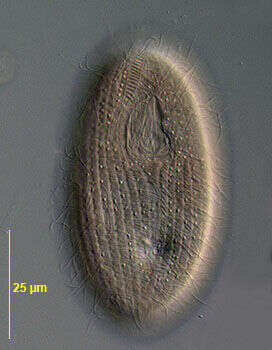
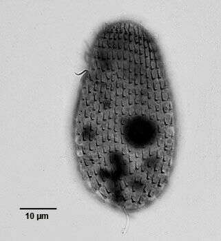
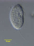
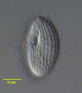
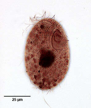
Portrait (ventral view) of the cinetochilid scuticociliate, Sphenostomella vernalis (Dragesco and Groliere, 1969) Jankowski, 1980 (synonym: Sathrophilus vernalis). The cell is elongate ovoid. The many large mucocysts (visible here between the somatic kineties)give the cell a yellowish-orange color when viewed by brightfield illumination. The roughly triangular peristome is in the anterior 1/3. There are 31-34 longitudinal somatic kineties. There are two left postoral kineties and one right postoral kinety (K1). There is a cluster of unciliated kinetids (scutica) adjacent to the posterior margin of the peristome. There is a short preoral suture and a longer broader postoral suture. There is a long caudal cilium. Groliere states that a long caudal cilium was absent in his population of S. vernalis (Groliere, C-A., J. Protozool. 20 (3): 369-376, 1973). Since all individuals collected from several different sites near Boise, Idaho had a long caudal cilium and otherwise were indistinguishable from the description by Groliere I have designated this species as S. vernalis. The cleft-like cytoproct is located in the ventral midline in the posterior suture between the excretory pore of the contractile vacuole and the posterior margin of the peristome. There are prominent transverse fibrils between the 2nd and 3rd right somatic kineties as they diverge anteriorly (seen well here). These are stained in silver nitrate preparations. There is a semicircular undulating membrane on the right margin of the peristome. There are three adoral membranelles. The M1 is an obliquely oriented group of 3 kineties. Its hook-shaped anterior end nearly reaches the anterior end of the undulating membrane. The M2 lies parallel to and is longer than M1 and also has a hook-shaped group of kinetids at its anterior end. M3 has broad âUâ shape directed anteriorly. Fibrils radiate from the posterior margin of the peristome toward the cytostome. There is a single central spherical macronucleus and an adjacent micronucleus. Collected from polysaprobic standing ditchwater at several different sites near Boise, Idaho March 2005. DIC.
-
-
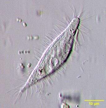
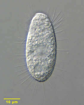
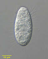
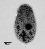
Cinetochilum (sigh-neat-owe-kai-lum) is a small bacterivorous ciliate, common and widely distributed. Differential interference contrast. Material from Nymph Creek and Nymph Lake, thermal sites within Yellowstone National Park, photograph by Kathy Sheehan and David Patterson.
-
Portrait (dorsal surface) of the cinetochilid scuticociliate ciliate, Sathrophilus muscorum (Kahl, 1931) Corliss, 1960. The ellipsoid cell is dorsoventrally flattened. The left side is slightly convex and the right side more straightened. The approximately triangular cytostome is at the junction of the first and middle 1/3. There are three obliquely oriented adoral membranelles. M1 is longest, M2 intermediate in length and M3 quite short. There is a short, slightly curved paraoral membrane bordering the right margin of the peristome. The oral apparatus is quite similar to that of Cinetochilum margaritaceum. The central spherical macronucleus is usually single but may be in as many as four parts. There are 12-17 longitudinal somatic kineties that run between prominent pellicular ridges. There is a single long caudal cilium that inserts on the dorsal surface (seen here). There is a short preoral and longer postoral suture. The slit-like cytoproct is in the postoral suture. There are inconspicuous wedge shaped peripheral extrusomes. Its ellipsoid shape and pellicular characteristics differentiate S. muscorum from the similar Cinetochilum and Platynematum both of which have more truncate notched posterior margins. Collected from sapropelic bottom sediments of a freshwater aquaculture tub near Boise, Idaho. DIC.
-
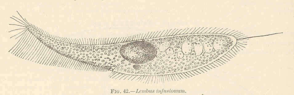
Lembus infusionum.
-
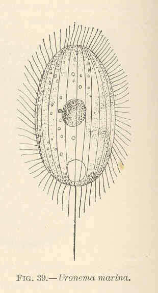
Uronema marina.
-
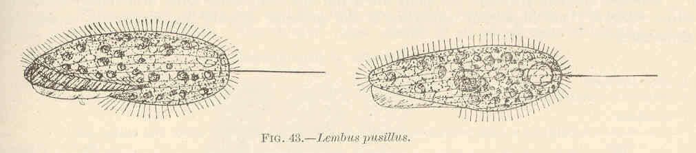
Lembus pusillus.
-
Peniscola, Valencia, Spain
-
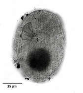
Detail view of Dexiotricha granulosa (Kent, 1881) Foissner, 1994 showing the prominent ring-shaped cytoplasmic glycogen granules. From freshwater pond near Boise, Idaho. DIC.
-
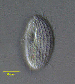
Portrait (lateral view) of the cinetochilid scuticociliate, Sphenostomella vernalis (Dragesco and Groliere, 1969) Jankowski, 1980 (synonym: Sathrophilus vernalis). The cell is elongate ovoid. The many large mucocysts give the cell a yellowish-orange color when viewed by brightfield illumination. The roughly triangular peristome is in the anterior 1/3 (not visible in this view). There are 31-34 longitudinal somatic kineties. There are two left postoral kineties and one right postoral kinety (K1). There is a cluster of unciliated kinetids (scutica) adjacent to the posterior margin of the peristome. There is a short preoral suture and a longer broader postoral suture. There is a long caudal cilium. Groliere states that a long caudal cilium was absent in his population of S. vernalis (Groliere, C-A., J. Protozool. 20 (3): 369-376, 1973). Since all individuals collected from several different sites near Boise, Idaho had a long caudal cilium (visible here)and otherwise were indistinguishable from the description by Groliere I have designated this species as S. vernalis.The genus Sphenostomella is monotypic to date. The cleft-like cytoproct is located in the ventral midline in the posterior suture between the excretory pore of the contractile vacuole and the posterior margin of the peristome. There are prominent transverse fibrils between the 2nd and 3rd right somatic kineties as they diverge anteriorly. These are stained in silver nitrate preparations. There is a semicircular undulating membrane on the right margin of the peristome. There are three adoral membranelles. The M1 is an obliquely oriented group of 3 kineties. Its hook-shaped anterior end nearly reaches the anterior end of the undulating membrane. The M2 lies parallel to and is longer than M1 and also has a hook-shaped group of kinetids at its anterior end. M3 has broad âUâ shape directed anteriorly. Fibrils radiate from the posterior margin of the peristome toward the cytostome. There is a single central spherical macronucleus and an adjacent micronucleus. Collected from polysaprobic standing ditchwater at several different sites near Boise, Idaho March 2005. DIC.
-
Dexiotricha tranquilla (Kahl,1926). DIC.
-
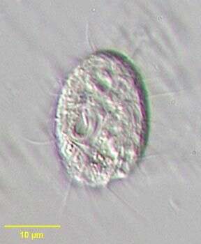
Cinetochilum margaritaceum (Ehrenberg, 1831) Perty, 1849, small rounded dorsoventrally flattened hymenostome ciliate. Cilia are located in shallow ventral furrows. Prominent oral aperture with small membranelles is seen on the organism's right posteriorly. Usually with long caudal cilia. Nucleus is central. Common. From freshwater pond near Boise, Idaho. Oblique illumination
-
Portrait (ventral surface) of the cinetochilid scuticociliate ciliate, Sathrophilus muscorum (Kahl, 1931) Corliss, 1960. The ellipsoid cell is dorsoventrally flattened. The left side is slightly convex and the right side more straightened. The approximately triangular cytostome is at the junction of the first and middle 1/3. There are three obliquely oriented adoral membranelles. M1 is longest, M2 intermediate in length and M3 quite short. There is a short, slightly curved paraoral membrane bordering the right margin of the peristome. The oral apparatus is quite similar to that of Cinetochilum margaritaceum. The central spherical macronucleus is usually single but may be in as many as four parts. There are 12-17 longitudinal somatic kineties that run between prominent pellicular ridges. There is a single long caudal cilium that inserts on the dorsal surface. There is a short preoral and longer postoral suture. The slit-like cytoproct is in the postoral suture. There are inconspicuous wedge shaped peripheral extrusomes. Its ellipsoid shape and pellicular characteristics differentiate S. muscorum from the similar Cinetochilum and Platynematum both of which have more truncate notched posterior margins. Collected from sapropelic bottom sediments of a freshwater aquaculture tub near Boise, Idaho. DIC.
-
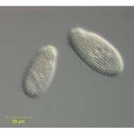
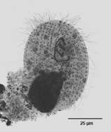
Portrait of the hymenostome ciliate, Dexiotricha granulosa (Kent, 1881) Foissner, 1994, synonymous with Loxocephulus granulosa. The cell is ovoid, broadly rounded posteriorly and truncate anteriorly. Regular longitudinal kineties terminate at a subapical band of circumferential kineties demarcating a cilia-free truncate apical area or frontal plate. There is a single long caudal cilium. The oral aperture is small and difficult to visualize in vivo. It is located in the anterior quarter with an undulating membrane on the right (seen faintly here) and 3 membranelles (not seen here). The macronucleus is spheroid and located in the mid-cell. The contractile vacuole is seen here to the left of the macronucleus. The spherical micronucleus is not seen here. The cytoplasm contains many small refractile ring-shaped glycogen granules, which are diagnostic for the species (see detail images). Dexitricha is bactiverous. From freshwater pond near Boise, Idaho. Differential interference contrast.
-
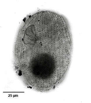
Ventral infraciliature of the cinetochilid scuticociliate, Sphenostomella vernalis (Dragesco and Groliere, 1969) Jankowski, 1980 (synonym: Sathrophilus vernalis). The cell is elongate ovoid. The many large mucocysts give the cell a yellowish-orange color when viewed by brightfield illumination. The roughly triangular peristome is in the anterior 1/3. There are 31-34 longitudinal somatic kineties. There are two left postoral kineties and one right postoral kinety (K1). There is a cluster of unciliated kinetids (scutica) adjacent to the posterior margin of the peristome. There is a short preoral suture and a longer broader postoral suture. There is a long caudal cilium. Groliere states that a long caudal cilium was absent in his population of S. vernalis (Groliere, C-A., J. Protozool. 20 (3): 369-376, 1973). Since all individuals collected from several different sites near Boise, Idaho had a long caudal cilium and otherwise were indistinguishable from the description by Groliere I have designated this species as S. vernalis. The cleft-like cytoproct is located in the ventral midline in the posterior suture between the excretory pore of the contractile vacuole and the posterior margin of the peristome. There are prominent transverse fibrils between the 2nd and 3rd right somatic kineties as they diverge anteriorly. These are stained in silver nitrate preparations. There is a semicircular undulating membrane on the right margin of the peristome. There are three adoral membranelles. The M1 is an obliquely oriented group of 3 kineties. Its hook-shaped anterior end nearly reaches the anterior end of the undulating membrane. The M2 lies parallel to and is longer than M1 and also has a hook-shaped group of kinetids at its anterior end. M3 has broad âUâ shape directed anteriorly. Fibrils radiate from the posterior margin of the peristome toward the cytostome. There is a single central spherical macronucleus and an adjacent micronucleus. Collected from polysaprobic standing ditchwater at several different sites near Boise, Idaho March 2005.Stained by the silver carbonate technic (see Foissner, W. Europ. J. Protistol., 27:313-330;1991). Brighfield.
-
Dexiotricha tranquilla (Kahl,1926). Phase contrast.
-
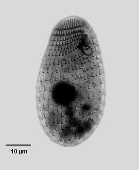
Dorsal view of the silverline system of the hymenostome ciliate, Cinetochilum margaritaceum (Ehrenberg, 1831) Pert, 1849.Stained by the dry silver nitrate technic (see Foissner, W. Europ. J. Protistol.27, 313-330; 1991). Collected from a freshwater pond near Boise, Idaho. Brightfield. Black and white.
-
Ventrolateral view of the infraciliature of the hymenostome ciliate, Dexiotricha granulosa (Kent, 1881) Foissner, 1994. Synonym of Loxocephulus granulosa. The cell is ovoid, broadly rounded posteriorly and truncate anteriorly. Regular longitudinal kineties terminate at a subapical band of circumferential kineties demarcating a cilia-free truncate apical area or frontal plate. Fibrils radiate anteriorly from the kinetids of the anteriormost paratene (seen here). There is a single long caudal cilium. The oral aperture is small and difficult to visualize in vivo. It is located in the anterior quarter with an undulating membrane on the right (seen here) and 3 membranelles (the posterior most seen here). The macronucleus is spheroid and located in the mid-cell. Single contractile vacuole. From freshwater pond near Boise, Idaho. Silver carbonate stain (see Foissner, W. Europ. J. Protistol., 27:313-330;1991).Brightfield. Black and white.
-

Ventral infraciliature of the cinetochilid scuticociliate, Sphenostomella vernalis (Dragesco and Groliere, 1969) Jankowski, 1980 (synonym: Sathrophilus vernalis). The cell is elongate ovoid. The many large mucocysts give the cell a yellowish-orange color when viewed by brightfield illumination. The roughly triangular peristome is in the anterior 1/3. There are 31-34 longitudinal somatic kineties.The anterior apex (frontal plate) is unciliated (seen here). There are two left postoral kineties and one right postoral kinety (K1). There is a cluster of unciliated kinetids (scutica) adjacent to the posterior margin of the peristome. There is a short preoral suture and a longer broader postoral suture. There is a long caudal cilium. Groliere states that a long caudal cilium was absent in his population of S. vernalis (Groliere, C-A., J. Protozool. 20 (3): 369-376, 1973). Since all individuals collected from several different sites near Boise, Idaho had a long caudal cilium and otherwise were indistinguishable from the description by Groliere I have designated this species as S. vernalis. The cleft-like cytoproct is located in the ventral midline in the posterior suture between the excretory pore of the contractile vacuole and the posterior margin of the peristome. There are prominent transverse fibrils between the 2nd and 3rd right somatic kineties as they diverge anteriorly. These are stained in silver nitrate preparations. There is a semicircular undulating membrane on the right margin of the peristome. There are three adoral membranelles. The M1 is an obliquely oriented group of 3 kineties. Its hook-shaped anterior end nearly reaches the anterior end of the undulating membrane. The M2 lies parallel to and is longer than M1 and also has a hook-shaped group of kinetids at its anterior end. M3 has broad âUâ shape directed anteriorly. Fibrils radiate from the posterior margin of the peristome toward the cytostome. There is a single central spherical macronucleus and an adjacent micronucleus. Collected from polysaprobic standing ditchwater at several different sites near Boise, Idaho March 2005.Stained by the silver carbonate technic (see Foissner, W. Europ. J. Protistol., 27:313-330;1991).Brightfield.
-
Dexiotricha tranquilla (Kahl,1926).Brightfield.
-
Ventral view of the silveline system (argyrome) of the hymenostome ciliate, Cintechilum margaritaceum (Ehrenberg, 1831) Perty, 1849. The oral apparatus is subequatorial. There is a semicircular undulating membrane on the right. The three obliquely oriented adoral membranelles (M1- M3 ) are seen here. M3 is small compared with M1 and M2. . Radial fibrils can be seen between the posterior part of the undulating membrane and the cytostome. The scutica (unciliated basal bodies associated with the stomatogenic field of kinetosomes) is seen as a short oblique line immediately posterior to the undulating membrane. Stained by the dry silver nitrate technic (see Foissner, W. Europ. J. Protistol.27, 313-330; 1991). Collected from a freshwater pond near Boise, Idaho. Brightfield. Black and white.
-
Dorsal view of the infraciliature of the hymenostome ciliate, Dexiotricha granulosa (Kent, 1881) Foissner, 1994. Synonym of Loxocephulus granulosa. The cell is ovoid, broadly rounded posteriorly and truncate anteriorly. Regular longitudinal kineties terminate at a subapical band of circumferential kineties demarcating a cilia-free truncate apical area or frontal plate. Fibrils radiate anteriorly from the kinetids of the anteriormost paratene (seen here). There is a single long caudal cilium. The oral aperture is small and difficult to visualize in vivo. It is located in the anterior quarter with an undulating membrane on the right (seen here) and 3 membranelles (the posterior most seen here). The macronucleus is spheroid and located in the mid-cell. Single contractile vacuole. From freshwater pond near Boise, Idaho. Silver carbonate stain (see Foissner, W. Europ. J. Protistol., 27:313-330;1991).Brightfield. Black and white.
-
Portrait (ventral view) of the cinetochilid scuticociliate, Sphenostomella vernalis (Dragesco and Groliere, 1969) Jankowski, 1980 (synonym: Sathrophilus vernalis). The cell is elongate ovoid. The many large mucocysts give the cell a yellowish-orange color when viewed by brightfield illumination. The roughly triangular peristome is in the anterior 1/3. There are 31-34 longitudinal somatic kineties. There are two left postoral kineties and one right postoral kinety (K1).the anterior apex (frontal plate) is unciliated.There is a cluster of unciliated kinetids (scutica) adjacent to the posterior margin of the peristome. There is a short preoral suture and a longer broader postoral suture. There is a long caudal cilium. Groliere states that a long caudal cilium was absent in his population of S. vernalis (Groliere, C-A., J. Protozool. 20 (3): 369-376, 1973). Since all individuals collected from several different sites near Boise, Idaho had a long caudal cilium and otherwise were indistinguishable from the description by Groliere I have designated this species as S. vernalis. The cleft-like cytoproct is located in the ventral midline in the posterior suture between the excretory pore of the contractile vacuole and the posterior margin of the peristome. There are prominent transverse fibrils between the 2nd and 3rd right somatic kineties as they diverge anteriorly. These are stained in silver nitrate preparations. There is a semicircular undulating membrane on the right margin of the peristome. There are three adoral membranelles. The M1 is an obliquely oriented group of 3 kineties. Its hook-shaped anterior end nearly reaches the anterior end of the undulating membrane. The M2 lies parallel to and is longer than M1 and also has a hook-shaped group of kinetids at its anterior end. M3 has broad âUâ shape directed anteriorly. Fibrils radiate from the posterior margin of the peristome toward the cytostome. There is a single central spherical macronucleus and an adjacent micronucleus. Collected from polysaprobic standing ditchwater at several different sites near Boise, Idaho March 2005.Stained by the silver carbonate technic (see Foissner, W. Europ. J. Protistol., 27:313-330;1991). Brighfield.