-
-
-
-
-
-
-
-
-
-
-
-
-
-
-
-
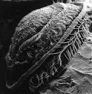
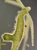
Description: English: A scanning electron micrograph of a trichodinid ciliate attached to the gills of an Australian mullet (Mugil cephalus). Date: 23 December 2007 (original upload date). Source: A.D.M. Dove. Author: A.D.M. Dove.
-

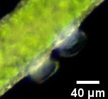
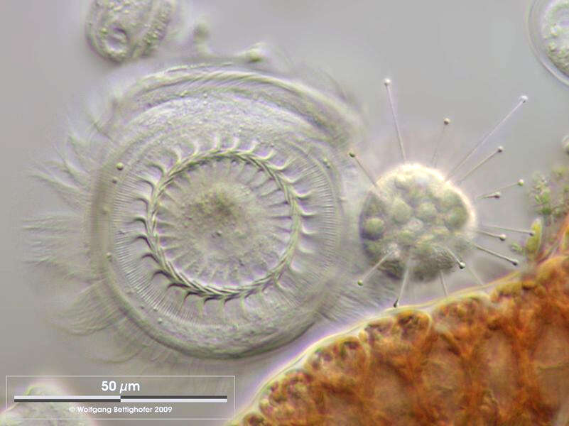
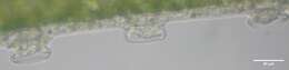
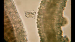
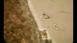
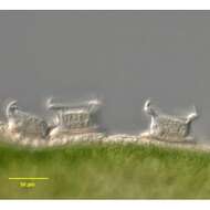
Trichodina steinii Trichodina steinii trapped by the Suctor Podophrya. There were lots of suctorial specimen in the Petri dish attached on red alga filaments. A number of them had captured prey. In every case Trichodina was the prey. Scale bar indicates 50 µm. Collected from Bodden, the brackish waters lying between the isles of Hiddensee and Ruegen (German Baltic Sea). This image was taken using Zeiss Universal with Olympus C7070 CCD camera.Image under Creative Commons License V 3.0 (CC BY-NC-SA). Place name: Hiddensee Bodden (Germany) Latitude: 54.582633 Longitude: 13.115051 Trichodina steinii, gefangen von einem Suctor der Gattung Podophrya. In der untersuchten Probe waren viele dieser Suctorien mit Beute zu sehen, sie saßen auf einem Rotalgenbüschel. Die Beute war in jedem Fall Trichodina. Der Messbalken markiert eine Länge von 50 µm. Probe aus dem Hiddenseer Bodden, der Brackwasserfläche zwischen den Inseln Hiddensee und Rügen. Mikrotechnik: Zeiss Universal, Kamera: Olympus C7070. Creative Commons License V 3.0 (CC BY-NC-SA). For permission to use of (high-resolution) images please contact postmaster@protisten.de.
-
Trichodina steinii Trichodina steinii trapped by the suctor Podophrya. Detail view. Scale bar indicates 50 µm. Collected from Bodden, the brackish waters lying between the isles of Hiddensee and Ruegen (German Baltic Sea). This image was taken using Zeiss Universal with Olympus C7070 CCD camera.Image under Creative Commons License V 3.0 (CC BY-NC-SA). Place name: Hiddensee Bodden (Germany) Latitude: 54.582633 Longitude: 13.115051 Trichodina steinii, gefangen von einem Suctor der Gattung Podophrya. Detailansicht. Der Messbalken markiert eine Länge von 50 µm. Probe aus dem Hiddenseer Bodden, der Brackwasserfläche zwischen den Inseln Hiddensee und Rügen. Mikrotechnik: Zeiss Universal, Kamera: Olympus C7070. Creative Commons License V 3.0 (CC BY-NC-SA). For permission to use of (high-resolution) images please contact postmaster@protisten.de.
-
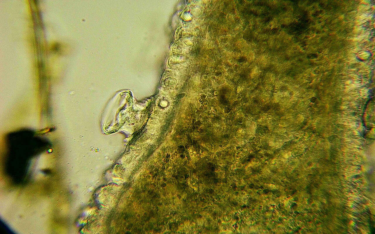
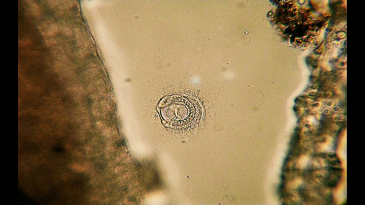
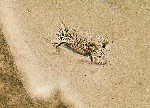
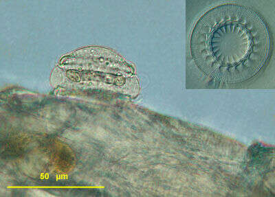
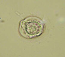
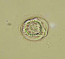
Description: Lateral view of the motile peritrich ciliate. Specimens were found on Eudiaptomus vulgaris, a common copepod. Compared with the bobbin-shaped Trichodina pediculus the lateral view of these cells is round, more dome-shaped, the size varying from 42 to 52 µm. The macronucleus is found in the transverse axis of the cell, almost circular. The large micronucleus lies at the outside of the c-shaped macronucleus. The conspicuous adhesive disk is located at the aboral side of the cell and, functioning as a holdfast organelle, attaches the ciliate to the surface of itâs host. The anatomy of the adhesive disk differs between species of the genus. The picture in the upper right angle shows an adhesive disk separated from itâs cell, counting 20 denticles.
-
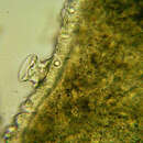
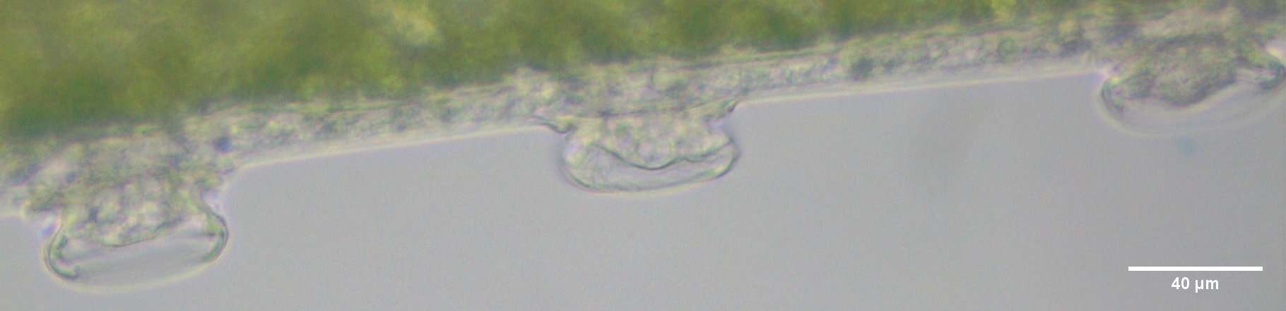
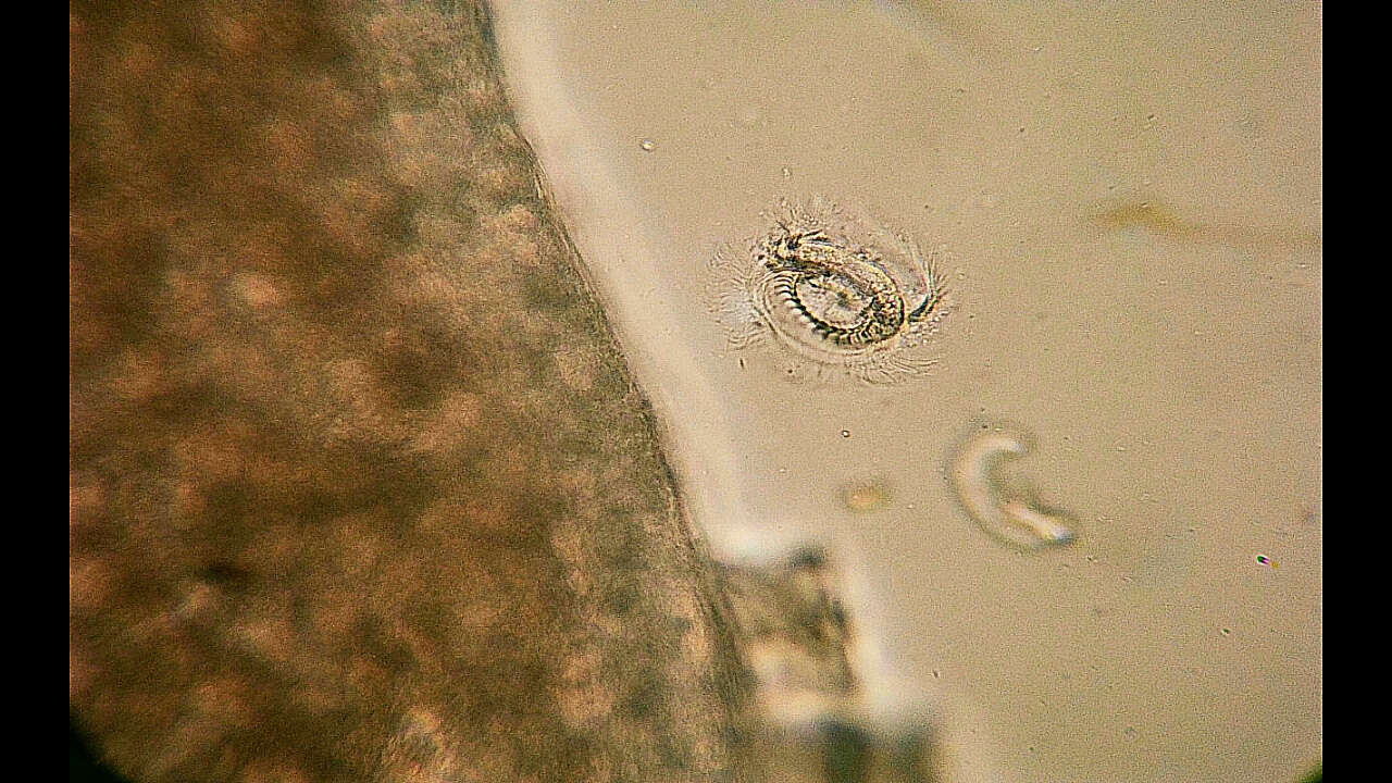
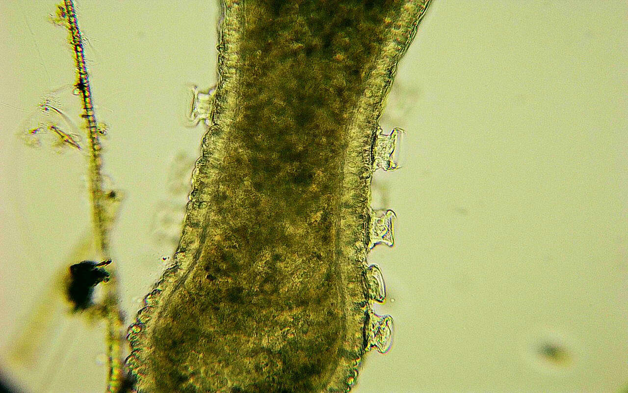
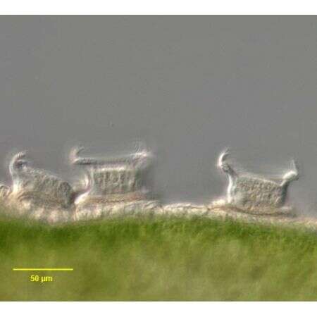
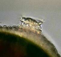
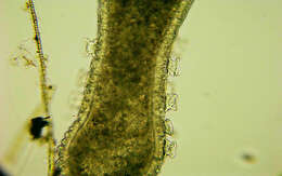
Lateral view of the mobiline peritrich ciliate, Trichodina pediculus (Ehrenberg,1851). These individuals are ectocommensals on Chlorohydra viridissimus. The bobbin-shaped cell body attaches to the epidermis of the host by an aboral adhesive disc or holdfast organelle that has a complex skeletal ring of denticles. When contracted the cells appear as flattened discs in lateral view. There is a ring of cilia around the aboral end. The peristomal ciliary field makes 1 1/4 turns around the anterior end. The pellicle between the anterior end and aboral end is bare of cilia. The C-shaped macronucleus lies in the transverse plane. There is one contractile vacuole.The cells scurry allong the surface of the host by means of the aboral cilia and adhesive disc. T. pediculus can be an important harmful parasite of freshwater fish. Collected from slow-flowing freshwater stream near Boise, Idaho February 2005. DIC.
-
Lateral view of the mobiline peritrich ciliate, Trichodina pediculus (Ehrenberg,1851). These individuals are ectocommensals on Chlorohydra viridissimus. The bobbin-shaped cell body attaches to the epidermis of the host by an aboral adhesive disc or holdfast organelle that has a complex skeletal ring of denticles .When contracted the cells appear as flattened discs in lateral view (e.g. individual to viewer's right). There is a ring of cilia around the aboral end. The peristomal ciliary field makes 1 1/4 turns around the anterior end. The pellicle between the anterior end and aboral end is bare of cilia. The C-shaped macronucleus lies in the transverse plane. There is one contractile vacuole.The cells scurry allong the surface of the host by means of the aboral cilia and adhesive disc. T. pediculus can be an important harmful parasite of freshwater fish. Collected from slow-flowing freshwater stream near Boise, Idaho February 2005. DIC.
-
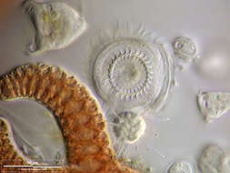
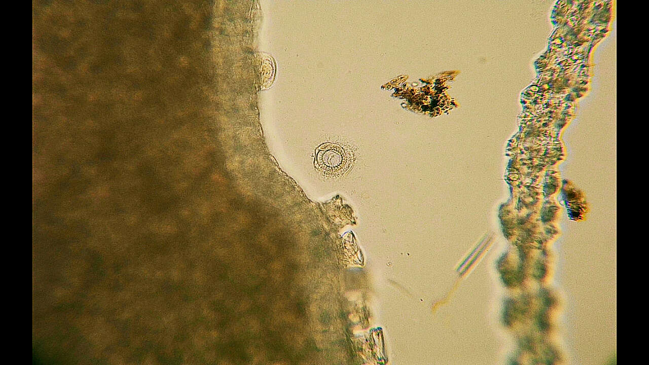
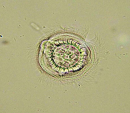
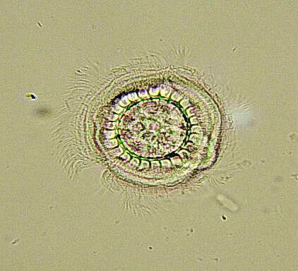
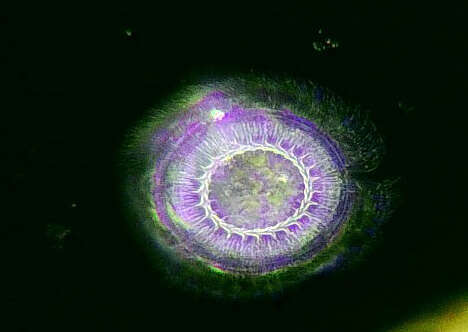
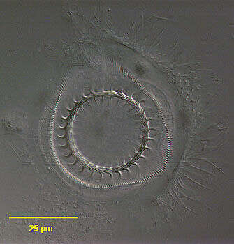
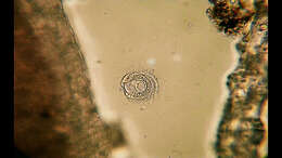
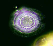
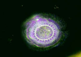
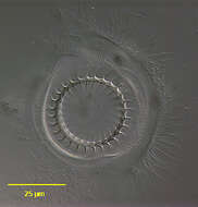
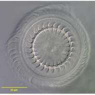
Aboral view of the mobiline peritrich ciliate, Trichodina pediculus (Ehrenberg,1851). T. pediculus is ectocommensal on the coelenterate, Chlorohydra viridissimus.This individual has been dislodged from its host. The bobbin-shaped cell body attaches to the epidermis of the host by an aboral adhesive disc or holdfast organelle that has a complex skeletal ring of denticles (seen here).When contracted the cells appear as flattened discs in lateral view. There is a ring of cilia around the aboral end. The peristomal ciliary field makes 1 1/4 turns around the anterior end. The pellicle between the anterior end and aboral end is bare of cilia. The C-shaped macronucleus lies in the transverse plane. There is one contractile vacuole.The cells scurry allong the surface of the host by means of the aboral cilia and adhesive disc. T. pediculus can be an important harmful parasite of freshwater fish. Collected from slow-flowing freshwater stream near Boise, Idaho February 2005. DIC.
-
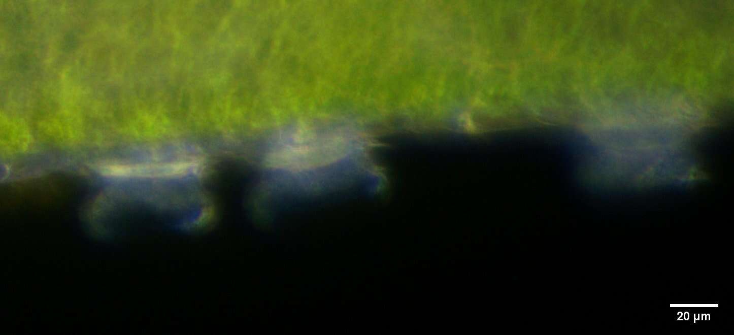
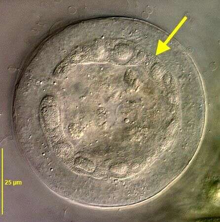
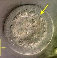
Optical section from anterior view showing C-shaped macronucleus (yellow arrow) of Trichodina pediculus (EHRENBERG,1851). DIC.
-
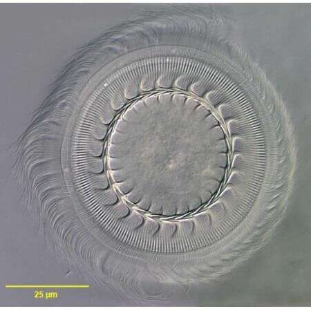
Aboral view of the mobiline peritrich ciliate, Trichodina pediculus (EHRENBERG,1851). T. pediculus is ectocommensal on the coelenterate, Chlorohydra viridissimus.This individual has been dislodged from its host. The bobbin-shaped cell body attaches to the epidermis of the host by an aboral adhesive disc or holdfast organelle that has a complex skeletal ring of denticles (seen here).When contracted the cells appear as flattened discs in lateral view. There is a ring of cilia around the aboral end. The peristomal ciliary field makes 1 1/4 turns around the anterior end. The pellicle between the anterior end and aboral end is bare of cilia. The C-shaped macronucleus lies in the transverse plane. There is one contractile vacuole.The cells scurry allong the surface of the host by means of the aboral cilia and adhesive disc. T. pediculus can be an important harmful parasite of freshwater fish. Collected on Hydra from bottom sediments of a freshwater pond near Boise, Idaho February 2005. DIC.