-
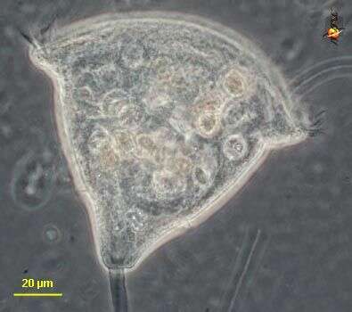
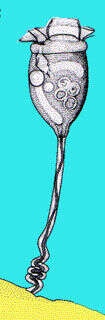
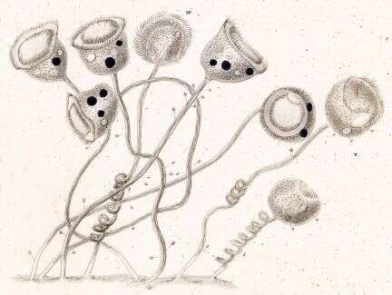
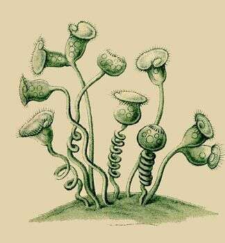
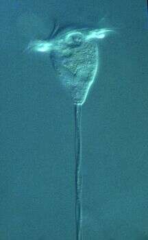
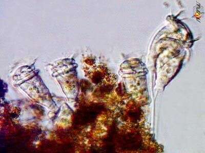
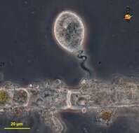
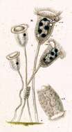
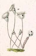
Vorticella (vort-ee-sell-a) the iconic peritrich ciliate. The feeding cells in sessile peritrich ciliates have lost all of the somatic cilia and only have the feeding cilia. The feeding cilia form a wreath which extends around the front of the cell and descends into a narrowing buccal cavity. This cavity ends at the cytostome where food is packaged into food vacuoles. If the cells become unhappy, they produce a temporary wreath of basal cilia (trochal cilia), break away from their stalk and use these to swim. The stalk contains a contractile filament, and when this contracts it coils up a bit like a spring. Cells with expanded form are usually referred to the species V. campanula. Phase contrast.
-
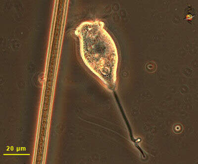
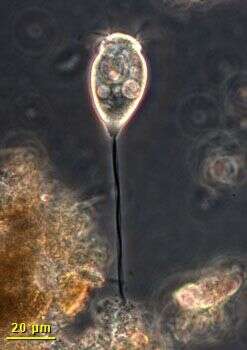
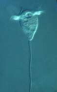
Vorticella (vort-ee-sell-ah) is a peritrich ciliate with a contractile stalk which attaches the cell to the substrate. This is a contracted cell and the feeding cilia have been withdrawn into the cell, Phase contrast. Material from Nymph Creek and Nymph Lake, thermal sites in Yellowstone National Park, photograph by Kathy Sheehan and David Patterson.
-
-
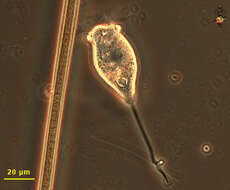
Vorticella, common peritrich ciliate. The stalk contains a contractile element called the spasmoneme - seen as a dark line in the picture. Oral cilia form a wreath around the broad flattened end of the cell. Phase contrast illumination.
-
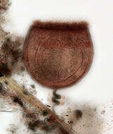
Image of the silverline system (argyrome) of Vorticella (Linnaeus, 1767). The fine parallel transverse striations have no interconnections. In the similar genera, Pseudohaplocaulus (Warren, 1988) and Pseudovorticella (Foissner and Schiffmann, 1975) the transverse argentophilic striations are connected by irregularly spaced vertical lines giving the silverline system a mesh-like appearance. In this instance the silverline system was tained by the silver carbonate technic technic (see Foissner, W. Europ. J. Protistol.27, 313-330; 1991). Usually silver carbonate and protargol methods do not stain the silverline sytem. The silverline sytem is most often stained using sliver nitrate methods. Collected from a freshwater pond near Boise, Idaho. Brightfield.
-
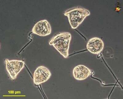
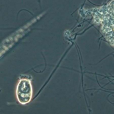
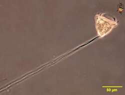
Single attached cell. The stalk can contract and this cell is extending itself. The cell feeds using the cilia at the broad end of the cone-shaped body. Phase contrast microscopy.
-
Vorticella: A common sessile protozoa with long stalk. This image was taken by Krishnakumar B. from a anaerobic bioreactor for organic rich wastewater treatment in the Regional Research Laboratory-Trivandrum (CSIR-India).
-
-
-
-
-
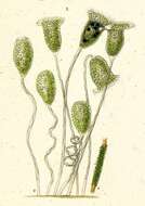
Vorticella convallaria.
-
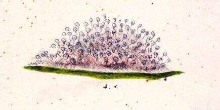
The heros of the process are the peritrich ciliates, like this Vorticella. Primarily through their efforts, the coliform and other suspended bacteria are filtered out of the sewage in the activated sludge tank. If the rate of throughout is increased to a rate where the growth of peritrichs is insufficient to keep up, then the ciliates are removed and the quality of the effluent plummets.
-
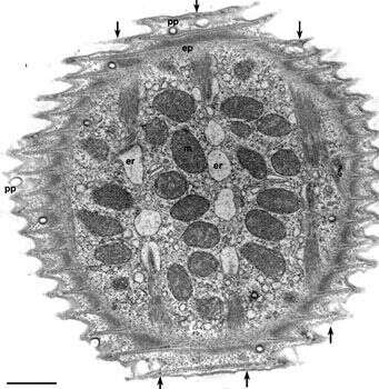
Electron micrograph of the surface of Vorticella convallaria. Like other peritrichs the pellicle is sculpted into ridges and grooves that circle the cell perpendicular to the aboral/adoral axis of the cell body. Pellicular pores (pp) penetrate the pellicle as cylindrical indentations of the plasma membrane (see Fig.3) that end as clathrin-coated pits. A system of alveoli underlies the plasma membrane and rod-like densities occupy the cytosolic tips of the pellicular ridges. A fibrous epiplasm (ep) covers the inside of the alveoli. Distinct myonemal bands run in the aboral to adoral direction (indicated by arrows) just inside the epiplasm. These bands are associated with the endoplasmic reticulum (er) by unique specializations of the ER (see Fig 17). In this EM preparation the ER has artifactually segmented into vesicles caused by the fixation process. Mitochondria (m) occupy the space between the bands. EM taken on 4/15/71 by R. Allen with Hitachi HU11A TEM. Neg. 9,250X. Bar = 1 micron.
This image is available in Richard Allen's collection.
-
-
-
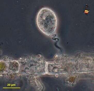
Phase contrast micrograph of a cell associated with detritus that attached to a submerged slide in the Lake.
-
-
-
-
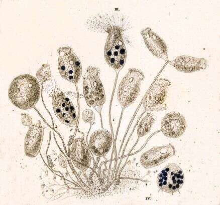
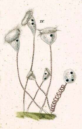
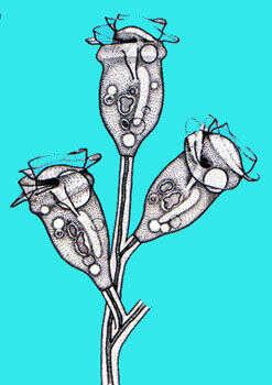
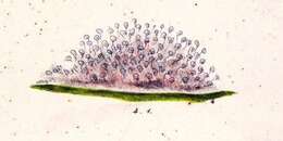
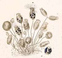
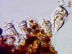
Zoothamnium (zoo-tham-knee-um) is a colonial peritrich ciliate. The feeding cells in sessile peritrich ciliates have lost all of the somatic cilia and only have the feeding cilia. The feeding cilia form a wreath which extends around the front of the cell and descends into a narrowing buccal cavity. This cavity ends at the cytostome where food is packaged into food vacuoles. If the cells become unhappy, they produce a temporary wreath of basal cilia (trochal cilia), break away from their stalk and use these to swim. The contractile elements of all associated cells of Zoothamnium colonies are interconnected so that if one cell contracts, all will tend to contract together. Differential interference contrast.
-
-
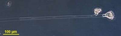
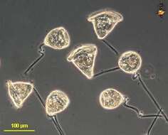
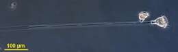
Small colony, two cells, attached to long stalk. The contractile spasmoneme is common to both cells.