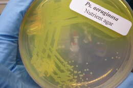
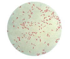
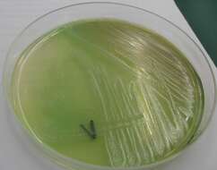
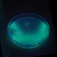
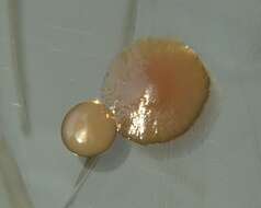
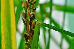
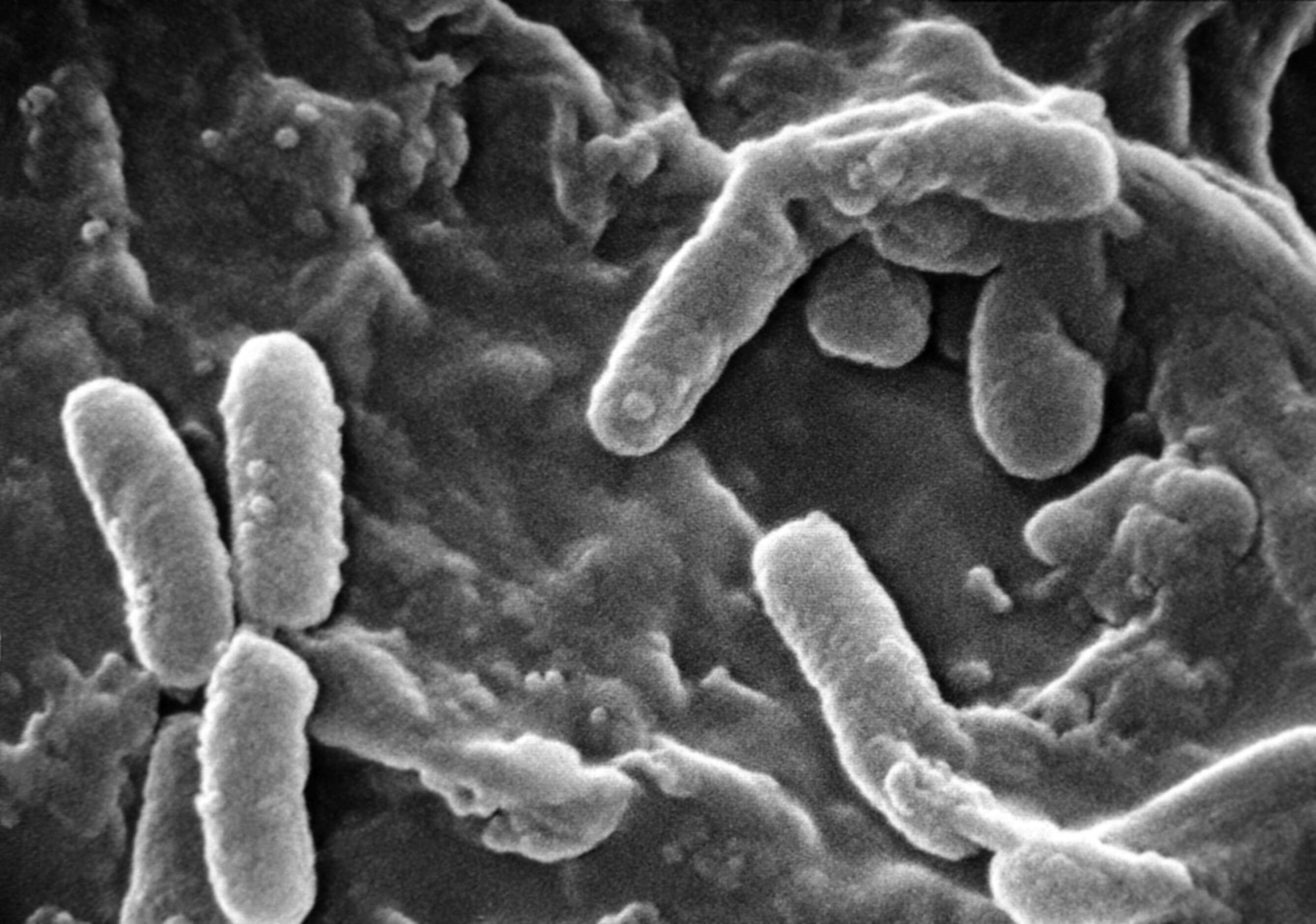
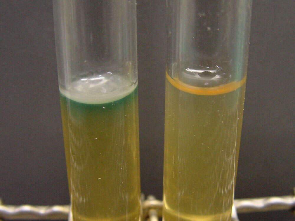
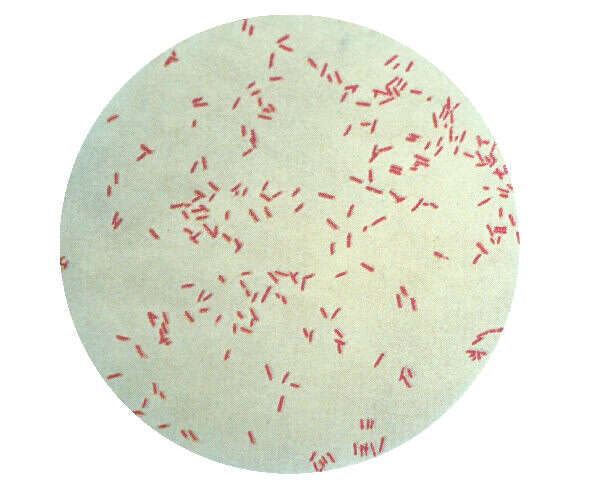
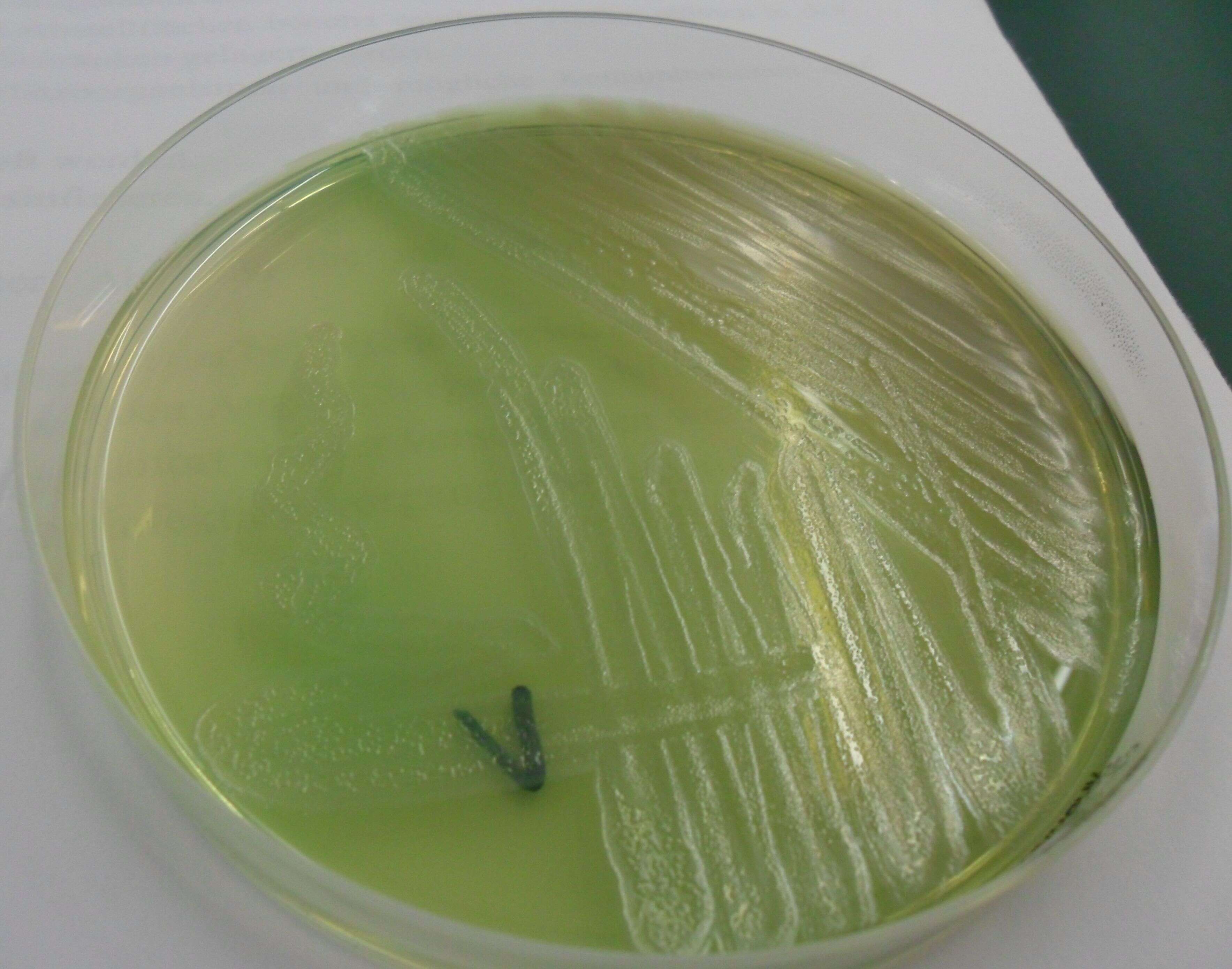
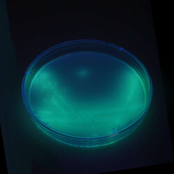
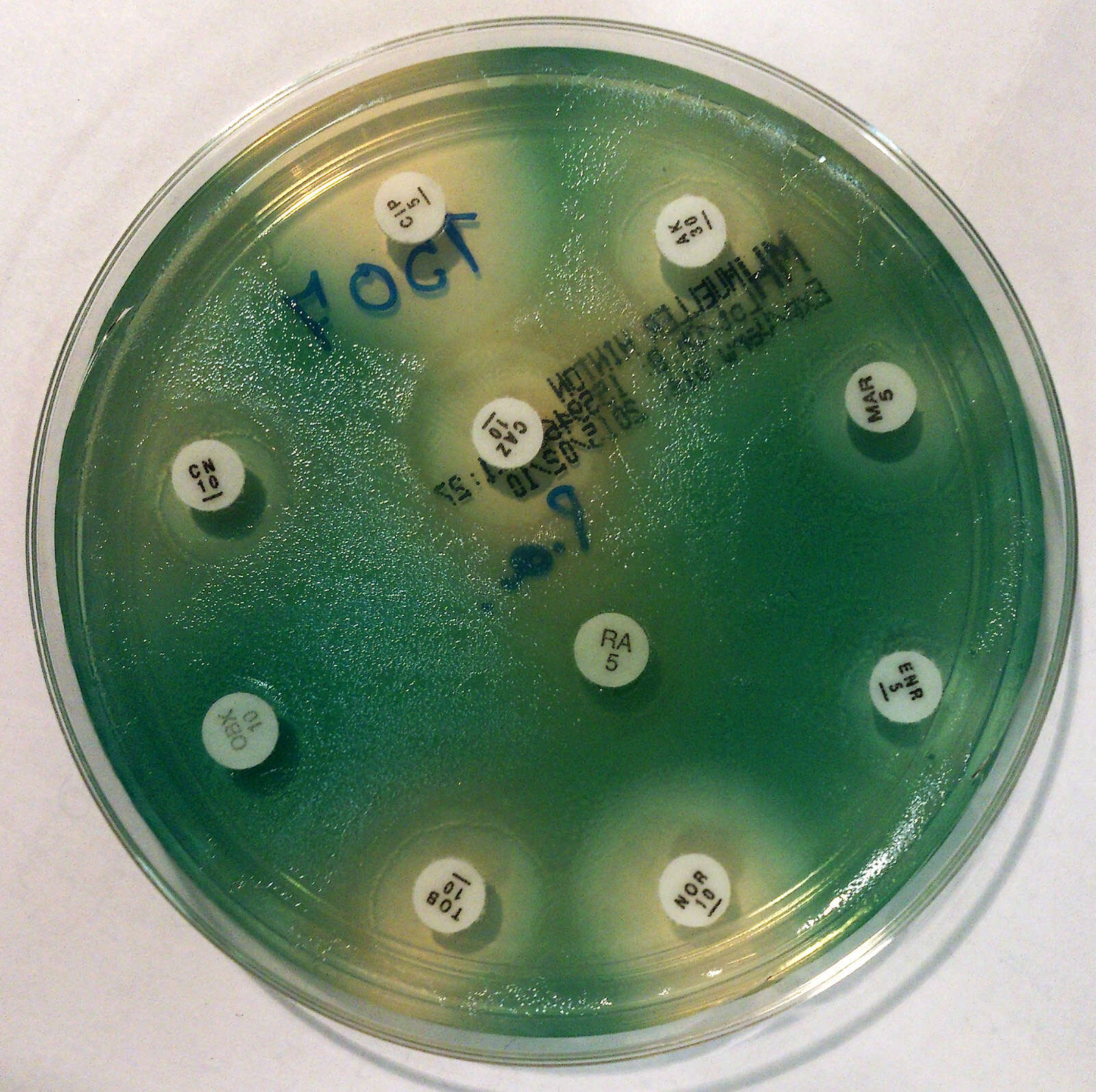
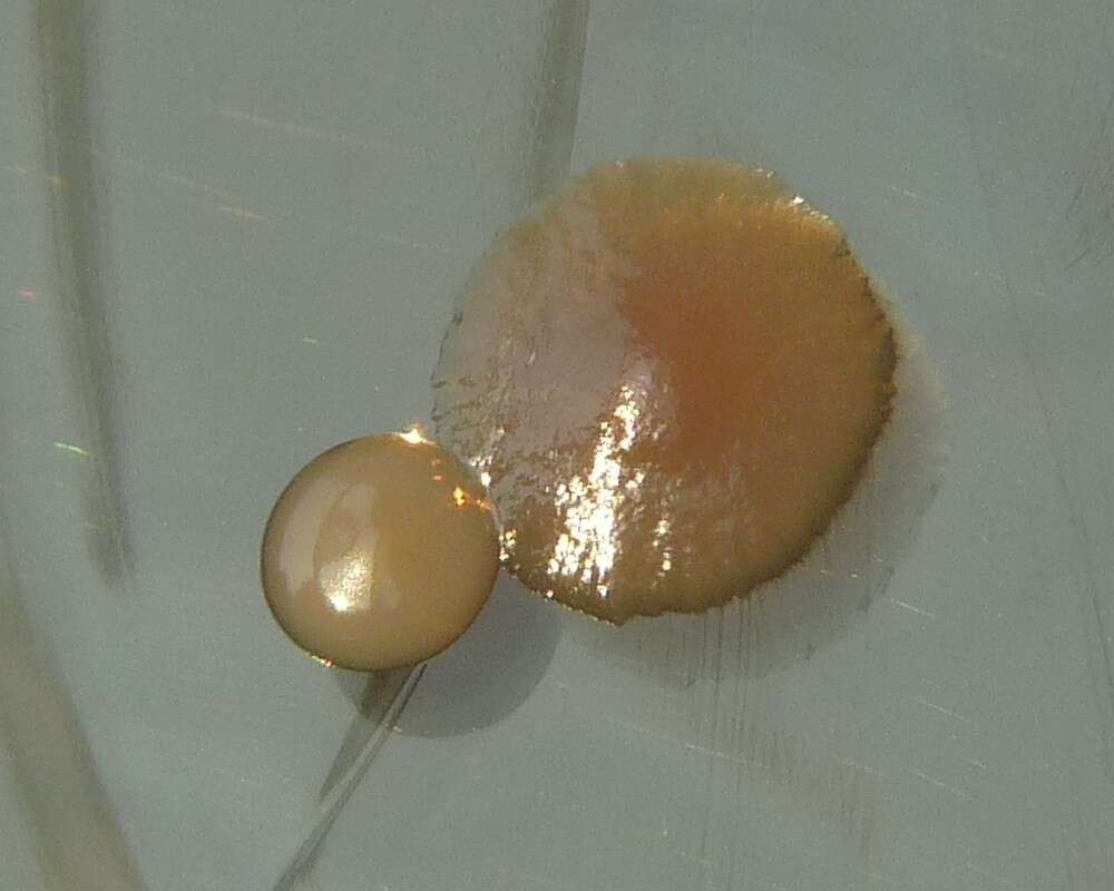
Description: English: Scanning Electron Micrograph of Pseudomonas aeruginosa Français : Pseudomonas aeruginosa colonise les poumons des personnes atteintes de mucoviscidose sous la forme de biofilm, qui diminue la réponse immunitaire des patients et confère à la bactérie une grande résistance aux antibiotiques. Pseudomonas aeruginosa vue au microscope électronique à balayage, est une bactérie pathogène et fréquemment rencontrée dans les infections nosocomiales. Polski: Pałeczka ropy błękitnej w elektronowym mikroskopie skaningowym. 日本語: 緑膿菌(ムコイド型)の走査型電子顕微鏡写真菌体の表面に分泌されたムコイドが付着し、さらに一部の菌(右部)はそれに埋もれて菌全体の形が判別しにくくなっている。. Español: Micrografía electrónica de barrido de la bacteria Pseudomonas aeruginosa, asociada con frecuencia a las infecciones pulmonares graves que complican la FQ. Català: Micrografia electrònica d'escombrat de bacteri Pseudomonas aeruginosa, associada amb freqüència a les infeccions pulmonars greus que compliquen la FQ. Source: : This media comes from the
Centers for Disease Control and Prevention's
Public Health Image Library (PHIL), with identification number
#232. Note: Not all PHIL images are public domain; be sure to check copyright status and credit authors and content providers.
العربية |
Deutsch |
English |
македонски |
slovenščina |
+/−. Author: Photo Credit: Janice Haney Carr Content Providers(s): CDC/ Janice Haney Carr. Permission(
Reusing this file): PD-USGov-HHS-CDC English: None - This image is in the public domain and thus free of any copyright restrictions. As a matter of courtesy we request that the content provider be credited and notified in any public or private usage of this image. Other versions:
Colorized version.


