NMNH Plagiodinium belizeanum type specimen
Descrission:
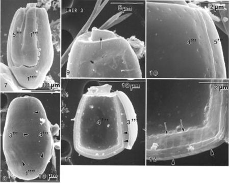
Figs 7-12. Plagiodinium belizeanum sp. nov. FIG. 7. Cell is in ventral view. Two narrow plates 1'" and 5'" are present slightly displaced. The left ventral plate (‘’’) has an anterior indentation limited to the left by a longitudinal ridge extending posteriorly, decreasing in height, and ending at the middle of the plate. The sulcus is very small. The antapical (1’’’’) plate is convexed posteriorly with a pointed tip in the center of the ventral margin. FIG. 8. Cell is in dorsal view. Hypotheca is oblong and bilaterally flattened. Plates 3'" and 4'" and antapical 1" are separated by wide intercalary bands (arrowheads). FIG. 9. Dorsal view of epitheca. The shape of the epitheca is oblong, undifferentiated (arrow), and reduced in size relative to the hypotheca. Cingulum is broad. FIG. 10. Thecal surface is smooth with scattered pores. Intercalary band delineates plates 4'" and 5'" in the form of a distinct suture. FIG. 11. Inside view of the theca. The inner cell surface is smooth; thecal plates are relatively thick with a distinct intercalary band. Dorsal plate 3'" is convex and attached to plate 4'" (arrowheads). FIG. 12. Inside view of the intercalary band. At this magnification the cell's intercalary band is broadly striated inside (arrows) and outside (arrowheads).
Ancludù an coste pàgine-sì:
- Life
- Cellular
- Eukaryota
- SAR (Stramenopiles, Alveolates, Rhizaria)
- Alveolata
- Dinophyceae
- Prorocentrales
- Prorocentraceae
- Plagiodinium
- Plagiodinium belizeanum
- Dinoflagellata
Costa plancia a compariss an gnun-e colession.
Anformassion an sla sorgiss
- licensa
- cc-by-nc-sa-3.0
- drit d'autor
- National Museum of Natural History, Smithsonian Institution
- original
- archivi ëd mojen original
- sit compagn
- NMNH Marine Dinoflagellates
- ID